Multispectral imaging for Bioscience with VideometerLab 3
•
1 like•635 views

Multispectral imaging provides valuable information on the quality and safety of a vast array of materials from pharmaceuticals to raw meat and burned biscuits. The same techniques can be used to measure skin sensitivity to sticking plaster, detect counterfeit drugs and packaging and gain insight into historical artefacts such as medieval manuscripts and weapons. The VideometerLab 3 takes 19 images at 19 specific wavelengths in ultraviolet, visible and infrared light. With multivariate statistical analysis we can reveal hidden patterns in our 19-dimension colour space and find information that would be otherwise 'invisible' to our human eyes.

Recommended
Recommended
More Related Content
What's hot
What's hot (11)
Similar to Multispectral imaging for Bioscience with VideometerLab 3
Similar to Multispectral imaging for Bioscience with VideometerLab 3 (20)
Recently uploaded
Recently uploaded (20)
Multispectral imaging for Bioscience with VideometerLab 3
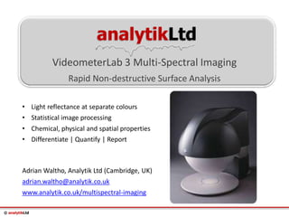
- 1. © analytikLtd analytikLtd VideometerLab 3 Multi-Spectral Imaging Rapid Non-destructive Surface Analysis Adrian Waltho, Analytik Ltd (Cambridge, UK) adrian.waltho@analytik.co.uk www.analytik.co.uk/multispectral-imaging • Light reflectance at separate colours • Statistical image processing • Chemical, physical and spatial properties • Differentiate | Quantify | Report
- 2. © analytikLtd Traditional colour imaging uses three broad bands of colour: Red, Green and Blue Normal Colour Imaging
- 3. © analytikLtd Normal Colour Imaging • RGB photographs have limited spectral resolution • Chlorophyll a and b give almost the same RGB signal and are not spectrally separated Chloro-a High Low High Chloro-b High Low High
- 4. © analytikLtd Multispectral Imaging Chloro-a High Low Low High Chloro-b Med High Med Low • Using just 4 wavelength bands with tightly defined ranges, Chlorophyll a and b can easily be distinguished • VideometerLab 3 uses 19 wavelength bands
- 5. © analytikLtd Multispectral Imaging • Many images obtained at selective wavelength bands • Each image pixel contains spectral data points • Spectral signature reveals chemo-specific information • See spatial location of surface chemical variation Ultraviolet Near-Infrared Infra-Red Red Yellow Green Blue Ultra-Violet
- 6. © analytikLtd VideometerLab 3 Schematic • Narrowband illumination by 19 LEDs at points between 375nm- 970nm (UV-Vis-VNIR), strobing sequentially with precise time control for known light output • Integrating sphere diffuses light onto sample from all angles • 5 mega-pixel monochrome CCD camera captures % reflectance at each LED wavelength for each image pixel, NIST calibration • Optional Emission filter wheel for fluorescence detection and Bright-field or Dark-field back lighting Camera LEDs Integrating sphere Emission filter wheel Sample
- 7. © analytikLtd VideometerLab 3 Schematic • Precise control at each separate point of the 375-970nm spectrum, only possible with LEDs, allows optimal illumination for best signal to noise • Provides calibrated data, suitable for statistical analysis and manipulation for discovery • Emission filters allow additional multispectral fluorescence imaging, with excitation at each LED wavelength • Powerful, user-friendly hardware setup and image analysis software is central to the system Camera LEDs Integrating sphere Emission filter wheel Sample
- 8. © analytikLtd VideometerLab 3 Schematic
- 9. © analytikLtd Simple Colony Counting Back to Index • The VideometerLab can be used as a simple visualisation aid – identify the strain of microorganism you are interested in and the VideometerLab will highlight all areas with a similar spectrum
- 10. © analytikLtd Simple Colony Counting Back to Index • By producing a binary image, the VideometerLab can count and quantify those colonies
- 11. © analytikLtd Simple Colony Counting Back to Index • VideometerLab has labeled each colony and generated size data in mm2 for each one
- 12. © analytikLtd Complex Colony Counting Back to Index • Because the VideometerLab uses spectral data to characterise colonies of interest, it is unique in its price band at being able to read plates with two or more microorganisms growing on them
- 13. © analytikLtd Complex Colony Counting Back to Index • These images show the VideometerLab picking out the ‘blue’ and ‘purple’ colonies respectively. This is a simple demonstration that is visually obvious, but the VideometerLab could be used to distinguish far finer detail (for example colonies that only differ in their UV absorption)
- 14. © analytikLtd Complex Colony Counting Back to Index • The RGB image is in the middle, and the left and right are masks created by the VideometerLab showing only those colonies it thinks are ‘blue’ on the left and ‘purple’ on the right
- 15. © analytikLtd • sRGB images of skin, little difference Skin Sensitisation Back to Index Before: No plaster After: Apply plaster ten times
- 16. © analytikLtd • nCDA mode, difference is great Skin Sensitisation Back to Index Before: No plaster After: Apply plaster ten times
- 17. © analytikLtd Skin Sensitisation Back to Index • Allergen testing
- 18. © analytikLtd Skin Sensitisation Back to Index
- 19. © analytikLtd Skin Sensitisation Back to Index
- 20. © analytikLtd Skin Sensitisation Back to Index • Increasing the contrast shows the irritation of JB Right