1. www.jkfas.org
pISSN 1738-3757 eISSN 2288-8551
J Korean Foot Ankle Soc 2015;19(1):27-31
http://dx.doi.org/10.14193/jkfas.2015.19.1.27
for the past 8 months. The pain was insidious in onset on the lat-
eral aspect of heel and was describes as dull aching type of pain.
Initially, it was aggravated only by walking especially on uneven
surface. However, this time the patient complained of pain dur-
ing rest, which had begun to progress significantly within the last
month. There was no diurnal variation in the pain, nor was there
any history of trauma or any systemic symptoms. On examination,
Benign lipomas are frequent, especially in adults and can affect
the bones, joints, tendon sheaths and other soft tissues. A lipoma
containing mature lipocytes and osseous materials is called osteo-
lipoma, which is very infrequent, and constitutes only 0.1% of all
the primary bone tumors.1)
Intraosseous lipoma of the calcaneus is
an even rarer subset of osteolipoma and is often identified inciden-
tally during radiologic examination, having no specific symptoms
besides heel pain at the worst.2)
To the best of ourknowledge, we
present one of the very rare case reports of Intraosseous lipoma
extending to the subtalar joint, and resulting in a pathological frac-
ture of the calcaneus. And the present report obtained informed
consent from the patient.
CASE REPORT
A 51-year-old female presented to our clinic with right heel pain
Received December 18, 2014 Revised January 29, 2015 Accepted February 9, 2015
Corresponding Author: Jin Soo Suh
Department of Orthopedic Surgery, Inje University Ilsan Paik Hospital, 170 Juhwa-
ro, Ilsanseo-gu, Goyang 411-706, Korea
Tel: 82-31-910-7968, Fax: 82-31-910-7967, E-mail: sjs0506@paik.ac.kr
Financial support: None.
Conflict of interest: None.
Case Report
CC This is an Open Access article distributed under the terms of the Creative Commons Attribution Non-Commercial License (http://creativecommons.org/licenses/
by-nc/3.0) which permits unrestricted non-commercial use, distribution, and reproduction in any medium, provided the original work is properly cited.
Copyright 2015 Korean Foot and Ankle Society. All rights reserved.ⓒ
Intraosseous lipoma is a benign tumor that originates from proliferating mature lipocytes. It often occurs in the metaphysis of long bones of
the lower extremity, and also in the calcaneus, humerus, mandible, sacrum, and rib bones. Frequently, it involutes spontaneously through
a process of infarction, calcification, and cyst formation. It can either present as pain, or be asymptomatic and only discovered through an
incidental radiological finding. In our case, the patient presented with heel pain. Intraoperatively, it was found that the intraosseous cavity
was filled with fat along with an adjacent but separate area of cystic degeneration. There was also a cortical perforation at the cystic lesion
which was communicating with the subtalar joint. This cortical breach is most likely the cause of diffuse lateral heel pain experienced by our
patient, and such a pathological fracture due to intraosseous lipoma has never been reported.
Key Words: Bone neoplasms, Calcaneus, Cortical perforation
Intraosseous Calcaneal Lipoma with Subtalar Perforation
through Cystic Degeneration: A Case Report
Abhishek Kumar, Stephanie Stephanie*, Jun Young Choi, Sunhee Chang†
, Jin Soo Suh
Department of Orthopedic Surgery, Inje University Ilsan Paik Hospital, Goyang, Korea, *Department of Surgery, Howard University
College of Medicine, Washington, DC, USA, †
Department of Pathology, Inje University Ilsan Paik Hospital, Goyang, Korea
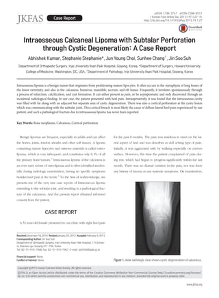
Figure 1. Axial radiologic view shows cystic degeneration of calcaneus.
2. 28 Vol. 19 No. 1, March 2015
the skin over the heel appeared to be normal. There was tender-
ness presented on the sinus tarsi area, but there was no swelling
or signs of inflammation. There was full flexion and extension
of ankle joint, which also appeared to be pain free. However,
inversion and eversion of the foot was restricted due to pain. The
hematological and biochemical studies did not show any abnor-
mality.
An axial radiographic view of the ankle showed three well-
defined cystic lesions in the body of the calcaneus. One was near
the lateral aspect of the calcaneus while the other two were just
anterior toit and divided by a septum. There was no reactive scle-
rosis around the cystic wall (Fig. 1). On the lateral view, a large
cystic lesion was presented over the calcaneal neutral triangle just
below the subtalar joint extending anterior to the calcaneo-cuboid
Figure 2. Lateral view of calcaneus shows superior and anterior exten-
sion of cystic change up to the subtalar joint and calcaneo-cuboid joint
respectively.
Figure 3. T1-weighted low to intermediate (A) and T2-weighted high mass like lesion on the sagittal (B) and coronal plane (C) are shown in the cal-
caneus.
A B C
Figure 4. Intraoperative image shows lesion centered between the two
peronei.
Figure 5. Flouroscopic image shows perforation of subtalar joint on the
joint weighted image-coronal cut.
3. www.jkfas.org
29Abhishek Kumar, et al. Intraosseous Calcaneal Lipoma with Subtalar Perforation
calcaneal wall.
Intraosseous deposition of fat almost in a liquefied state extend-
ing all the way anterior to the calcaneo-cuboid joint was found
inside the cyst. Superiorly, the cavity extended all the way up to
the subtalar joint, causing a pathological fracture and breakage of
calcaneal articular surface (Fig. 5). Once the fat was curetted, a
cystic cavity was seen exactly over the window touching the me-
dial aspect of the calcaneal wall and two more cavities were found
on the posterior aspect of the calcaneus, which was separated by
a septa.
All cysts were curetted, the walls cauterized and the margin
of the cysts grinded with a diamond burr. Following a thorough
irrigation, the cavity was packed with cancellous allograft and
the window was fixed with two 3.5 mm cancellous screws since
joint (Fig. 2).
A magnetic resonance imaging (MRI) of the right foot suggested
a well-marginated mass in the right calcaneus with high signal in-
tensity on T1-weighted image. There was two cystic lesions inside
the mass which showed a low signal intensity on the fat suppres-
sion image (Fig. 3).
The patient was scheduled for curettage of the cystic lesion and
bone grafting. A well-padded tourniquet was placed on the proxi-
mal aspect of the right thigh, and a lateral approach was used with
the patient on the left lateral decubitus position. Intraoperatively,
the authors found the center of the lesion to be exactly in between
the two peronei tendons (Fig. 4). Sural nerve did not encounter
in the field as region between two peronei approached directly. A
window was made under fluoroscopic guidance over the lateral
Figure 6. Postoperative radiograph shows cancellous graft and fixation
of window using two screws.
Figure 7. The trabecular bone is intermixed with the adipocytes and
small blood vessels. Hematopoietic elements are absent. These find-
ings are consistent with intraosseous lipoma (H&E stain, x100).
A B
Figure 8. Complete consolidation of the
lesion (arrows) is shown on the plain ra-
diograph taken at 15 months postopera-
tively (A) and after screw removal (B).
4. 30 Vol. 19 No. 1, March 2015
radiograph. Stage 3 contains extensive fat necrosis with variable
degrees of cysts formation, calcifications and formations of reactive
bone.
Computed tomography (CT) demonstrates a well-defined lytic
lesion with negative Hounsfield unit equivalent to those of fat.
MRI shows a high signal intensity on T1- and T2-weighted images
similar to that of subcutaneous fat. Thus, surgical biopsy for the
diagnosis of such tumors is not significant.8)
The differential diagnosis of osteolipoma includes solitary bone
cyst, fibrous bone infarct, enchondroma, fibroma,aneurismal bone
cyst, chondrosarcoma, osteoid osteoma, liposarcoma and eosin-
ophillic granuloma. Solitary bone cyst occurs at the base of the
calcaneal neck but is typically seen in adolescence. Aneurysmal
bone cyst is extremely rare before the age of fiveand after the
age of thirty. In the case of liposarcoma, CT scan shows a less
homogenous lesion without typical negative Hounsfield unit.1)
En-
chondroma typically shows endosteal scalloping and calcification
that ranges from punctuate to rim type lesions. Osteoid osteoma
is characterized by the typical night pain and dense sclerotic cen-
tral nidus on radiograph imaging. Bone infarct in the calcaneus
is always seen at the dorsal aspect, with calcification usually seen
in the periphery. On the contrary, there is almost always a central
calcificationin lipoma.6,7)
The most frequent treatment of osteolipoma consists of curet-
tage of the lesion through an ample bone window and filling of
the defect with autologous bone mixed with a frozen, dried al-
lograft or Polymethylmethacrylatecement.
Malignant transformation of pre-existing bone lipoma in the
femur and tibia have been reported but there has been no studies
on calcaneal lipoma.9)
Pathological fracture or cortical perforation
has been reported by several authors10)
but in the authors’case,
cortical perforation opening into the subtalar joint was one of the
intraoperative findings that leads to secondary fracture of the cal-
caneus. This pathological fracture might be the reason for the heel
pain.
In summary, calcaneal Intraosseous lipoma with cystic degen-
eration is a very infrequent benign tumor. Such lesions can be
asymptomatic or may present with heel pain. Diagnosis is usually
made accidentally on radiograph images, and CT or MRI scans are
used to confirm the fatty nature of the lipoma. This condition has
a very good prognosis and its symptoms improve with rest and the
use of analgesics. Long standing cases may complicate into patho-
logical fractures and become symptomatic. At the very least, these
cases require surgical intervention at the earliest.
it was huge and strong (Fig. 6). And the screw purchase was
good. The wound was closed and a short leg splint was applied.
The tissues taken out from the bone was sent for the pathologic
determination and the report came back consistent with that of
lipoma (Fig. 7). Postoperative pain management was done only
by intravenous analgesic agent. The patient was allowed to do
partial weight-bearing exercise with the splint for four weeks and
then full-weight bearing was allowed after that. At postoperative
15 months, patient was symptom-free state and complete bony
consolidation was confirmed with radiographs. All devices were
removed without any associated complications (Fig. 8).
DISCUSSION
Intraosseous lipoma has an incidence of 0.1%∼2.5% of all
primary bone tumors.3)
The etiology of the tumor still remains un-
known. Three theories have been put forward: 1) Traumatic origin
and later fat degeneration, 2) infection or osseous fat infarction
with metaplasia, and 3) primary tumor of fat marrow. The third
theory seems to be the most probable one.4)
Intraosseous lipoma has no gender predilection and can oc-
cur at any age, often occurring in the fourth decade.4)
It affects
long bones of lower extremity more frequently than the upper
extremity by 6 to 1 ratio. The most common occurrence is in the
calcaneus, which is only 8%, followed by the skull, jaw, and then
ribs.5)
Clinical presentation varies significantly, leaving almost two
thirds of the patient asymptomatic. The most frequent symptom in
calcaneal pain, which is often related to prolonged standing or ex-
ercise.4)
When asymptomatic, most of the diagnosis are incidental.
Radiologically, intra-osseus lipoma is characterized by cystic,
radiolucent lesion with thin, sclerotic and well-defined borders.
In long bones, the lesion is more expansile without any perios-
teal reaction or cortical breach in the metaphysis or epiphysis of
the bones. In short tubular bones, the lesion shows a geographic
pattern with sclerotic rim. In the calcaneus, it appears as a radiolu-
cent cystic image with sclerotic borders and a central calcification
(Bull’s eye image), which is almost always located in the neutral
triangle at the base of the neck of calcaneus, versus bone infarcts
when it occurs in the dorsal part of calcaneus.6,7)
Milgram,5)
Chow
and Lee3)
classified a lipoma into three separate categories: Stage 1
contains mature lipocytes without necrosis, and osteolytic lesions
seen radiologically. Stage 2 contains partial fat necrosis (due to
pressure on capillaries) with focal calcification, live lipocytes still
present, and radiolucent lesion with central calcification seen on
5. www.jkfas.org
31Abhishek Kumar, et al. Intraosseous Calcaneal Lipoma with Subtalar Perforation
study of nine cases. Am J Surg Pathol. 1992;16:401-10.
444 Revenga Martínez M, Bachiller Corral FJ, Rubio García J, Muñoz
Beltrán M, Zea Mendoza AC. Cystic lesion of the calcaneus. In-
traosseous lipoma. Reumatol Clin. 2007;3:139-42.
555 Milgram JW. Intraosseous lipomas. A clinicopathologic study of
66 cases. Clin Orthop Relat Res. 1988;(231):277-302.
666 Abrahim-Zadeh R, Klein RM, Leslie D, Norman A. Characteris-
tics of calcaneal bone infarction: an MR imaging investigation.
Skeletal Radiol. 1998;27:321-4.
777 Van Linthoudt D, Lagier R. Calcaneal cysts. A radiologi-
cal and anatomico-pathological study. Acta Orthop Scand.
1978;49:310-6.
888 Blacksin MF, Ende N, Benevenia J. Magnetic resonance imaging
of intraosseous lipomas: a radiologic-pathologic correlation.
Skeletal Radiol. 1995;24:37-41.
999 Milgram JW. Malignant transformation in bone lipomas. Skeletal
Radiol. 1990;19:347-52.
1111 Pappas AJ, Haffner KE, Mendicino SS. An intraosseous lipoma of
the calcaneus: a case report. J Foot Ankle Surg. 2014;53:638-42.
Intraosseous lipoma has been thought of as an infrequent occur-
ring benign tumor. In the past, pathological fracture secondary to
Intraosseous calcaneal lipoma has never been reported most likely
because the tumor occurs in the non-weight bearing areas of the
calcaneus such as the neutral triangle.5)
In the authors’case, we
experienced a cortical perforation through the cystic degeneration
of the tumor, which are important concerns and thus merits spe-
cial attention.
REFERENCES
111 Bertram C, Popken F, Rütt J. Intraosseous lipoma of the calca-
neus. Langenbecks Arch Surg. 2001;386:313-7.
222 Campbell RS, Grainger AJ, Mangham DC, Beggs I, Teh J, Davies
AM. Intraosseous lipoma: report of 35 new cases and a review
of the literature. Skeletal Radiol. 2003;32:209-22.
333 Chow LT, Lee KC. Intraosseous lipoma. A clinicopathologic