Acute Appendicitis, Fatty Liver, Thrombus, and Atherosclerosis Slides
•Download as PPTX, PDF•
1 like•599 views


Recommended
More Related Content
Viewers also liked
Similar to Acute Appendicitis, Fatty Liver, Thrombus, and Atherosclerosis Slides
Similar to Acute Appendicitis, Fatty Liver, Thrombus, and Atherosclerosis Slides (20)
More from Alaa Radwan
Recently uploaded
Recently uploaded (20)
Acute Appendicitis, Fatty Liver, Thrombus, and Atherosclerosis Slides
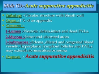
- 1. Slide (1):-Acute suppurative appendicitis Naked eye :-Circular structure with bluish wall Organ :-T.S. of an appendix Comment :- 1-Lumen :-Necrotic debris,intact and dead PNLs, 2-Mucosa :-Intact and ulcerated areas 3-Submucosa :-Edema ,dilated and congested blood vessels , hyperplastic lymphoid follicles and PNL,s may extends to musculosa or serosa Diagnosis :- Acute suppurative appendicitis
- 3. Slide (2):- Chronic inflammation Organ :- Section in the skin Comment :- 1-Epidermis :- Thickened epidermis and keratin 2-Dermis :- -Arterioles :- Thickened and narrowed (EAO) -Peri-vascular cuffing by (lymphocytes ,plasma cells and macrophages) -Increased fibroblasts and collagen. Diagnosis :- Chronic inflammation in skin
- 5. Slide(3):-Myocardial scarring Organ :- Section in heart Comment :- Intact cardiac muscles :- Running in different directions Infarction area :- Pale pink,fibrous tissue area ,composed of wavy collagen,few fibroblasts and few dilated thin walled capillaries Diagnosis:- Myocardial scarring
- 7. Cell injury (degeneration) Water accumulation :- e.g Cloudy swelling of kidney Fat accumulation :- e.g fatty change of liver
- 8. Cloudy swelling :- Reversible cell damage characterized by mild accumulation of water inside cells. Oragns affected :-Proximal convoluted tubules of kidney ,liver and heart Pathogenesis:-swollen mitochondria is fragmented with resultant decreased production of ATP and consequent distrubed Na/K pump retention of Na and H2O inside cells . Pathology Grossly :- The organ size :- Increased Colour :- Pale Consistency:- Soft
- 9. Cloudy swelling of kidney
- 10. Section in the kidney :- Glomeruli :- Normal Tubules :- -Lumen :- Narrowed and star shaped -Lining epithelial cells *Swollen *Conical (pyramidal ) shaped.Its apex directed inward. *Pink and granular cytoplasm (mitochondrial damage. *Intact rounded nuclei . Diagnosis :- Cloudy swelling of the kidney
- 12. Fatty change ,liver A condition characterized by accumulation of triglyceride inside hepatocytes Causes:- 1-Toxins->diphtheria 2-Chemicals as phosphorus and CCL4 Etiology:- 1-Increased entrance of FFAheptocytes 2-Increased synthesis of FA and decreasd its oxidation 3-Increased estrification of FA 4-Decreased formation of phospholipids 5-Decreased excretion of phospholipids.
- 13. Specimen :- Section of liver Size :- enlarged D13 Surface :-Smooth Capsule :-Thin ,stretched easily Streped Colour :- Yellowish Borders :- Rounded Consistency:- Soft
- 14. Slide(5):-Fatty changes of liver Section in liver :- Liver cells :- Cytoplasm :- Contain large clear vacuoles Nuclei :-Peripheral located nuclei(signet ring appearance. Some intact liver cells are seen. Diagnosis :- Fatty changes of liver.
- 16. Slide(6):-Recent thrombus Naked eye :-Rounded structure with intra-luminal thrombus (2nd rounded structure) Section in blood vessel :- 1- The lumen of blood vessel :- -Filled by thrombus mass attached to the vessel wall at one point (head of the thrombus ) 2-The thrombus mass containing :- pale pink lines radiating from the head (lines of Zahn) and contain in between meshwork of fibrin ,RBC,s and WBC,s. Diagnosis :- Recent thrombus
- 17. 2nd of circular lesions
- 18. Slide(7):-Atherosclerosis Naked eye :- 3rd rounded structure Section in an artery :- Intima :- Degenerated and elevated Subintima :- Fibro-fatty mass containing , needle like crystals of cholesterol. and haylinized C.T ± calcification Elastic lamina :-Fragmented Media opposite the lesion :- Atrophic . Diagnosis :- Atherosclerosis of an artery