2. 2 | P a g e
1- Structures posterior to the kidney:
a- 4 muscles: - diaphragm (upper 1/3)
- Psoas major
- Quadratus lumborum
- Transversus abdominus
b- 4 structures: - subcostalvessels
- Subcostalnerve
- Iliohypogastric nerve
- Ilioinguinalnerve
2- Embryonic origin of male urethra:
a- Constriction of the urogenital sinus gives = upper partof
prostatic urethra.
b- Definitive urogenital sinus divides into 2 parts:
- Pelvic part gives lower prostatic urethra and
membranous urethra
- Phallic part gives penile urethra
c- Urethra in the Glans arises from ectodermal invagination of the
glans.
3- 4 factors affecting GFR:
a- Diameter of arterioles:
- Afferent arteriole : its vasodilation increases the
GFR while its vasoconstriction decreases the
GFR.
- Efferent arteriole: its vasodilation decreases the
GFR while its mild vasoconstriction increases the
GFR “ Moderate to severe vasoconstriction
decreases the GFR “
b- Renal blood flow : when it increases it causes rise in the GFR.
c- Sympathetic stimulation : it causes vasoconstriction of both
afferent and efferent arterioles but it affects mainly the
afferent arteriole decreasing the GFR.
3. 3 | P a g e
d- Arterial blood pressure: it is regulated by the autoregulation
mechanisms.
4- Role of loop of Henle in the concentrating mechanism of the
kidney:
Itshares in creating the hyperosmolarity of the renal medulla by 2
mechanisms:
a- Active reabsorption of Na-K-2Clin thick ascending limb of loop
pf Henle .
b- Passivereabsorption of NaCl in thin ascending limb of loop of
Henle which depends mainly on previous reabsorption of
water in thin descending limb of loop of Henle.
5- Glucosetubular transport:
- Glucoseis completely reabsorbed from the
tubules in the proximal convoluted tubule
through secondary activetransportwith Na.
- There is what is known as tubular transport
maximum for glucose : when blood glucose level
reaches threshold value 180mg/dl, glucose
begins to appear in urine as some nephrons
become saturated .
With progressiveincreasein blood glucoselevel ,
its secretion in urine also increases gradually till
all nephrons becomesaturated reaching their
maximum absorping capacity which is 375
mg/min for men and 300 mg/min for women.
6- Proteinuria :
Itis mainly due to increase albumin in urine ( normal level 30-200
mg/ml)
Itmay be due to physiologicalor pathological:
- Physiological( not more than 500 mg/ml) may
be due to pregnancy or severemuscular exercise
or prolonged standing.
4. 4 | P a g e
- Pathological ( more than 500 mg/ml) it may be;
Prerenal as in Heart Failure
Renal as in nephritis or nephrosis
Postrenalas in cystitis
Globulins may appear in urine and known as Bence Jonse proteins.
7- Structuralelements in the wall of urinary bladder that adapt it to
varying urinevolume:
1- Lining epithelium of the inner surfaceis transitional epithelium
“urothelium” which can change its number of layers according
to changes of volume .
2- The dome shped cells of the urothelium “ umbrella cells” their
luminal border is thickened by membranous plaques which are
attached to microfilaments inside the cells “ it also can increase
surfacearea or decrease it according to changes in volume”
3- The lamina propria is rich in elastic fibers.
4- The musculosa contain large amount of connective tissue
between the muscle fibers to adapt changes in volume.
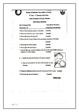
8- A- The glomerulus
B- Intraglomerular mesangialcell
Its main functions are : -supportivefunction
- Phagocytic function
- Secretory function
- Contractile function
5. 5 | P a g e
9-
Point of comparison Renal cell carcinoma Wilms’ tumor
Incidence th
It is common in the 6
decade of life
It occurs mainly in
children(2-5 years)
Origin Arises from tubular
epithelium
Arises from blastema
cells
Gross picture Mass about 3:15 cm
sometimes with
satellites
C.S: variegated
appearance
Large solitary well
circumscribed mass
Soft homogenous and
fleshy like.
Microscopic picture Rounded cells with
clear cytoplasm (
glycogen and lipid
content)
Areas of hemorrhage
and necrosis
Triphasic appearance :
Blastemal cells
Epithelial cells
Stromal cells
Clinical picture 1- Painless total
hematuria
2- Pain in the
costovertebral
region.
1- Large Palpable
mass in the
abdomen.
10 – causes of hematuria:
1- Acute nephritic syndrome.
2- Tumors as renal cell carcinoma.
3- Stones.
4- Acute and chronic pyelonephritis.
5- Bilharziasis.
16. 16 | P a g e
1. Four causes decrease GFR:
-Decreaserenal blood flow
-Afferentglomerular arteriolar constriction
-Efferent glomerular arteriolar dilatation
-Sympathetic stimulation
-Increaseintra pelvic pressure
-Increasecolloid osmotic pressure
-Decreasepermeability of glomerular capillaries
2. Na reabsorbtionby renal tubules :
Site: fromproximal convulated tubule on the
basolateral surfaceof tubular epithelial cell
Percentage : 65% of Na in glomerular filterate is
actively reabsorbed from proximal tubules
Mechanisms : cell membrane of basolateral surface
of proximal tubule epithelial cell contain Na K ATPase
that cleave ATP and release energy that transportNa
ions from the cell into interstitium
Na outward diminish Na concentration and increase
the negativity inside to -70 ml v that causes diffusion
of Na ions from tubular lumen into the cell by a
carrier protein in the membrane according to
electrochemical gradient
17. 17 | P a g e
3. Ammonia buffer system:
p.46
ammonia synthesized from glutamine by glutaminase giving
Glutamic acid and NH3
then Glutamic acid by dehydrogenasegives NH3 and
alpaketoglutarate
(also glutamine metabolized giving 2 NH4 + 2 HCO3)
IncreaseNH3 concentration inside the cell that diffuseinto either
tubular lumen or peritubular capillaries but it favours diffusion into
lumen to react with H to form NH4 that combind with cl forming
NH4Cl but Na exchanged with H and absorbed to blood forming new
NaHco3
4. Hyperosmolar medulla
-Na ions active transportwith K and cl secondary active transportout
of thick acsinding limb into outer medullary interstitial fluid
18. 18 | P a g e
- Na and Cl passivereabsorption from the thin ascending limb .
5-EXPLAIN HOW MACULA DENSA RESPONDS TO LOWTUBULAR NACL
LEVEL (2 MARKS).
Juxtaglomerular apparatus including macula densa cell has a very
important role in regulation both GFR and renal blood flow
1-regulation the GFR:
A-Afferentarteriolar vasodilatation:
In caseof hypotension and decreased blood flow with its content
of Na and CL ions this is felt by macula densa cell which will send
stimulus to the afferent arteriole causing its vasodilatation
After that increasing amount of blood and increasing the GFR back
to normal
B-Efferent arteriolar vasoconstriction:
In caseof hypotension and decreased blood flow with its content
of Na and CL ions this is felt by macula densa cell and then it will
send a stimulus to JG cell to release renin from their granules
Renin will act on stimulation of angiotensin aldosteronesystem
,aldosteronewill act on the efferent arteriole causing its
vasoconstriction
This will help the presence of the filtrate long period helping large
amount of Na and Cl back to normal.
2- Regulation of renal blood flow:
Act mainly on the afferentarteriole:
In caseof hypotension and decreased blood flow with its content
of Na and CL ions this is felt by macula densa cell which will send
stimulus to the afferent arteriole causing its vasodilatation
19. 19 | P a g e
6-Describerectanguleof Morris as regard
boundaries and clinical importance (3 marks)
1-it is a rectangle fromthe back describing the
anatomical position (surfaceanatomy or vertebral
level) of the kidney fromthe back for clinical
purposes as corebiopsy taking.
2-it is a rectangle with 2 vertical lines parallel to
each other and 2 horizontallines parallel to each
other
1. The vertical lines :
One extending 1 inche from the midline and the other extends 3.5
inches fromthe midline
2. The horizontal lines:
One extends at the level of spinal process of T12 and the other at the
level of L3 spinal process
The hilum is presentat the level of L1 AND extending about 5 cm or 2
inches fromthe midline
7-describe the positionof ureteric andejaculatory duct openings and
give embryological explanationfor these positions(3marks)
1-ureteric opening:
Itis located at the posterior superior angleof the UB (originates from
ureteric bud frommesonephric tubules
2-ejaculatory ductopening:
Itis located at the prostatic urethra opening at the base of the prostate
(originate frommesonephric duct its end)
Embryologicalexplanation:
1. DUE TO UNEQUAL GROWTH of the posterior wall of the UB (the
upper part grow more than the lower part leading to displace to
20. 20 | P a g e
opening of the ureters upward and the opening of the urethra
downward
2. Due to traction of the kidney of the ureters upward and that of
the prostatedownward.
8-Describethe structural component of the filtrationbarrier andits
function (2 marks)
Filtration barrier is formed of 3 components:
1. Fenestrated endothelium:
Itis formed of the fenestrations presentbetween the capillary
endothelium (it act as initial physicalbarrier that preventthe passage
of blood components as RBCS WBCS AND OTHERS)
2. THE BASEMENT MEMBRANE:
This is a fused basallamina of the capillary endothelial cells and
podocytes (microscopically it is formed of 2 electron lucent lamina
rara and one electron denselamina densa in-between
Functions:
Itis the principle part of the barrier
Lamina densa formed of collagen type 1 that act to preventthe
passageof large proteins (physicalbarrier)
Laminae rara act as electrical barrier that prevent the passage
of negatively charged proteins
3. Filtration slits
Itis formed of glycoprotein and glycocalyxand it is about 25nm only
small protein less than 10nm can pass
9- The picture of DCT p74
A. NAME THE SEGMENT:
DCT
B. ULTRASRRUCTURES = EM PICTURE
The ultrastructureshowing numerous mitochondria and
organelles of active ion transportation and reabsorption:
21. 21 | P a g e
1. APICAL SURFACE:
Apical surfaceshow long numerous closely packed microvilli
(brush border = narrow lumen)
This brush border bosh the nuclei towards the basal border
There are coated pits and numerous vacuoles and vesicles these
fused with lysosomes for degradation of the absorbed substance
2. Basolateral border;
At the basalsurfacethere are many columns of mitochondria
longitudinally arranged forming basal infolding eosinophilic
striation.
At the lateral border there are many interdigitations with the
adjacent cells.
10-Discuss briefly the types of proteinuria (2marks).
Normal albumin in urine is about (30-200mg/day) and if increased it
causealbuminuria
a. Physiologic:
This occur if albumin present in urine is less than 500mg/day
Can be seen in:
Pregnancy, long standing, exercise and after a high protein meal
b. Pathologic:
1-prerenal: the cause is beforethe kidney (heart failure)
2-renal: the causeis in the kidney (nephritis, nephrosis)
3-postrenal: cystitis UB infection
Another abnormaltype of protein can be seen as Bence Jones Proteins
which is abnormalglobulin.
11-Enumerate 4 causes of chronic renal failure (2MARKS)
1. Chronic glomerulonephritis
2. Bilateral chronic pyelonephritis
3. Bilateral hydro-ureters and hydronephrosis
4. Polycystic kidneys
5. Amyloidosis and malignant hypertension.
23. 23 | P a g e
12-tabulate the differences between renal cell carcinoma and
Wilium's disease (2 marks)
renal cell carcinoma Wilium's tumor
Incidence 90% of malignant tumors
Male: female is 2:1
6th
decade and rare in children
Mainly children 4 years
Rare occur in adults
Peak incidence 2-5 years
origin Tubular epithelial cells Primitive renal plasma
cells
Cause Genetic factor may play a role
Pipe cigar cigarette smoking
Genetic factor
C.S Variegated appearanceyellow grey
and white distorts with areas of
hemorrhageand necrosis
Homogenous softgrey to
tan in color with
occasionalfoci of
hemorrhageand necrosis
Size and
number
Occur in any part may be spherical(3-
15 cm in diameter)or stellate and may
be multiple
Large solitary well
circumscribed and may be
multifocal
Microscopically Arranged in compartments with
delicate highly vascular stroma
cells are rounded or polygonal
with cleared cytoplasm and round
regular nucleus
areas of hemorrhage and necrosis
can be seen
Different stages of
nephrogenesis
Classically triphasic
asprimetive blastema
cell epithelial cells and
stroma cells:
Epithelial cells
differentiated into
abortive tubules
Stromal cells may be
fibrocystic or myxoid
Clinically Painless terminal hematuria
Pain in the costolumber region
Palpable mass
May come with lung and bone
metastasis
Extra-renal manifestation as
polycythemia fever
hypertension and
hypercalcemia
Large palpable mass
Fever, abdominal
pain intestinal
obstruction
spread Direct,blood,lymphatics Direct,blood,lymphatics
24. 24 | P a g e
13-Discuss the pathogenesis of crescent formation (2mark)
1. Inflammation occur leading to endothelial injury
2. Escape of proteins and fibrils and inflammatory cells
3. Fibrils cause irritation to the epithelial cells leading to their
proliferation
4. Inflammatory cells causechemotactic agents and so many
inflammatory cells come as fibroblast, neutrophils, mesangialcells
and leukocytes
5. This causeobliteration of Bowman's capsuleand oliguria
14-microbiology case (2 marks)
This is a case of Abacteria pyuria (PRACTICAL PART
NEEDED IN NAZARY)
CAUSES:
A. M.Tuberculosis
B. Chlamydia trachomatis
C. Mycoplasma
D. Anaerobic bacteria
E. Effect of antibiotics
F. Urinary stones.
15-MENTION 2 DIFFERENCES BETWEEN THIAZIED AND LOOP
DIURETICS (2 MARKS)
THIAZIED LOOP DIURETICS
EXAMPLES CLOROTHIAZIDE
HYDROCLOROTHIAZIDE
FUROSEMIDE
BUTINAMIDE
SITE OF
ACTION
Act on the DCT,CT Act onTALH
Kinetics Freely absorbed from the gut
Oralexcept CLOROTHIAZIDE
absorbed from the gut
oral IV infusion
MECANISM OF
ACTION
Bind Na/Cl transportinhibiting it
Low ceiling diuretics with
maximal natriuresis
InhibitNa ,K ,2Cl in TALH
Increasethe urinary
excretion of Ca+2 ,Mg
25. 25 | P a g e
IncreaseCa+2 in the blood
decreasing its urinary output
USES Hypertension
Mild edema
Nephrolithiasis
Nephrogenic DI
MARKED EDEMA
Acute renal failure
Anion over dose
Duration of
action
8-12 hrs 2-4hrs
SIDE EFFECTS Hypokalemia
Hypercalcemia, hyperuricemia
Hyperglycemia , hyperlipidemia
Allergic reactions sulphonamides
Ototoxicity,
Hypokalemia, Alkalosis
Hypomagnesemia
16- Mention 1 therapeutic use of:
1-thiazied diuretics:
Hypertension
Mild edema
Nephrolithiasis
Nephrogenic DI
2-eplerenone
Primary aldosteronism
Edema of liver cirrhosis
Hypertension
HF
Added to thiazide to replace K+ loss
3-Osmotic diuretics
Mountain sickness, eliminate toxins and decreaseintracranial and
intraocular pressure
4-carbonic anhydraseinhibitors:In caseof drugs needed alkaline urine as
aspirin, hypophosphatemia, glaucoma, metabolic acidosis and epilepsy