Ef310 unit 08 client assessment matrix fitt pros 3
reflex arc (pns)
1. Gabriel Wigington
08/10/11
Reflex Arc Assignment
The human body has two different types of reflexes. We have inborn or intrinsic
reflexes and learned or acquired reflexes. The inborn ones are a rapid response
to a stimulus which and the reflex are involuntary and is not planned. These
prevent us from having to think all the time about whatwe are doing to maintain
simple life functions. The learned reflexes are from repetition or practice. This
type of reflex usually requires time and effort put in to develop a skill. The reflex
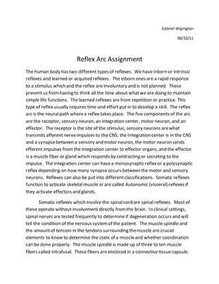
arc is the neural path wherea reflex takes place. The five components of the arc
are the receptor, sensory neuron, an integration center, motor neuron, and an
effector. The receptor is the site of the stimulus, sensory neurons arewhat
transmits afferent nerveimpulses to the CNS, the Integration center is in the CNS
and a synapsebetween a sensory and motor neuron, the motor neuron sends
efferent impulses from the integration center to effector organs, and the effector
is a muscle fiber or gland which responds by contracting or secreting to the
impulse. The integration center can havea monosynaptic reflexor a polysynaptic
reflex depending on how many synapseoccurs between the motor and sensory
neurons. Reflexes can also be put into differentclassifications. Somatic reflexes
function to activate skeletal muscle or are called Autonomic (visceral) reflexes if
they activate effectors and glands.
Somatic reflexes which involve the spinalcord are spinal reflexes. Most of
these operate without involvement directly fromthe brain. In clinical settings,
spinal nerves are tested frequently to determine if degeneration occurs and will
tell the condition of the nervous systemof the patient. The muscle spindle and
the amountof tension in the tendons surrounding themuscle are crucial
elements to know to determine the state of a muscleand whether coordination
can be done properly. The muscle spindle is made up of three to ten muscle
fibers called intrafusal. These fibers are enclosed in a connective tissue capsule.
2. There are also extra-fusalfibers that areonly ¼ the sizeof effector fibers in the
muscle. The intra-fusaldoes not havemyofilaments but are wrapped by two
types of afferent endings that send sensory inputto CNS. Primary sensory
endings innervate the spindle center while being stimulated by the rate and
degree of the stretch. Secondary sensory endings makeup the end of the spindle
and are only stimulated by the degree of stretch. The intra-fusalfibers have
regions which are innervated by gamma efferent fibers. These gamma come from
small motor neurons within the ventral horn in the spinalcord. There are also
alpha efferent fibers of the alpha motor neurons that stimulate the contraction of
the extra-fusalfibers. There are different ways a muscle spindle can stretch. Itis
stretched either by applying an external forcewhich lengthens the muscle or by
activating y motor neurons which stimulate the distal ends of intrafusalfibers
causing contraction and stretch of the middle of the spindle. The stretch reflex
makes the musclestays at a certain length. A knee jerk reflex is one wherethe
knee does not buckle when you are standing straight. When the large quadriceps
muscle elongates and knee is buckling, the reflex causes contraction of the
quadriceps muscle. This is a great example of a knee jerk reflex. This is really
important in the trunk muscles for postureand the large extensors as well.
Reciprocal Inhibition is a result of afferentfibers synapsing with interneurons that
inhibit the motor neurons which control antagonistmuscles. All stretch reflexes
are monosynaptic and ipsilateral. This means that they involve one synapseand
motor activity is on the same side of the body. The stretch reflex is actually
monosynaptic, butthe reflex arc is polysynaptic. Stretched muscle spindles are
what create a stretch reflex. This causes the stretched muscle to contractand
inhibit the antagonist muscle. This process begins with the muscle spindle being
activated by the stretch the associated sensory neurons transmitting afferent
impulses at high frequency to the spinal cord. Next, sensory neurons synapse
with the Alpha neurons exciting extrafusalfibers of the muscle. Afferent fibers
synapsewith interneurons which inhibit the motor neurons along with the
antagonistmuscle. Third, efferent impulses of the alpha neurons causemuscle to
contract reversing the stretch. Finally, efferent impulses of the alpha motor
neurons are sentto the antagonist muscles are reduced resulting in the
Reciprocal inhibition.
3. As speed and difficulty of a movement increases, the brain will increasey
motor output to increase the sensitivity of the musclespindle. Some athletes will
want to suppress thestretch reflex like before a baseball player winds up to pitch,
but other athletes will want to stretch justbeforethe muscle action because of
the need to generate maximum forceas in jumping or running. Efferent and
afferent fibers are critical to the muscle spindle in order to havegreat muscle
tone and coordination. Golgi tendon reflexes, which are polysynaptic, will
producethe oppositeeffect of the stretch reflex. When muscle tension increases
during contraction or passivestretching, the Golgi tendon organs become
activated. Afferent impulses are now sent to the spinal cord and on to the
cerebellum wherethe information can be used to adjustmuscle tension.
Reciprocal Activation is when the antagonist musclebecomes activated because
the spinalcord circuits which are supplying the contraction are inhibited.
The flexor reflex is initiated by a painful stimulus which then causes a withdrawal
fromthe body. These reflexes are classified as ipsilateral and polysynaptic. These
are protective reflexes that overridethe spinal pathways so that no other reflexes
can operate in these pathways atthe sametime. Many muscles are recruited
when these reflexes are active because the body is trying to protect itself and
survive. Thesereflexes can also be overridden by the brain though. An example
of this is a prick of the finger froma nurse. The cross-extensor reflexis very
important in keeping balance in the body. This is a complex spinal reflex that
made of an ipsilateral withdrawalreflex and a contralateral extensor reflex.
Afferent fibers will synapsewith interneurons which control the withdrawal
responseon the same sideof the body and with interneurons that control
extensor muscles fromthe opposite side. The ipsilateral responsecauses an
immediate rapid lifting of the foot when stepping on broken glass. The
contralateral responsewould be when the weight is shifted to the oppositeleg
activating the extensor muscles.
Superficial reflexes are caused by a slightcutaneous stimulation. They
depend on the function of upper motor pathways and on cord-levelreflex arcs.
Plantar reflex is an example of a superficialreflex. The area on the spinal cord
fromL4-S2 is tested during the Plantar reflex. Itwill determine right away if the
4. corticospinaltracts are working properly. Thetesting of a plantar reflex is to
strikethe sole of the foot with a blunt object on the lateral part of the foot. The
responsein resultshould be downward flexion of the toes. Babinski’s sign is an
abnormalreflex that happens when the primary motor cortex or corticospinal
tract is damaged. In this scenario, the great toe (hallux) dorsiflexes while the
smaller toes point laterally. The physiologicalaspect of Babinski’s sign is not
totally understood. An abdominal reflex happens when the umbilicus causes a
reflex contraction of the abdominal muscles. These reflexes will check the
integrity of the spinalcord and the ventral ramifrom T8-T12 of the brachial
plexus. When a person has a corticospinaltract lesion these reflexes are absent.