Autonomic Nervous System: Sympathetic vs Parasympathetic Functions
•
249 likes•146,142 views

Autonomic nervous system

Recommended
More Related Content
What's hot
What's hot (20)
Similar to Autonomic Nervous System: Sympathetic vs Parasympathetic Functions
Similar to Autonomic Nervous System: Sympathetic vs Parasympathetic Functions (20)
More from Subramani Parasuraman
More from Subramani Parasuraman (20)
Recently uploaded
Recently uploaded (20)
Autonomic Nervous System: Sympathetic vs Parasympathetic Functions
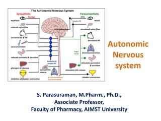
- 1. Autonomic Nervous system S. Parasuraman, M.Pharm., Ph.D., Associate Professor, Faculty of Pharmacy, AIMST University
- 2. Learning Outcomes • At the end of this session, the student would be able to: – Briefly describe Sympathetic and parasympathetic outflow and its functions. – List the differences between Sympathetic and Parasympathetic division. – Explain the adrenergic and cholinergic receptors
- 3. Nervous System Central nervous system Peripheral nervous system Afferent division (Sensory) Efferent division (Motor) Somatic system (voluntary) Autonomic nervous (involuntary) Sympathetic system (thorcolumbar outflow) Come from the thoracic and lumbar regions (T1 to L2/3) of the spinal cord Parasympathetic system (craniosacral outflow) Come from brainstem (Cranial Nerves III, VII, IX, X) or the sacral spinal cord (S2, S3, S4) Enteric nervous system
- 4. Spinal nerves • There are 31 pairs of spinal nerves – 8 cervical – 12 thoracic – 5 lumbar – 5 sacral – 1 coccygeal The human spinal column is made up of 33 bones. • 7 - cervical region • 12 - thoracic region • 5 - lumbar region • 5 - sacral region • 4 - coccygeal region
- 5. Peripheral Nervous System • Most of the nerves of the peripheral nervous system are composed of sensory nerve fibres conveying afferent impulses from sensory end organs to the brain and motor nerve fibres conveying efferent impulses from the brain through the spinal cord to the effector organs. Peripheral nervous system Efferent division (Motor) Afferent division (Sensory) Somatic system (voluntary) Autonomic nervous (involuntary)
- 6. Somatic Nervous System • The somatic nervous system (SNS or voluntary nervous system) is the part of the peripheral nervous system. • The somatic nervous system includes both sensory (afferent nerves) and motor (efferent nerves) neurons. • Sensory neurons convey input from receptors for somatic senses (tactile, thermal, pain, and proprioceptive sensations) and from receptors for the special senses (sight, hearing, taste, smell, and equilibrium)
- 7. Autonomic Nervous System • The autonomic nervous system is involved in a complex of reflex activities, which depend on sensory input to the brain or spinal cord, and on motor output. • The majority of the organs of the body are supplied by both sympathetic and parasympathetic nerves which have opposite effects that are finely balanced to ensure the optimum functioning of the organ.
- 8. Autonomic Nervous System • The autonomic nervous system (ANS) is a complex set of neurons that mediate internal homeostasis without conscious intervention or voluntary control. • The ANS maintains blood pressure, regulates the rate of breathing, influences digestion, urination, and modulates sexual arousal. • There are two main branches to the ANS – the sympathetic nervous system and the parasympathetic nervous system. The effects of autonomic control are rapid and essential for homeostasis
- 9. Sympathetic nervous system • Sympathetic nervous system otherwise called as thoracolumbar system. • Sympathetic stimulation prepares the body to deal with exciting and stressful situations, e.g. strengthening its defences in danger. sympathetic stimulation mobilises the body for 'fight or flight'. • Neurones convey impulses from their origin in the hypothalamus, reticular formation and medulla oblongata to effector organs and tissues. The first neurone has its cell body in the brain and its fibre extends into the spinal cord.
- 10. Sympathetic nervous system • Structure of the Sympathetic Division – Pathway from Spinal Cord to Sympathetic Trunk Ganglia – Organization of Sympathetic Trunk Ganglia – Pathways from Sympathetic Trunk Ganglia to Visceral Effectors
- 11. Structure of the sympathetic division of the autonomic nervous system Trachea and bronchi: Bronchodilation Liver: Glycogen glucose conversion increased Iris muscle: Pupil dilated Blood vessels in heart : Vasoconstriction Heart: Rate and force of contraction increased Salivary glands: Secretion inhibited Stomach: Peristalsis reduced Sphincters closed Intestines: Peristalsis and tone decreased Vasoconstriction Kidney: Urine secretion decreased Bladder: Smooth muscle wall relaxed Sphincter closed Sex organs: Generally Vasoconstriction
- 12. Parasympathetic nervous system • Parasympathetic nervous system otherwise called as craniosacral outflow. • Parasympathetic stimulation has a tendency to slow down body processes except digestion and absorption of food and the functions of the genitourinary systems. Its general effect is that of a 'peace maker' allowing restoration processes to occur quietly and peacefully. • Cell bodies of parasympathetic preganglionic neurons are found in nuclei in the brain stem.
- 13. Parasympathetic nervous system • Structure of the Parasympathetic Division – The cranial parasympathetic outflow consists of preganglionic axons that extend from the brain stem in four cranial nerves. The cranial outflow has four pairs of ganglia and the ganglia associated with the vagus (X) nerve. – The sacral parasympathetic outflow consists of preganglionic axons in anterior roots of the second through fourth sacral spinal nerves.
- 14. Structure of the parasympathetic division of the autonomic nervous system Trachea and bronchi: Bronchoconstriction Liver: Blood vessels dilated Secretion of bile increased Iris muscle: Pupil constricted Heart: Rate and force of contraction decreased Salivary glands: Secretion increased Stomach: Secretion of gastric juice and peristalsis increased Intestines: Digestion and absorption increased Kidney: Urine secretion increased Bladder: Muscle of wall contracted Sphincters relaxed Sex organs: Male: erection; Female: variable
- 17. Autonomic Motor Pathways • Each division of the ANS has two motor neurons (preganglionic and postganglionic neuron). • Preganglionic Neurons – In the sympathetic division (thoracolumbar division/ thoracolumbar outflow), the preganglionic neurons have their cell bodies in the lateral horns of the gray matter in the 12 thoracic segments and the first two (and sometimes three) lumbar segments of the spinal cord. – In the parasympathetic division (craniosacral division/ craniosacral outflow), the preganglionic neurons have their cell bodies in in the nuclei of four cranial nerves in the brain stem (III, VII, IX, and X) and in the lateral gray matter of the second through fourth sacral segments of the spinal cord.
- 18. Autonomic Motor Pathways Preganglionic Neurons • Autonomic Ganglia – There are two major groups of autonomic ganglia • sympathetic ganglia • parasympathetic ganglia • Sympathetic Ganglia: – The sympathetic ganglia are the sites of synapses between sympathetic preganglionic and postganglionic neurons. – There are two major types of sympathetic ganglia: • sympathetic trunk ganglia (also called vertebral chain ganglia or paravertebral ganglia) • prevertebral ganglia (collateral)- Five types of prevertebral ganglia are celiac ganglion, superior mesenteric ganglion, inferior mesenteric ganglion, aorticorenal ganglion and renal ganglion.
- 19. Autonomic Motor Pathways Preganglionic Neurons • Autonomic Ganglia – There are two major groups of autonomic ganglia • sympathetic ganglia • parasympathetic ganglia • parasympathetic ganglia: – Preganglionic axons of the parasympathetic division synapse with postganglionic neurons in terminal (intramural) ganglia. They are the ciliary ganglion, pterygopalatine ganglion, submandibular ganglion, and otic ganglion
- 20. Autonomic Motor Pathways Postganglionic Neurons • Once axons of sympathetic preganglionic neurons pass to sympathetic trunk ganglia, they may connect with postganglionic neurons. • A single sympathetic preganglionic fiber has many axon collaterals (branches) and may synapse with 20 or more postganglionic neurons.
- 21. Autonomic Motor Pathways Postganglionic Neurons • Axons of preganglionic neurons of the parasympathetic division pass to terminal ganglia near or within a visceral effector. In the ganglion, the presynaptic neuron usually synapses with only four or five postsynaptic neurons, all of which supply a single visceral effector, allowing parasympathetic responses to be localized to a single effector.
- 23. ANS Neurotransmitters and Receptors ANS Receptor Receptor Sub-type Parasympathetic nervous system Nicotinic cholinergic receptors Nn, Nm Muscarinic cholinergic receptors M1, M2, M3, M4, M5 Sympathetic nervous system α adrenergic receptor α1, α2 β adrenergic receptor β1, β2, β3 Sympathetic nervous system Parasympathetic nervous system
- 24. Comparison of Somatic and Autonomic Motor Neurons
- 25. Comparison of Somatic and Autonomic Motor Neurons Somatic Autonomic Voluntary effectors: striated muscles Involuntary effectors: smooth & cardiac muscles, glands single motor neuron from spinal cord to target organ usually 2 neurons with synapse (ganglion) between from spinal cord to target organ Neurotransmitter always stimulatory Neurotransmitter stimulatory or inhibitory ACh released at synapse ACh and NE released at synapses No firing at rest Baseline firing – speeds up when Stimulated Effector at rest is flaccid Effector at rest has intrinsic tone
- 26. Comparison of Somatic and Autonomic Motor Neurons Motor neuron pathways in the (a) somatic nervous system and (b) autonomic nervous system
- 27. Thank you
Editor's Notes
- Cervical spine: 7 vertebrae (C1–C7) Thoracic spine: 12 vertebrae (T1–T12) Lumbar spine: 5 vertebrae (L1–L5) Sacrum: 5 (fused) vertebrae (S1–S5) Coccyx: 4 (3–5) (fused) vertebrae (Tailbone)