Neonatal respiratory distress- surgical perspective
•
31 likes•4,980 views

The surgical causes for neonatal respiratory distress are life threatening and challenging. Early diagnosis and immediate timely surgical intervention are the key for the final successful outcome.

Recommended
More Related Content
What's hot
What's hot (20)
Viewers also liked
Viewers also liked (20)
Similar to Neonatal respiratory distress- surgical perspective
Similar to Neonatal respiratory distress- surgical perspective (20)
More from Selvaraj Balasubramani
More from Selvaraj Balasubramani (20)
Recently uploaded
Recently uploaded (20)
Neonatal respiratory distress- surgical perspective
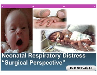
- 1. L/O/G/O Neonatal Respiratory Distress “Surgical Perspective” Neonatal Respiratory Distress “Surgical Perspective” M M M C Dr.B.SELVARAJ
- 3. www.themegallery.com Neonatal Respiratory DistressNeonatal Respiratory DistressNeonatal Respiratory DistressNeonatal Respiratory Distress “Surgical Perspective”“Surgical Perspective”“Surgical Perspective”“Surgical Perspective”
- 4. www.themegallery.com Dr.B.SELVARAJ MS;Mch;FICS; • PEDIATRIC SURGEON • ASSOCIATE PROFESSOR • MELAKA MANIPAL MEDICAL COLLEGE • MELAKA- 75150 • MALAYSIA
- 5. Neonatal Respiratory Distress Recognise various conditions Make early& accurate diagnosis Prompt Life Saving treatment Immediate surgical referral4 1 2 3 Surgical Perspective Objectives M M M C
- 6. A NeonateA NeonateA NeonateA Neonate’s request to Surgeons request to Surgeons request to Surgeons request to Surgeon “Please exercise the greatest gentleness with my diminutive tissues and try to correct the deformity at first operation; give me blood and proper amount of fluid and electrolytes; add plenty of oxygen to anesthesia, and I will show you that I can tolerate a terrific amount of surgery. You will be surprised at the speed of my recovery, and I shall be grateful to you” --Dr. Willis Potts M M M C
- 7. Neonatal Respiratory DistressNeonatal Respiratory DistressNeonatal Respiratory DistressNeonatal Respiratory Distress— Surgical CausesSurgical CausesSurgical CausesSurgical Causes Causes B E C D AEsophageal Atresia Diaphragmatic Hernia Congenital Lobar Emphysema Posterior Choanal Atresia Pierre Robin Sequence S V M C
- 8. Embryology Of Esophageal Atresia S V M C
- 9. Esophageal AtresiaEsophageal AtresiaEsophageal AtresiaEsophageal Atresia EA Challenging& Fascinating Problem Team Work Approach Post op Ventilator Care VACTERL Anomaly Incidence 1 in 3500 livebirths Epitome of Modern Surgery S V M C
- 10. www.themegallery.com Esophageal AtresiaEsophageal AtresiaEsophageal AtresiaEsophageal Atresia TypesTypesTypesTypes S V M C
- 11. Esophageal AtresiaEsophageal AtresiaEsophageal AtresiaEsophageal Atresia---- Associated AnomaliesAssociated AnomaliesAssociated AnomaliesAssociated Anomalies Vertebral Anorectal Cardiac- commonest Tracheo Esophageal Fistula Renal Limb S V M C
- 12. Esophageal AtresiaEsophageal AtresiaEsophageal AtresiaEsophageal Atresia Clinical FeaturesClinical FeaturesClinical FeaturesClinical Features Clinical Features Drooling of salivaMaternal Polyhydramnios Inability to pass NGT into Stomach In atresia with TEF Aspiration of gastric contents Chemical Pneumonitis Feeding Cough, choking & Cyanosis In pure atresia Gasless Abdomen Scaphoid Abd S V M C
- 13. Esophageal AtresiaEsophageal AtresiaEsophageal AtresiaEsophageal Atresia----Drooling of salivaDrooling of salivaDrooling of salivaDrooling of saliva S V M C
- 14. Physiological Effect of Distal TEF S V M C • 1. Hyaline membrane disease may necessitate higher ventilator pressures, which encourage air to pass through the distal fistula. • 2. A distended abdomen elevates and "splints" the diaphragm. • 3. Gastric distension may result in gastric rupture and pneumoperitoneum. • 4. Passage of air through a distal tracheoesophageal fistula diminishes the effective tidal volume. (B) 1. Aspiration of gastric juices leads to soiling of the lungs and pneumonia • 2. Gastroesophageal reflux • 3. Direction of gastric fluid proximally through distal fistula. • 4. Overflow of secretions or inadvertent feeding may contribute to aspiration and contamination of the airway.
- 15. Esophageal Atresia Imaging Studies AXR Gasless in pure Atresia CXR Atelectasis&Pneumonia Antenatal MRI of Fetus USG Abd to R/O Urogenital anomaly Echo to R/O cardiac anomaly&Rt Aortic arch AXR &CXR Curledup NGT in blind upper pouch Imaging StudiesS V M C
- 17. Esophageal Atresia Clinical Diagnosis S V M C • . (A) Diagnosis of esophageal atresia is confirmed when a 10- gauge (French) catheter cannot be passed beyond 10 cm from the gums. (B) A smaller-caliber tube is not used because it may curl up in the upper esophageal segment, giving a false impression of esophageal atresia.
- 19. Esophageal Atresia With TE Fistula- Bronchoscopy S V M C TE Fistula Rt Bronchus
- 20. Esophageal Atresia Pre op Management Pre opProximal pouch Decompression NPO If for staged repair Do Gastrostomy Head up position In pure atresia Stretch proximal pouch daily I V AntibioticsS V M C
- 21. Esophageal Atresia Pre op Management Pre opProximal pouch Decompression NPO If for staged repair Do Gastrostomy Head up position In pure atresia Stretch proximal pouch daily I V AntibioticsS V M C
- 22. Esophageal Atresia Waterston’s Risk Categories ●Birth weight >2.5 Kgs ●No Anomalies ●No Pneumonitis ●Primary Repair 100%survival ●Birth weight 1.8 to 2.5 Kgs ●Non life threatening anomalies ●Mild Pneumonitis ●Delayed Primary Repair 80%survival ●Birth weight < 1.8 Kgs ●Life threatening anomalies ●Severe Pneumonitis ●Staged Repair 40%survival Risk Categories Category A Category B Category C S V M C
- 23. Esophageal Atresia Operative Management 1 2 3 Lanman’s Rt posterolateral retropleural thoracotomy Ligation & division of Azygos vein Disconnect TEF; Repair tracheal defect 4 Liberally mobilise the upper pouch for tension free anastomosis S V M C
- 24. Esophageal Atresia Operative Management 5 6 7 In wide gap Livaditi’s circular myotomies Never mobilise distal pouch much Extra pleural drain S V M C 8 Transanastomotic feeding tube for early gavage feeding
- 26. Normal Mediastinum- Rt side S V M C
- 27. Esophageal Atresia Immediate Primary Repair S V M C
- 31. Esophageal atresia Post op Management in NICU Gastrograffin swallow on 7th POD; If no leak oral Feeding & remove chest drain Feeding through transanastomotic feeding Tube from 2nd POD Regular chest Physio&Nasopharyngeal suction Otherwise exubate in 1st POD S V M C Electively paralyse&mechanically ventilate For 3 to 5 days in tension anastomosis
- 32. Esophageal Atresia Complications LATE Tracheomalacia GE Reflux EARLY Anastomotic Leakage Anastomotic Stricture Recurrent TEF Esophageal Dysmotility S V M C
- 33. Clinical Features Operation Preop Trt •VACTERL •Maternal Poly Hydramnios •Drooling of saliva in baby •Inability to pass NGT into stomach •NPO •Headup position •IV Antibiotics •Upper pouch suction Complica tions Associ Anomaly EA& TEF Esophageal Atresia TE Fistula Recap Imaging CXR •Curledup NGT in blind upper pouch •Echo to R/O cardiac Anomaly • USG Abd to R/O Urogenital anomaly •Immediate primary Repair •Delayed primary Repair •Staged Repair •Anastomotic leak •Anastomotic stricture •Tracheomalacia •GE Reflux S V M C
- 35. Congenital Diaphragmatic Hernia & Eventration of diaphragm Herniation of abdominal contents into the thorax through a defect in diaphragm In Eventration, diaphragm is replaced by a thin membrane Incidence 1 in 4000 live births S V M C
- 37. Congenital Diaphragmatic Hernia Pathology Posterolateral Bochdalek Hernia: Commonest; Lt side 80% Rtside 19% Bilateral 1% Anteromedial Morgagni’s Hernia: 2% Hypoplastic ipsilateral lung due to compression by herniated viscera Persistent pulmonary hypertension(PPHN) due to abnormal pulmonary vasculature S V M C
- 38. Congenital Diaphragmatic Hernia Associated Anomalies Neural tube defects PDA,VSD,Coarctation of Aorta Midgut Malrotation Cleft palate Exomphalos S V M C
- 39. Congenital Diaphragmatic Hernia Clinical Features Respiratory distress Tachypnea, Cyanosis,Tachycardia,Etc Scaphoid Abdomen Mediastinal shift to opposite side In Lt sided CDH: Pseudodextrocardia Absence of breath sounds & presence of bowel sounds in ipsilateral hemithorax S V M C
- 40. Congenital Diaphragmatic Hernia— Imaging Studies Antenatal USG & MRI Chest of the fetus Postnatal CXR Presence of bowel shadows in the hemithorax; Mediastinal shift to the opposite side;Diaphragmatic margin is absent In Eventration Frontal&lateral CXR show elevated diaphragm with smooth unbroken outline Flouroscopy:Paradoxical movement of diaphragm in eventration S V M C
- 41. Congenital Diaphragmatic Hernia— Antenatal Fetal USG Chest
- 42. Congenital Diaphragmatic Hernia— Antenatal Fetal MRI
- 43. Congenital Diaphragmatic Hernia— Imaging Studies
- 44. Congenital Diaphragmatic Hernia— Imaging Studies
- 45. Diaphragmatic Hernia Pre op Management In Utero transfer to a maternity unit close to Pediatric surgical centre Electively deliver, promptly resuscitate & operate NGT aspiration to decompress GIT ET tube ventilation; “No face mask ventilation” “EXIT” Extrauterine Intrapartum Treatment Correct acidosis with NaHCO3 & Tromethamine S V M C
- 46. Diaphragmatic Hernia Pre op Management Delayed Repair after preop stabilisation aimed at correcting hypoxia, hypoperfusion& PPHN Correct PPHN with Tolazoline or Nitric oxide Pre&post ductal O2 saturation to assess the degree of arterial oxygenation &ductal shunting In intractable PPHN ECMO may be tried S V M C
- 47. Diaphragmatic Hernia Bad Prognostic Indicators Infants born to mothers with Polyhydramnios Stomach in thorax Lung Head ratio: LHR < 1 Presentation within first 6Hrs of birth Very low blood pH & high pCo2 Rt sided hernia S V M C
- 48. Diaphragmatic Hernia Prognostic Indicators Alveolar-arterial O2 tension difference (AaDo2) :If>500mmHg bad prognosis Ventilatory Index: VI=RRxMAPxPaco2;When Paco2 is <40mmHg & VI is <1000 all babies survive O2Index:OI=MAPXFiO2X100/Pao2 OI<6 Survival rate 98% OI>17 No Survivors S V M C
- 52. Diaphragmatic Hernia Post op Management in NICU Ventilatory support to maintain postductal Po2 80 to 100mmHg;Pco2 < 30mmHg & pH > 7.4 This can be achieved by high frequency ventilation with low airway pressures & low tidal volume Weaning from ventilator should be meticulous & slow as small variation in pH, Po2& Pco2 will lead to PPHN S V M C
- 53. CDH is a congenital anomaly with a high mortality. Usually associated with pulmonary hypoplasia and pulmonary hypertension. Surgical repair is the only treatment. Delayed surgery until the patient is stable is associated with better outcomes. Congenital cardiac and renal diseases, hypoxemia and hypercapnia increases mortality. HFOV, ECMO, iNO has improved the survival of CDH. S V M C Congenital Diaphragmatic Hernia Recap
- 54. Permissive hypercapnia with acceptable pO2 has shown to improve survival. Long term follow up is necessary to detect complications. Tracheal occlusion in utero, keeps lung expanded but it is an abnormal lung. Primary repair if small defect, patch if large defect, to prevent tension S V M C Congenital Diaphragmatic Hernia Recap
- 56. Congenital Lobar Emphysema S V M C •Congenital lobar overinflation –Normal architecture with overdistension of the alveoli –In true emphysema permanent distension of airspaces distal to terminal bronchiole with destruction of their walls •Etiology –Not found in up to 50% –Bronchial obstruction found in ~25% •Allows collapse on exhalation (ball-valve mechanism) •Air trapping leads to alveolar overinflation •Intrinsic obstruction (more common) –Intramural: Defect in the bronchial wall •Defective quantity or quality of cartilage –Intraluminal: Lesion in the lumen of the bronchus •Redundant bronchial folds, mucous plugs •Extrinsic obstruction –Compression of the bronchus from a lesion outside the bronchial wall •Cardiovascular: PDA, vascular sling •Mass: Lymph node, bronchogenic cyst, oncologic mass
- 57. Congenital Lobar Emphysema S V M C •Males:Females= 3:1 •Upper lobes are predominantly involved –LUL: 42% –RML: 35% –RUL: 21% –Lower lobes: <1% –Bilateral involvement: <20% •Associated congenital malformations: 14-21% –Congenital heart disease •PDA •VSD –Of those with CHD, >10% have additional anomalies •Rib cage defects •Renal defects
- 58. Congenital Lobar Emphysema S V M C •Presentation –Age •At birth: 33% •By 1 m.o.: 50% •After 6 m.o.: 5% –Symptoms (in order of decreasing frequency) •Moderate respiratory distress (most) •Cyanosis (half) •Mild respiratory distress (less than half) •Asymptomatic (infrequent) •Severe life-threatening distress (least common)
- 59. Congenital Lobar Emphysema S V M C •Radiographic evaluation –CXR: PA and lateral •Large, space-occupying air-filled lobe •Collapse of ipsilateral lung •Mediastinal shift •Atelectasis of contralateral lung •Clear anterior mediastinum –CT •May identify point of obstruction, intraluminal or extrinsic –Ventilation-Perfusion scan •Decreased perfusion of affected lobe (secondary to vessel compression) •Increased perfusion of unaffected lobe (secondary to shunting)
- 60. Congenital Lobar Emphysema Imaging Studies
- 63. Pierre-Robin Sequence Micrognathia/ Retrognathia Glossoptosis High arched Palate and/or Cleft Palate Occur sporadic or syndromic Stickler’s & Velocardiofacial syndromes Respiratory distress aggravated during feeding S V M C
- 64. Pierre-Robin Sequence- Grading Grade1: Eats & breaths well in supine position Grade 2: Breaths well but obstructs when fed by mouth Grade 3: Cannot breathe or eat without obstruction and desaturation S V M C
- 65. Pierre-Robin Sequence Most neonates responds to 2-3 months of conservative treatment Nursing the baby in prone position with head tilted to one side prevent tongue fall back Feeding the baby in lateral position with jaw pushed forward Rarely in severe cases may require anterior fixation of the tongue Anterior Glossopexy S V M C
- 68. Posterior Choanal Atresia Absence of posterior nasal openings; Atresia can be a membrane or bony overgrowth Common in Rt nostril Neonates are obligatory nasal breathers; in bilateral cases “ Cyclical Cyanosis”relieved by crying Respiratory distress aggravated during feeding Difficulty in feeding & failure to thrive S V M C
- 69. Posterior Choanal Atresia Failure to pass NGT through nostrils CT Skull Reveal bony block unilateral or bilateral Respiration is not a problem in unilateral cases but it is in bilateral cases oral airway Feeding by a spoon or a long nipple directly into the pharynx Transpalatal or transnasal surgical correction S V M C
- 70. Posterior Choanal Atresia Associaed Anomaly S V M C Coloboma and/or CNS abnormalities Heart abnormality Atresia choanae Retardation of growth Genital defects (males) Ear anomalies/deafness
- 74. Posterior Choanal Atresia S V M C With the advent of miniaturized endoscopic equipment and powered instrumentation, the most popular and successful method over the past decade has been the endoscopic transnasal technique.
- 76. NEONATAL RESPIRATORY DISTRESS Surgical Perspective Sl No Symptoms Signs Workup Diagnosis Treatment 1 Attacks of choking, coughing and cyanosis during feeding Drooling of saliva Failure to pass NGT into the stomach CXR:Curled up NGT in upper esophagus AXR:Gasless abdomen in pure atresia Esophageal Atresia with or without TEF •Primary repair •Delayed Primary repair •Staged repair 2 Tachypnea Tachycardia Cyanosis Scaphoid abdomen Mediastinal shift Bowel sound in the hemithorax CXR:Bowel loops in hemithorax Mediastinal shift Congenital diaphragmatic hernia/ Eventration Repair/ Plication of Diaphragm
- 77. NEONATAL RESPIRATORY DISTRESS Surgical Perspective Sl No Symptoms Signs Workup Diagnosis Treatment 3 Cough, tachypnea and breathlessness Shift of trachea/ Mediastinum Area of hyper resonance Decreased breath sounds CXR:Mediastin al shift Hyperlucent area of lung Adjacent normal lung Congenital Lobar Emphysema Bronchoscopy if needed Lobectomy/ Segmental resection 4 Respiratory distress aggravated during feeding Micronathia Glossoptosis Cleft palate ----- Pierre-Robin sequence Nurse baby in prone position Anterior glossopexy
- 78. NEONATAL RESPIRATORY DISTRESS Surgical Perspective Sl No Symptoms Signs Workup Diagnosis Treatment 5 Respiratory distress aggravated during feeding “ Cyclical cyanosis” Failure to pass a catheter through the nostril CT Skull Bony block unilateral or bilateral Posterior Choanal Atresia Surgical correction Transnasal or Transpalatal approach
- 79. “ Practice without theory is blind” “Theory without practice is sterile”
- 80. CARRY HOME MESSAGE S V M C “EYES CANNOT SEE WHAT THE MIND DOES NOT KNOW”