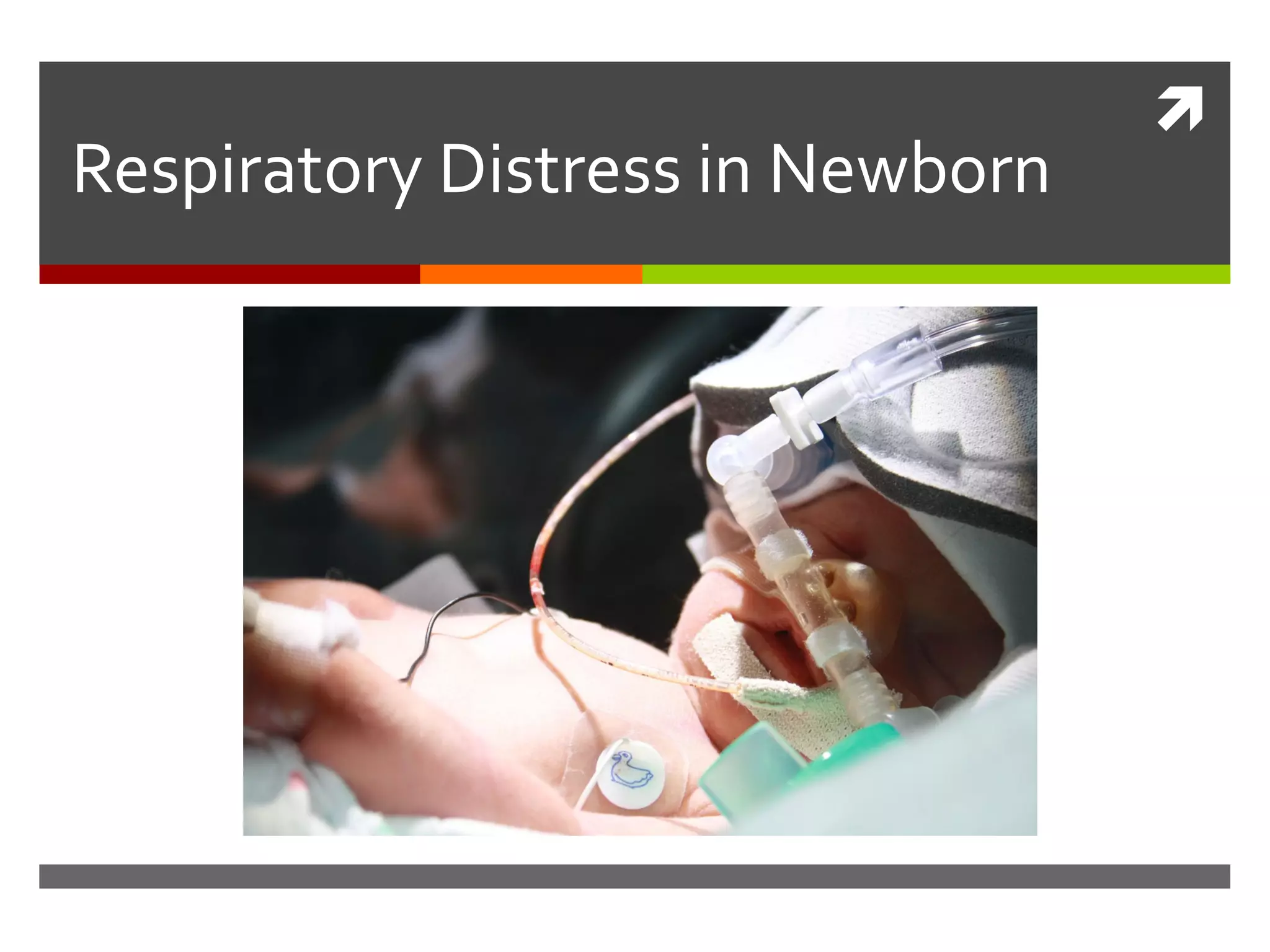

Respiratory Distress in Newborn
Neonatal Respiratory Distress
Signs and symptoms
 Tachypnea (RR > 60/min)
 Nasal flaring
 Retraction
 Grunting
 +/- Cyanosis
 +/- Desaturation
 Decreased air entry

Down score

Neonatal Respiratory Distress Etiologies
Pulmonary
Transient tachypnea
of the newborn (TTN)
Respiratory distress
syndrome (RDS)
Pneumonia
Meconium aspiration
syndrome (MAS)
Air leak syndromes
Pulmonary
hemorrhage
Systemic
Metabolic (e.g.,
hypoglycemia, hypothermia
or hyperthermia)
metabolic acidosis
anemia, polycythemia
Cardiac
• Congenital heart disease;
cyanotic or acyanotic
• Congestive heart failure
• Persistent pulmonary
hypertension of the newborn
(PPHN)
Neurological (e.g., prenatal
asphyxia, meningitis)
Anatomic
Upper airway
obstruction
Airway
malformation
Rib cage anomalies
Diaphragmatic
disorders
(e.g., congenital
diaphragmatic
hernia,
diaphragmatic
paralysis)
Pulmonary
 1- Transient tachypnea of newborn
 2- Hyaline membrane disease
 3- Meconium aspiration syndrome (MAS)
 4- Pneumonia
 5- Air Leak Syndromes
TransientTachypnea of Newborn
 TTN (known as wet lung) is a relatively mild,
self limiting disorder of near-term or term
 Delay in clearance of fetal lung fluid results in
transient pulmonary edema. The increased
fluid volume causes a reduction in lung
compliance and increased airway resistance.
TransientTachypnea of Newborn
Risk factors:
 Maternal asthma
 C- section
 Macrosomia, maternal diabetes
 Prolonged labor, Excessive maternal sedation
 Fluid overload to the mother,Delayed clamping of the
umbilical cord .
TransientTachypnea of Newborn
 Usually near-term or term
 Tachypnea immediately after birth or within 6 hrs
after delivery, mild to moderate respiratory distress.
 These manifestations usually persist for 12-24 hrs,
but can last up to 72 hrs
 Auscultation usually reveals good air entry with or
without crackles
 Spontaneous improvement of the neonate is an
important marker of TTN.
TransientTachypnea of Newborn
Chest x-ray :
 Prominent perihilar streaking (due to engorgement of
periarterial lymphatics)
 Fluid in the minor fissure
 Prominent pulmonary vascular markings
 Hyperinflation of the lungs, with depression of
diaphragm
 ► Chest x-ray usually shows evidence of clearing by 12-
18 hrs with complete resolution by 48-72 hrs
chest X-ray:TransientTachypnea of Newborn
Fluid in the
fissure
Fluid in the
fissure
General Management of Respiratory Distress
 Supplemental oxygen or MV, if needed.
 Continuously monitor with pulse oximeter.
 Obtain a chest radiograph.
 Correct metabolic abnormalities
(acidosis,hypoglycemia).
 Obtain a blood culture & begin an antibiotic
coverage (ampicillin + gentamicin)
General Management
 Provide an adequate nutrion. Infants with
sustained RR >60 breaths/min should not be fed
orally & should be maintained on gavage feedings
for RR 60-80 breaths/min, and NPO with IV fluids
or TPN for more severe tachypnea
Pulmonary
 1- Transient tachypnea of newborn
 2- Hyaline membrane disease
 3- Meconium aspiration syndrome (MAS)
 4- Pneumonia
 5- Air Leak Syndromes
Respiratory Distress Syndrome
 Also called as hyaline membrane disease
 Most common cause of respiratory distress in
premature infants, correlating with structural &
functional lung immaturity.
 primarily affects preterm infants; its incidence is
inversely related to gestational age and
birthweight.
 15-30% of those between 32-36 weeks‘ gestation,
in about 5% beyond 37 weeks' gestation
Physiologic abnormalities
 Surfactant deficiency- increase in alveolar
surface tension.
 Lung compliance decreased to 10-20% of
normal
 Atelectasis…areas not ventilated
 Decrease alveolar ventilation
 Reduce lung volume
 Areas not perfused
Normal Expiration
With Surfactant
Surfactant Function
Abnormal Respiration
Without Surfactant
17
Compliance
Pressure
Volume
Opening pressures
Maximal volume
Risk factors
 Prematurity
 Maternal diabetes
 Multiple births
 Elective cesarean section without labor
 Perinatal asphyxia
 Cold stress
 Genetic disorders
Decreased risk
Chronic intrauterine stress
Prolonged rupture of membranes
Antenatal steroid prophylaxis
Clinical Manifestations
 Appear within minutes of birth may not be recognized for several
hours in larger preterm
 Tachypnea (>60 breaths/min), nasal flaring, subcostal and intercostal
retractions, cyanosis & expiratory grunting
 Breath sounds may be normal or diminished and fine rales may be
heard
 Progressive worsening of cyanosis & dyspnea. BP may fall; fatigue,
cyanosis and pallor increase & grunting decreases.
 Apnea and irregular respirations are ominous signs
 In most cases, symptoms and signs reach a peak within 3 days, after
which improvement occurs gradually.
Findings can be graded according to the severity:
 Grade 1 (mild cases): the lungs show fine
homogenous ground glass shadowing
 Grade 2: widespread air bronchogram become
visible
 Grade 3: confluent alveolar shadowing
 Grade 4: complete white lung fields with obscuring
of the cardiac shadow
Chest x-ray:
Grade 1
Grade 2
Grade 3
Grade 4
Management
Prevention:
 Lung maturity testing: lecithin/sphingomyelin (L/S) ratio
 Tocolytics to inhibit premature labor.
 Antenatal corticosteroid therapy:
► They induce surfactant production and accelerate fetal lung
maturation.
► Are indicated in pregnant women 24-34 weeks' gestation at
high risk of preterm delivery within the next 7 days.
► Optimal benefit begins 24 hrs after initiation of therapy and
lasts seven days.
Prevention
 Antenatal corticosteroid therapy consists of either :
□ Betamethasone 12 mg/dose IM for 2 doses, 24 hrs apart, or
□ Dexamethasone 6 mg/dose IM for 4 doses, 12 hrs apart
 Early surfactant therapy: prophylactic use of
surfactant in preterm newborn <27 weeks'
gestation.
 Early CPAP administration in the delivery room.
Treatment
 Administer oxygen
 Initiate CPAP as early as possible in infants with mild
RDS
 Start MV if respiratory acidosis (PaCO2 >60 mmHg,
PaO2 <50 mmHg or SaO2 <90%) with an FiO2 >0.5, or
severe frequent apnea.
 Administer surfactant therapy: early rescue therapy
within 2 hrs after birth is better than late rescue
treatment when the full picture of RDS is evident.
Types of Surfactant
Natural Surfactants: contain appoproteins SP-B & SP-C
 Curosurf (extract of pig lung mince)
 Survanta (extract of cow lung mince)
 Infasurf (extract of calf lung)
Synthetic Surfactants:do not contain proteins
 Exocerf
 ALEC
 Lucinactant (Surfaxin)
30
SurfactantTherapy for RDSSurfactantTherapy for RDS
Improvement in compliance, functional
residual capacity, and oxygenation
Reduces incidence of air leaks
Decreases mortality
Mode of administration of Surfactant
 Dosing may be
divided into 2
alliquots and
adminitered via
a 5-Fr catheter
passed in the
ET
Insure technique
 Intubation-
 surfactant-
 extubation to CPAP
Pulmonary
 1- Transient tachypnea of newborn
 2- Hyaline membrane disease
 3- Meconium aspiration syndrome (MAS)
 4- Pneumonia
 5- Air Leak Syndromes
Risk Factors:
 Post-term pregnancy
 Pre-eclampsia, eclampsia, maternal hypertension,
 Maternal diabetes mellitus
 IUGR
 Evidences of fetal distress (e.g.,abnormal biophysical
profile)
Meconium Aspiration Syndrome
Clinical Manifestations
 Meconium staining amniotic fluid (meconium stained
nails, skin & umbilical cord )
 Some infants may have mild initial respiratory
distress, which becomes more severe hours after
delivery.
 Pneumothorax and/or pneumomediastinum
 PPHN in severe cases
 Hypoxia to other organs (e.g., seizures, oliguria)
Pathophysiology
Chest x-ray: Areas of hyperexpansion mixed with patchy
densities and atelectasis
Management
In the DR or OR:
 Visualization of the vocal cords & tracheal suctioning before
ambu-bagging should be done only if the baby is not vigorous
In the NICU:
 Empty stomach contents to avoid further aspiration.
 Suction frequently & perform chest physiotherapy.
Management
 Consider CPAP, if FiO2 requirements >0.4; however CPAP
mayaggravate air trapping and must be used cautiously.
 Mechanical ventilation: in severe cases (paCO2 >60 mmHg
orpersistent hypoxemia (paO2 <50 mmHg).
 Correct systemic hypotension (hypovolemia, myocardial
dysfunction).
 Manage PPHN, if present
 Manage seizures or renal problems, if present.
 Surfactant therapy in infants whose clinical status continue
todeteriorate.
Pulmonary
 1- Transient tachypnea of newborn
 2- Hyaline membrane disease
 3- Meconium aspiration syndrome (MAS)
 4- Pneumonia
 5- Air Leak Syndromes
PneumoniaPneumonia
Common organisms:
 GBS
 gram–ve organisms (e.g. E.Coli,
Klebsiella,Pseudomonas)
 , Staph. aureus, Staph. epidermidis
 Candida.
 acquired viral infections (e.g., HSV, CMV).
Clinical Manifestations
 Early manifestations may be nonspecific (e.g., poor
feeding, lethargy, irritability, cyanosis, temperature
instability
 Respiratory distress signs may be superimposed upon RDS
or BPD.
 In a ventilated infant, the most prominent change may be
the need for an increased ventilatory support.
 Signs of pneumonia (dullness to percussion, change in
breathsounds, rales or rhonchi) are difficult to appreciate.
Chest x-rays: infiltrates or effusion
44
Chlamydia pneumonia with features of an interstitial
pneumonitis and characteristic widespread interstitial changes.
Management
Initiate ampicillin and gentamicin IV;
modify according to culture results
and continue therapy for 14 days.
If there is a fungal infection, an
antifungal agent is used.
Pulmonary
 1- Transient tachypnea of newborn
 2- Hyaline membrane disease
 3- Meconium aspiration syndrome (MAS)
 4- Pneumonia
 5- Air Leak Syndromes
Air Leak Syndromes
Risk Factors:
 MV,MAS, surfactant therapy without
decreasing pressure support in ventilated
infants
 vigorous resuscitation,
 prematurity
 pneumonia
Clinical Manifestations
 Spontaneous pneumothorax may be asymptomatic or
only mildly symptomatic (i.e., tachypnea and ↑O2
needs).
 In unilateral cases, chest asymmetry is noted,
mediastinum shift to the opposite side.
 If the infant is on ventilatory support will have sudden
onset of clinical deterioration (i.e., cyanosis,
hypoxemia, hypercarbia & respiratory acidosis
associated with decreased breath sounds and shifted
heart sounds).
Tension pneumothorax
 (a life-
threatening
condition) →
↓cardiac
output and
obstructive
shock; urgent
drainage prior
to a radiograph
is mandatory.
Chest x-ray: Right-sided pneumothorax
Right-sided tension pneumothorax with mediastinal shift. Both
lungs demonstrate opacification of alveolar collapse.
Left-sided pneumothorax under tension.There is pulmonary interstitial
emphysema in the right lung and a small basal right pneumothorax.
Others
Pneumomediastinum
 It can occur with aggressive ETT insertion, Ryle's feeding tube
insertion, lung disease, MV, or chest surgery (e.g., TEF).
Pneumopericardium
Pneumoperitoneum
Subcutaneous emphysema
Systemic air embolism
Chest x-ray with Pneumomediastinum
Massive Pneumoperitoneum in MV neonate
Chest x-ray with pneumopericardium
Severe bilateral PIE affecting the right more than the left lung; there is gross
cardiac compression. A chest drain is in situin the right hemithorax.
Management
 Conservative therapy: close observation of the
degree of respiratory distress as well as oxygen
saturation, without any other intervention aiming
at spontaneous resolution and absorption of air.
 Needle aspiration should be done for suspected
cases of pneumothorax with deteriorating general
condition until intercostal tube is inserted.
 Decompression of air leak according to the type
(intercostal tube insertion in case of pneumothorax).
Thank You …
Thank You …

Neonatal Respiratory Distress

  • 1.
  • 2.
    Neonatal Respiratory Distress Signsand symptoms  Tachypnea (RR > 60/min)  Nasal flaring  Retraction  Grunting  +/- Cyanosis  +/- Desaturation  Decreased air entry
  • 3.
  • 4.
     Neonatal Respiratory DistressEtiologies Pulmonary Transient tachypnea of the newborn (TTN) Respiratory distress syndrome (RDS) Pneumonia Meconium aspiration syndrome (MAS) Air leak syndromes Pulmonary hemorrhage Systemic Metabolic (e.g., hypoglycemia, hypothermia or hyperthermia) metabolic acidosis anemia, polycythemia Cardiac • Congenital heart disease; cyanotic or acyanotic • Congestive heart failure • Persistent pulmonary hypertension of the newborn (PPHN) Neurological (e.g., prenatal asphyxia, meningitis) Anatomic Upper airway obstruction Airway malformation Rib cage anomalies Diaphragmatic disorders (e.g., congenital diaphragmatic hernia, diaphragmatic paralysis)
  • 5.
    Pulmonary  1- Transienttachypnea of newborn  2- Hyaline membrane disease  3- Meconium aspiration syndrome (MAS)  4- Pneumonia  5- Air Leak Syndromes
  • 6.
    TransientTachypnea of Newborn TTN (known as wet lung) is a relatively mild, self limiting disorder of near-term or term  Delay in clearance of fetal lung fluid results in transient pulmonary edema. The increased fluid volume causes a reduction in lung compliance and increased airway resistance.
  • 7.
    TransientTachypnea of Newborn Riskfactors:  Maternal asthma  C- section  Macrosomia, maternal diabetes  Prolonged labor, Excessive maternal sedation  Fluid overload to the mother,Delayed clamping of the umbilical cord .
  • 8.
    TransientTachypnea of Newborn Usually near-term or term  Tachypnea immediately after birth or within 6 hrs after delivery, mild to moderate respiratory distress.  These manifestations usually persist for 12-24 hrs, but can last up to 72 hrs  Auscultation usually reveals good air entry with or without crackles  Spontaneous improvement of the neonate is an important marker of TTN.
  • 9.
    TransientTachypnea of Newborn Chestx-ray :  Prominent perihilar streaking (due to engorgement of periarterial lymphatics)  Fluid in the minor fissure  Prominent pulmonary vascular markings  Hyperinflation of the lungs, with depression of diaphragm  ► Chest x-ray usually shows evidence of clearing by 12- 18 hrs with complete resolution by 48-72 hrs
  • 10.
    chest X-ray:TransientTachypnea ofNewborn Fluid in the fissure Fluid in the fissure
  • 11.
    General Management ofRespiratory Distress  Supplemental oxygen or MV, if needed.  Continuously monitor with pulse oximeter.  Obtain a chest radiograph.  Correct metabolic abnormalities (acidosis,hypoglycemia).  Obtain a blood culture & begin an antibiotic coverage (ampicillin + gentamicin)
  • 12.
    General Management  Providean adequate nutrion. Infants with sustained RR >60 breaths/min should not be fed orally & should be maintained on gavage feedings for RR 60-80 breaths/min, and NPO with IV fluids or TPN for more severe tachypnea
  • 13.
    Pulmonary  1- Transienttachypnea of newborn  2- Hyaline membrane disease  3- Meconium aspiration syndrome (MAS)  4- Pneumonia  5- Air Leak Syndromes
  • 14.
    Respiratory Distress Syndrome Also called as hyaline membrane disease  Most common cause of respiratory distress in premature infants, correlating with structural & functional lung immaturity.  primarily affects preterm infants; its incidence is inversely related to gestational age and birthweight.  15-30% of those between 32-36 weeks‘ gestation, in about 5% beyond 37 weeks' gestation
  • 15.
    Physiologic abnormalities  Surfactantdeficiency- increase in alveolar surface tension.  Lung compliance decreased to 10-20% of normal  Atelectasis…areas not ventilated  Decrease alveolar ventilation  Reduce lung volume  Areas not perfused
  • 16.
    Normal Expiration With Surfactant SurfactantFunction Abnormal Respiration Without Surfactant
  • 17.
  • 18.
    Risk factors  Prematurity Maternal diabetes  Multiple births  Elective cesarean section without labor  Perinatal asphyxia  Cold stress  Genetic disorders
  • 19.
    Decreased risk Chronic intrauterinestress Prolonged rupture of membranes Antenatal steroid prophylaxis
  • 20.
    Clinical Manifestations  Appearwithin minutes of birth may not be recognized for several hours in larger preterm  Tachypnea (>60 breaths/min), nasal flaring, subcostal and intercostal retractions, cyanosis & expiratory grunting  Breath sounds may be normal or diminished and fine rales may be heard  Progressive worsening of cyanosis & dyspnea. BP may fall; fatigue, cyanosis and pallor increase & grunting decreases.  Apnea and irregular respirations are ominous signs  In most cases, symptoms and signs reach a peak within 3 days, after which improvement occurs gradually.
  • 21.
    Findings can begraded according to the severity:  Grade 1 (mild cases): the lungs show fine homogenous ground glass shadowing  Grade 2: widespread air bronchogram become visible  Grade 3: confluent alveolar shadowing  Grade 4: complete white lung fields with obscuring of the cardiac shadow Chest x-ray:
  • 22.
  • 23.
  • 24.
  • 25.
  • 26.
    Management Prevention:  Lung maturitytesting: lecithin/sphingomyelin (L/S) ratio  Tocolytics to inhibit premature labor.  Antenatal corticosteroid therapy: ► They induce surfactant production and accelerate fetal lung maturation. ► Are indicated in pregnant women 24-34 weeks' gestation at high risk of preterm delivery within the next 7 days. ► Optimal benefit begins 24 hrs after initiation of therapy and lasts seven days.
  • 27.
    Prevention  Antenatal corticosteroidtherapy consists of either : □ Betamethasone 12 mg/dose IM for 2 doses, 24 hrs apart, or □ Dexamethasone 6 mg/dose IM for 4 doses, 12 hrs apart  Early surfactant therapy: prophylactic use of surfactant in preterm newborn <27 weeks' gestation.  Early CPAP administration in the delivery room.
  • 28.
    Treatment  Administer oxygen Initiate CPAP as early as possible in infants with mild RDS  Start MV if respiratory acidosis (PaCO2 >60 mmHg, PaO2 <50 mmHg or SaO2 <90%) with an FiO2 >0.5, or severe frequent apnea.  Administer surfactant therapy: early rescue therapy within 2 hrs after birth is better than late rescue treatment when the full picture of RDS is evident.
  • 29.
    Types of Surfactant NaturalSurfactants: contain appoproteins SP-B & SP-C  Curosurf (extract of pig lung mince)  Survanta (extract of cow lung mince)  Infasurf (extract of calf lung) Synthetic Surfactants:do not contain proteins  Exocerf  ALEC  Lucinactant (Surfaxin)
  • 30.
    30 SurfactantTherapy for RDSSurfactantTherapyfor RDS Improvement in compliance, functional residual capacity, and oxygenation Reduces incidence of air leaks Decreases mortality
  • 31.
    Mode of administrationof Surfactant  Dosing may be divided into 2 alliquots and adminitered via a 5-Fr catheter passed in the ET
  • 32.
    Insure technique  Intubation- surfactant-  extubation to CPAP
  • 33.
    Pulmonary  1- Transienttachypnea of newborn  2- Hyaline membrane disease  3- Meconium aspiration syndrome (MAS)  4- Pneumonia  5- Air Leak Syndromes
  • 34.
    Risk Factors:  Post-termpregnancy  Pre-eclampsia, eclampsia, maternal hypertension,  Maternal diabetes mellitus  IUGR  Evidences of fetal distress (e.g.,abnormal biophysical profile) Meconium Aspiration Syndrome
  • 35.
    Clinical Manifestations  Meconiumstaining amniotic fluid (meconium stained nails, skin & umbilical cord )  Some infants may have mild initial respiratory distress, which becomes more severe hours after delivery.  Pneumothorax and/or pneumomediastinum  PPHN in severe cases  Hypoxia to other organs (e.g., seizures, oliguria)
  • 36.
  • 37.
    Chest x-ray: Areasof hyperexpansion mixed with patchy densities and atelectasis
  • 38.
    Management In the DRor OR:  Visualization of the vocal cords & tracheal suctioning before ambu-bagging should be done only if the baby is not vigorous In the NICU:  Empty stomach contents to avoid further aspiration.  Suction frequently & perform chest physiotherapy.
  • 39.
    Management  Consider CPAP,if FiO2 requirements >0.4; however CPAP mayaggravate air trapping and must be used cautiously.  Mechanical ventilation: in severe cases (paCO2 >60 mmHg orpersistent hypoxemia (paO2 <50 mmHg).  Correct systemic hypotension (hypovolemia, myocardial dysfunction).  Manage PPHN, if present  Manage seizures or renal problems, if present.  Surfactant therapy in infants whose clinical status continue todeteriorate.
  • 40.
    Pulmonary  1- Transienttachypnea of newborn  2- Hyaline membrane disease  3- Meconium aspiration syndrome (MAS)  4- Pneumonia  5- Air Leak Syndromes
  • 41.
    PneumoniaPneumonia Common organisms:  GBS gram–ve organisms (e.g. E.Coli, Klebsiella,Pseudomonas)  , Staph. aureus, Staph. epidermidis  Candida.  acquired viral infections (e.g., HSV, CMV).
  • 42.
    Clinical Manifestations  Earlymanifestations may be nonspecific (e.g., poor feeding, lethargy, irritability, cyanosis, temperature instability  Respiratory distress signs may be superimposed upon RDS or BPD.  In a ventilated infant, the most prominent change may be the need for an increased ventilatory support.  Signs of pneumonia (dullness to percussion, change in breathsounds, rales or rhonchi) are difficult to appreciate.
  • 43.
  • 44.
    44 Chlamydia pneumonia withfeatures of an interstitial pneumonitis and characteristic widespread interstitial changes.
  • 45.
    Management Initiate ampicillin andgentamicin IV; modify according to culture results and continue therapy for 14 days. If there is a fungal infection, an antifungal agent is used.
  • 46.
    Pulmonary  1- Transienttachypnea of newborn  2- Hyaline membrane disease  3- Meconium aspiration syndrome (MAS)  4- Pneumonia  5- Air Leak Syndromes
  • 47.
    Air Leak Syndromes RiskFactors:  MV,MAS, surfactant therapy without decreasing pressure support in ventilated infants  vigorous resuscitation,  prematurity  pneumonia
  • 48.
    Clinical Manifestations  Spontaneouspneumothorax may be asymptomatic or only mildly symptomatic (i.e., tachypnea and ↑O2 needs).  In unilateral cases, chest asymmetry is noted, mediastinum shift to the opposite side.  If the infant is on ventilatory support will have sudden onset of clinical deterioration (i.e., cyanosis, hypoxemia, hypercarbia & respiratory acidosis associated with decreased breath sounds and shifted heart sounds).
  • 49.
    Tension pneumothorax  (alife- threatening condition) → ↓cardiac output and obstructive shock; urgent drainage prior to a radiograph is mandatory.
  • 50.
  • 51.
    Right-sided tension pneumothoraxwith mediastinal shift. Both lungs demonstrate opacification of alveolar collapse.
  • 52.
    Left-sided pneumothorax undertension.There is pulmonary interstitial emphysema in the right lung and a small basal right pneumothorax.
  • 53.
    Others Pneumomediastinum  It canoccur with aggressive ETT insertion, Ryle's feeding tube insertion, lung disease, MV, or chest surgery (e.g., TEF). Pneumopericardium Pneumoperitoneum Subcutaneous emphysema Systemic air embolism
  • 54.
    Chest x-ray withPneumomediastinum
  • 55.
  • 56.
    Chest x-ray withpneumopericardium
  • 57.
    Severe bilateral PIEaffecting the right more than the left lung; there is gross cardiac compression. A chest drain is in situin the right hemithorax.
  • 58.
    Management  Conservative therapy:close observation of the degree of respiratory distress as well as oxygen saturation, without any other intervention aiming at spontaneous resolution and absorption of air.  Needle aspiration should be done for suspected cases of pneumothorax with deteriorating general condition until intercostal tube is inserted.  Decompression of air leak according to the type (intercostal tube insertion in case of pneumothorax).
  • 59.

Editor's Notes

  • #45 RDS, H influenza pneumonia