This presentation has been made easy to make understand to the students by emphasizing more on the visual than the words.I hope it is helpful to the students and followers as well.
4. Introduction
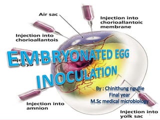
• Cultivation of viruses in chick embryo
• different type of approach.
• For all practical purposes they behave as
tissue cultures
4
5. History
Burnet
First used for cultivation of viruses
by Ernest GoodPasteur and Burnet
(1931)
F.M. Burnet in the
laboratory in the
early 1950's, was
experimenting
on influenza
virus genetics,
using the
developing hen's
egg.
6. EMBRYONATED EGG
6
state of a fertilized egg
containing an embryo
foetus in its early
stages of developments
especially before it has
reached a distinctively
recognizable form).
7. EGGS USED IN VIROLOGY
• HEN EGG
• DUCK EGG
• TURKEY’S EGG
7
8. Selection of egg
• must be sterile
• shell should be intact and healthy.
• should be obtained from non-vaccinated,
disease-free flocks
8
9. Process of artificial incubation
• incubation - 38 – 39°C and 60 – 70% humidity.
• need to be turned at least twice a day
or
• rolled continually in a specially designed egg
incubator.
9
11. Egg incubator
• artificially hatched - controlled and favourable conditions
• maintain favourable incubation/ environment - constant
temperature over a specified period.
• electrically heated – thermostat
• intelligent control system - correct measurement
of heat quantity ,
• - adjusting hatching control
temperature constantly
• variation of temperature - ambient to 70° C
• controlled by “JUMO”/ EGO”
German Capillary thermostat
having accuracy of + 0.5° c.
• Capacity:
• 50 to 2000 eggs
•
11
12. ADVANTAGES OF EMBRYONATED EGG
Ideal for viruses to grow, offers several sites for virus cultivation
Isolation and cultivation of many avian viruses and few mammalian viruses
Sterile and wide range of tissues and fluids
Economical and Readily available
Maintenance easier
Less labour (not need feeding and caging)
They do not have immune mechanism like animals to counteract virus
infection.
12
13. DISADVANTAGES
Some viruses do not show growth on primary
inoculation into the egg.
Slight amount of bacterial contamination in
the inoculum may kill the embryo.
Eggs may be contaminated with mycoplasma
and latent fowl viruses which may interfere
with the growth of other virus.
13
14. CANDLING OF EGG:
14
process of holding a strong beam of light
above or below the egg
to observe the embryo.
done in a
darkened room
or
area shielded by curtains
Use candling box
15. CANDLING BOX
15
consists of
A candling lamp
a strong electric bulb
covered by a plastic or
aluminium container
with handle and aperture.
18. • Under the candling lamp, the
embryo appears as a dark
shadow with the head as a
dark spot (eye).
• Incubated eggs are
candled daily to see the
chicken embryos inside
18
21. MARKING OF AIRSAC
1. Hold the blunt end of the egg against
the aperture of the candling lamp and
note the position of the head of the
embryo.
2.Draw a line on the shell marking the
edge of the air sac.
3.Draw an x approximately 2mm above
this line.
21
28. Puncture the shell
over the centre
of the air sac.
Insert a 23- gauge
needle, 1-1/2 inches
in length on a 1 ml
syringe, into the egg
through the puncture
in the shell at a 45
angle to the long axis
of the egg and away
from the embryo.
28
29. • Inject O.2ml of fluid into the egg
• Seal the puncture with
- Nail polish/cellophone tape
• Position the eggs and incubate at 37o C.
29
31. Harvesting of Allantoic Fluid
• Eggs must be chilled to obtain allantoic fluid
free of RBCs.
• Clean the upper half of the shell with 70%
alcohol.
• Cut away the shell above the air space.
• Peel away the white opaque shell membrane
lining the air space, exposing the transparent
allantoic membrane directly beneath.
31
32. • Tear the allantoic membrane with sterile
forceps.
• Attach a ballpoint needle to a syringe and
insert the needle into the cavity.
• Remove the fluid by suction.
• Culture the harvested fluid in a suitable
medium for a sterility check.
32
33. YOLK SAC INOCULATION
• 6-8 days old eggs required.
33
Virus and bacteria which
can be harvested
Rickettsiae
Chlamydia
trachomatis
C. psittaci
HERPES
SIMPLEX
VIRUS
36. 3. Insert a 22 gauge
needle,2 inches in
length on a syringe,
into the egg via the
puncture
4. Point the needle
straight down for
depth of about 1-1/2
inches.
5. Express 0.5 ml of
inoculum into
the yolk sac.
36
38. HARVESTING OF YOLK SAC
• Disinfect the upper half of the shell.
• Remove the shell, shell membrane and
underlying chorio-allantoic membrane.
• Lift the embryo up with sterile forceps to
expose the attached yolk sac.
• Pull the yolk sac free with another pair of
forceps and place it in a sterile petridish.
38
43. • Place a drop of sterile physiological saline on the side hole and
gently tease apart the fibers of the shell membrane with a 27-
gauge needle.
• When the shell membrane has been penetrated, the drop of
saline will be drawn into the egg as a result of separation of
the CAM and shell membrane.
43
44. Apply negative pressure to
the air space opening by
means of mouth suction
with a rubber tube.
As the air is removed the
CAM will drop from the
shell around the side hole,
creating an artificial
airspace , outline the limits
of artificial airspace.
Express 0.2 ml of inoculum
through the side opening
onto the CAM.
44
46. • Seal openings with cellophane tape.
• Gently rotate the egg to spread the
inoculum over the entire CAM under the
false air space.
• Incubate the eggs on side with false air space
upward.
46
47. Harvesting the CAM Membrane
• Disinfect the shell.
• With sterile scissors, cut through the shell
along the longitudinal axis, about 1/3 down
from the upper surface.
• Gently remove the shell to a discard pan.
• With sterile forceps,lift the CAM, cut free.
• Place the CAM in sterile saline and float free
for examination.
47
49. STEPS
• Candle the egg and mark the position of the
embryo and the outline of the airspace on the
shell.
• Punch a hole through the shell at the edge of
the airspace directly above the embryo.
• Using a 23- gauge, 1 inch needle on a syringe
make a short jab through the punched hole,
towards the embryo.
49
51. • When the amniotic membrane is penetrated, the
embryo will be seen to follow the movements of
the needle.
• Express upto 0.2 ml of inoculum.
• Seal the puncture with nail polish or cellophane
tape and incubate at 37o C.
51
52. HARVESTING OF AMNIOTIC FLUID
• Remove the shell and shell membrane below
the air space.
• Remove the fluid from the allantoic cavity ,
the amnion should then be clearly visible.
• Remove the amniotic fluid with a 20- gauge
needle and syringe.
52
53. death of the embryo
embryo cell damage
• formation of typical
pocks or lesions
• on the egg membranes
oedema of the
developing membranes
inclusion bodies
Presence of viral antigen
in egg fluids
53
Viral growth
and
multiplication
in the egg
embryo is
indicated by
1
2
3
4
5
6
55. GROWTH OF VIRUS ON THE CAM
55
• Formation of characteristic pocks.
• Variola produces small circular pocks, dome shaped, no
surrounding necrosis or haemmorrhage whereas
• Vaccinia virus larger lesions , flattened with necrosis and
haemmorrhage.
• Herpes simplex virus-
-small , oval shaped with
- no evidence of necrosis
56. GROWTH OF A VIRUS IN THE YOLK SAC
PRESENCE OF
BASOPHILIC
INCLUSION BODIES
56