Test bank for critical care nursing a holistic approach 11th edition morton f...
Placental Stem Cells Promote Tissue Regeneration and Angiogenesis
1. Placental MSC Support In Vitro and In Vivo Bone Formation and Angiogenesis
Jiawei He2, Sivan M. Oyserman1, Naomi J. Walker3, Dori L. Borjesson3, J. Kent Leach2, Michael S. Friedman1
Thermogenesis Corporation1, University of California Davis Biomedical Engineering2, University of California Davis School of Veterinary Medicine3
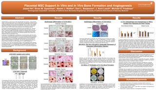
Abstract Results Results Results
Placental tissue is a unique source of stem cells with MSC-like properties. In these scientific Multilineage Differentiation of UCM MSCs Multilineage Differentiation of UCM MSCs In Vivo Osteogenesis and Angiogenesis by MSCs
studies, we examined the in vitro and in vivo differentiation and regenerative potential of equine
umbical cord matrix (UCM) MSC, umbilical cord perivascular (UCPV) MSC, or from umbilical Adipocyte: Normoxia A. Chondrocyte B.
Isolated from Placental and Adult Tissues
cord blood (UCB) MSC and adult bone marrow (BM) MSC. MSC from UCM, UCB, and BM
differentiate to osteoblasts, chondrocytes, and adipocytes as determined by cytochemical Bone Mineral Density Vessel Counts
staining. However, UCPVC failed to demonstrate any in vitro multilineage potential.
Control Control
To evaluate the transcriptional profiles of these cells, we used quantitative PCR. Placental
MSC express very low to undetectable levels of the primitive stem cell markers Oct3, and
nanog. In response to BMP6, adult BM MSC upregulate the osteoblast master transcription
factor, osterix, as well as Dlx5. Placental MSC express low to undetectable levels of these
factors. Similar patterns were observed for the osteoblast associated matrix proteins bone
sialoprotein (Bsp) and osteomodulin (Omd).
Induced Induced
To determine if in vitro differentiation correlated with in vivo bone formation, MSC from UCB,
UCM, UCPVC, and adult BM were seeded onto PLGA-hydroxyapatite scaffolds and implanted
subcutaneously in immunocompromised rats. At 8 weeks, bone formation and angiogenesis
were evaluated by micro CT and histology. Interestingly, the levels of bone formation were
greatest in scaffolds seeded with adult BM MSC and UCM MSC (bone mineral denstiy of Adipocyte: Hypoxia Chondrogenic differentiation of UCM MSC was evaluated using the micromass culture system.
923±40 and 910±23 mg HA/cm3, respectively). The levels of angiogenesis were also very The micromasses were evaluated after 21 days of differentiation in normoxia in medium No significant differences were detected in vessel density between cell types and regardless of
similar for BM and UCM MSC (27.8±6.8 vessels/mm2 vs. 28.6±6.5 vessels/mm2). containing TGF beta 3 and BMP6. The micromasses were harvested, fixed, embedded, BMP treatment, although we observed more vessels on average for cells not treated with BMP.
sectioned, and stained with Alcian Blue or Masson’s Trichrome stain. UCM MSC expanded in On average, control cells derived from bone marrow (27.8±6.8 vessels/mm2) or cord matrix
In conclusion, UCB, UCM, and adult BM MSC display in vitro multilineage potential, although normoxia or hypoxia show similar levels of chondrogenesis (data not shown). (28.6±6.5 vessels/mm2) generated the highest vascular density, while vessel-derived MSCs
the transcriptional program for osteoblast differentiation is substantially different between adult Control yielded the fewest vessels, regardless of BMP treatment (approximately 21 vessels/mm2). We
and placental cell types. In vivo, adult BM MSC and UCM MSC are the most supportive of BM MSCs, But Not UCM MSCs Upregulate Expression of observed similar values for bone volume fraction (BVF) for all cell types, regardless of BMP-
bone formation and angiogenesis. Future studies will focus on characterizing the regenerative treatment (approximately 0.22). Interestingly, we observed the greatest bone mineral density
potential of placental MSC. Osteoblast Differentiation Markers (BMD) for control MSCs derived from bone marrow and cord matrix (923±40 and 910±23 mg
HA/cm3, respectively), while cells treated with BMP yielded similar or lower BMDs compared to
Fold Change Gene Expression
Induced their control counterparts.
Background
Discussion
UCM MSC Isolation and Culture Osteoblast: Normoxia In these studies, we determined that equine UCB MSCs could be readily isolated from fresh as
well as cryopreserved and thawed umbilical cord tissue. UCM MSCs isolated from tissue
Normoxia 0 Hours Normoxia 24 Hours Normoxia 6 Days cryopreserved in the BioArchive® demonstrate MSC proliferation and differentiation capacities
that are equivalent to fresh UCM MSCs (data not shown).
Control BM MSCs and UCM MSCs were induced to osteoblast differentiation with ascorbate, beta
Digest glycerol phosphate, and BMP6. RNA was harvested at day 6 of osteo-induction and Culture in hypoxia enhances UCM MSC outgrowth in at least 50% of the samples. UCM MSCs
quantitative rt-PCR was performed. Expression of the osteoblast transcription factors Osterix, cultured in hypoxia, moved to normoxia, and placed in differentiation conditions, show levels of
Dlx5, as well as the osteoblast ECM proteins Bsp and Osteomodulin was evaluated. UCM differentiation that are similar to UCM MSCs cultured in normoxia.
MSCs express low to undetectable levels of all 4 markers of osteoblast differentiation before Interestingly UCM MSCs induced to osteoblast differentiation with BMP6 fail to upregulate
and after osteo-induction. expression of Osterix, Dlx-5, bone sialoprotein, and osteomodulin—markers associated with
Induced terminal osteoblast differentiation. By contrast, adult BM MSCs readily upregulate expression
3-D Micro CT Evaluation of In Vivo Bone Formation of these genes. Thus, while UCM MSCs are able to mineralize the extracellular matrix, their
transcriptional differentiation program is characteristically different than adult bone marrow
BM MSCs UCM-MSCs MSCs.
Hypoxia 0 Hours Hypoxia 24 Hours Hypoxia 6 Days Osteoblast: Hypoxia Surprisingly, when we evaluated the in vivo bone formation and angiogenesis potential of adult
bone marrow relative to different placental MSC types, we found that scaffolds seeded with BM
MSCs and UCM MSCs demonstrated the greatest levels of bone formation and angiogenesis.
UCM MSC Outgrowth BMP pre-treatment of these different cell populations had no substantial effect.
Control In conclusion, UCM MSCs are a unique MSC type with multi lineage potential. In vivo, these
cells contribute to both angiogenesis and bone formation at levels that are similar to adult BM
MSCs. Future experiments will focus on further characterizing the regenerative potential of this
unique cell type.
Induced
For processing, the chorion, amnion, and cord vessels were dissected away from the umbilical
Acknowledgements
cord. The umbilical cord was rinsed with novalsan, sprayed with 70% ethanol, and then rinsed
with DPBS containing fungizone and penicillin streptomycin. A small section section of the cord UCB-MSCs UC-PVCs 1. These studies were funded by Thermogenesis Corporation, Rancho Cordova, CA
was finely minced with surgical scissors. The minced cord was cryopreserved in a BioArchive®,
subsequently thawed, and digested for 1.5-2 hours in PBS containing .5% BSA and The multilineage potential of UCM MSCs expanded in normoxia or hypoxia was evaluated in 3 Biodegradable PLGA-hydorxyapatite scaffolds were seeded with BM MSCs, UCM MSCs, UCB 2. All umbilical cord blood samples were provided by the UCD Center for Equine Health, the
collagenase, or processed fresh without prior cryoprservation. The digestate was centrifuged, different donors (left to right). UCM MSCs cultured in normoxia or hypoxia until passage 6-9 MSCs, and UC perivascular cells (UC_PVCs) (control or BMP-treated), and one scaffold Harris Ranch, R Ranch, and LTR Ranch.
rinsed in PBS, and then passed through a 3” diameter wire mesh sieve. The cells were were placed in normoxia under differentiation conditions to generate adipocytes (Oil Red O) or containing each cell type was implanted into the dorsum of immunocompromised rats (4 3. Thank you to John Chapman, Junzhi Li, Sean Owens, Larry Galuppo, and Greg Ferraro for
subsequently plated in parallel on fibronectin coated plates and cultured in hypoxia or osteoblasts (Alizarin Red S). UCM MSCs expanded in normoxia showed slightly greater levels scaffolds/rat; n=4 per group). Scaffolds were explanted 8 weeks after implantation. The your tireless efforts on this project.
normoxia. The time to first passage of a 70% cofluent well (6 well plate) was recorded. of adipocyte and osteoblast differentiation relative to UCM MSCs expanded in hypoxia (with presence of mineralized tissue within the scaffolds was evaluated using microCT.
4. BioArchive® is a registered trademark of Thermogenesis Corporation.
subsequent differentiation in normoxia).