Recommended

Recommended
More Related Content
Similar to Cord Blood MSC Isolation and Expansion Enhanced by Fibronectin and Hypoxia
Similar to Cord Blood MSC Isolation and Expansion Enhanced by Fibronectin and Hypoxia (20)
Recently uploaded
Recently uploaded (20)
Cord Blood MSC Isolation and Expansion Enhanced by Fibronectin and Hypoxia
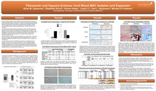
- 1. Fibronectin and Hypoxia Enhance Cord Blood MSC Isolation and Expansion Sivan M. Oyserman1, Elizabeth Schuh2, Darren Heeke1, Junzhi Li1, Dori L. Borjesson2, Michael S Friedman1 Thermogenesis1, University of California School of Veterinary Medicine2 Abstract Results Results Results Attempts to isolate mesenchymal stem cells (MSCs) from equine umbilical cord blood (UCB) have met with limited success. In these studies, we defined critical parameters correlating with Nucleated Cell Concentration Using the AXP System Phenotype of Proliferative and Senescent UCB MSC UCB MSC Exhibit Multilineage Potential successful UCB MSC isolation and expansion. 55 equine UCB units were processed using the AutoXpress (AXP) system to reduce volume and deplete red blood cells (RBCs). Total NC Pre-VXP Proliferative Senescent Adipocyte Osteoblast Chondrocyte recovery averaged 87.0% (SE 1.57%) with mononuclear cell recovery at 95.4% (SE 4.3%). Pre- and post-AXP processing viability averaged 94.5% (SE 1.5%) and 92.3% (SE 2.3%), Post-VXP respectively. Vimentin A. UCB MSCs were successfully isolated and expanded from >80% of the UCB samples. Post- 30 35 processing UCB NC number was the single critical parameter predictive for successful MSC 30 WB 25 expansion. UCB-derived MSC lines proliferated for > 20 passages before senescence. Early Cx 25 passage MSCs were negative for CD18, pancytokeratin, and factor VIII. Approximately 20% of 103/ 20 CD18 % cells expressed vimentin and smooth muscle actin. Conversely, MSCs that were poorly µl 20 15 proliferative or senescent, expressed vimentin (>80%), actin (>50%), osteonectin, and 15 osteocalcin (sporadic staining). 10 10 Culture of UCB MNC on fibronectin plates increased MSC CFU numbers nearly 14 fold relative 5 Actin 5 to standard or modified (Corning CellBind) tissue culture treated plastic (TCT). The use of fibronectin also increased the expansion success rate of UCB MSC to 100% compared to 40% Nucleated Cells Hematocrit for standard TCT. To further enhance the success of UCB MSC culture expansion, we evaluated the use of The effect of AutoXpress™ (MXP) processing on equine UCB. MXP processing significantly (A) hypoxia, which is known to have many beneficial effects. 5 out of 5 samples successfully increased nucleated cell number and B) decreased red blood cell number, as measured by hematocrit percent (p< 0.001, both measurements). Columns indicate the average of 56 Osteocalcin expanded in hypoxia (5% O2) compared to 2 out 5 under normoxia. UCB MSCs cultures Umbilical cord blood MSCs were cultured to passage 5-7 and plated at 20,00 cells per well of a initiated in noromoxia, hypoxia, or on fibronectin coated plates, all demonstrated similar levels samples, bars represent the standard error of the mean. 12 well plate. Adipogenesis was induced in media supplemented with rabbit serum, insulin, of multilineage differentiation to adipocytes, osteoblasts, and chondrocytes, as determined by IBMX, dexamethasone, and troglitazone. The cells were harvested at day 16-22 and stained cytochemical staining. In conclusion, equine UCB is a rich source of readily obtainable, highly Antibodies with known cross-reactivity to equine antigens were used to evaluate markers of with Oil Red O. Osteoblast differentiation was induced with ascorbate, beta glycerol phosphate, proliferative MSCs that could be banked for therapeutic use. Cord Blood Processing and Cord Blood MSC Culture mesenchymal origin (vimentin) as well as markers of differentiated cell lineages including dexamethasone, and BMP6. The cells were harvested at days 16-22 and stained with Alizarin endothelial cells (factor VIIIra, aka VonWillebrand factor), epithelial cells (pan cytokeratin), Red S. Chondrogenesis was induced in micromass cultures with TGF beta 3, BMP6, L-proline, leukocytes (CD18), smooth muscle (actin) and bone (osteocalcin and osteonectin). All equine insulin, transferrin, selenium, ascorbate, and L-proline. Micromasses were harvested at day 21, Interestingly, early passage, highly proliferative MSCs were negative for all markers except embedded, sectioned, and stained using Masson’s Trichrome technique. Umbilical cord blood Background sporadic expression of vimentin and actin. Conversely MSCs that were poorly proliferative and/or senescent expressed markers of differentiation. MSCs cultured in hypoxia, or initiated on fibronectin plates showed similar levels of differentiation. Summary of Immunocytochemistry Data Discussion Cord blood was collected from quarterhorses or thoroughbreds in a 250 mL collection bag, and anticoagulated with CPDA. The sample was shipped overnight in a SafeCellTM thermal isolation chamber. Upon arrival, the containers were opened and the temperature recorded. Approximately 5 mL of the sample was removed for pre-processing testing. For processing, In these studies, we determined that equine umbilical cord blood nucleated cells could be hetastarch (20% v/v) was added to the sample. The sample was split into two MXPTM bagsets, reliably concentrated using the AutoXpressTM platform. Furthermore, MSC could be reliably and loaded into the MXPTM mononuclear cell concentration device. After processing, one isolated and expanded from cord blood that was previously concentrated using the bagset was frozen in the BioarchiveTM and one was used for experimental evaluation. A Comparison of viability, days to first passage and successful MSC colony isolation and AutoXpressTM platform. MSC could be isolated from cord blood samples that were previously number of parameters were evaluated, including microbiology, differential blood counts, and expansion between fresh equine UCB-derived nucleated cells; cryopreserved, UCB-derived frozen in the BioArchiveTM as well. culture of cord blood derived mesenchymal stem cells. nucleated cells and equine UCB-derived NCs that were expanded to MSCs prior to Interestingly, UCB MSC from some donors exhibited a stellate morphology. These cells were cryopreservation then thawed and re-expanded. highly proliferative, but failed to differentiate to osteoblasts, adipocytes, or chondrocytes (data not shown). The lineage of these cells is unclear, and confounded by the lack of equine Sample Receipt-----MXP Processing-------------------------Experimental Conditions------------------ antibodies for specific surface markers. UCB MSCs Exhibit Different Morphologies Tissue Culture Plastic and Normoxia (21%O2 ) Fibronectin and Hypoxia Enhance UCM MSC Expansion During the course of cell culture, UCB MSC from approximately 1/3 of donors became senescent at relatively early passages (passage 2-3), and began to display markers associated A B with differentiation, such as actin and osteocalcin. We found no correlation between initial First 24 samples colony number and successful outgrowth to late passage. The lack of appropriate phenotypic markers for the equine species also make further analysis of this observation difficult. Initial plating on fibronectin, as well as culture in hypoxia, dramatically increased the successful outgrowth and expansion of UCB MSCs. Future studies will explore the effects of hypoxic Next 5 Tissue Culture Plastic and Hypoxia (5%O2 ) culture on early senescence, differentiation, and therapeutic efficacy of UCB MSC. We will also samples focus our efforts on identifying factors that increase the success of UCB MSC outgrowth. C D Acknowledgements Next 9 samples Fibronectin Substrate and Normoxia (21%O2 ) Average volume = 216 mLs Volume reduction to 21 mLs 1. These studies were funded by Thermogenesis RBC reduction to Cord blood MSCs from different donors exhibited strikingly different morphophologies at early MXP concentrated UCB from different donors was plated on tissue culture treated plastic (TCT) 2. All umbilical cord blood samples were provided by the UCD Center for Equine Health, the hematocrit of 11% Fibronectin Substrate and Normoxia (21%O2 ) passages (1-3), ranging from spindle (A), to stellate/flattened (B). At later passages (4-6), cord or on fibronectin coated TCT. Upon reaching ~70% confluence, the adherent UCB MSC were Harris Ranch, and LTR Ranch. NC enrichment Final samples blood MSCs from approximately 33% of donors became senscent and adopted a jagged passaged to a new vessel that was not coated with fibronectin. In a separate study (data not 3. Thank you to Margaret Nguyen, Dorian Lara, Danielle Carrade, Naomi Walker, Sean (n=13) morphology (C) while MSCs from other donors had a classic spindle morphology, and were shown), MXP concentrated UCB was also cultured in normoxia or 2% hypoxia on TCT. 2 out 5 Owens, Larry Galuppo, and Greg Ferraro for your tireless efforts in placenta, cord blood highly proliferative past passage 10 (D). donors expanded in normoxia, while 5 out of 5 expanded in hypoxia. collection, and cord blood processing.