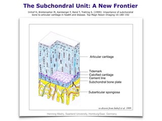
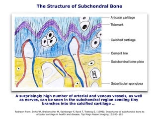
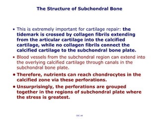
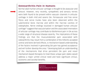
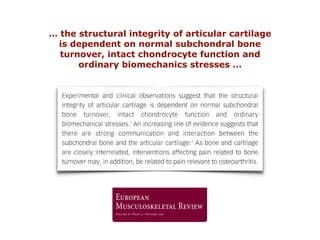
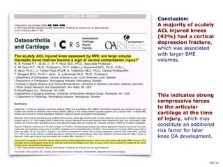
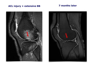
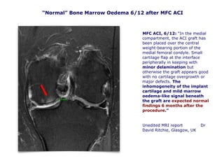
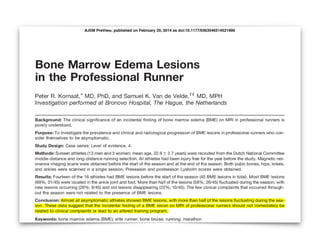
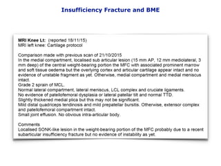
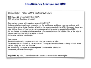
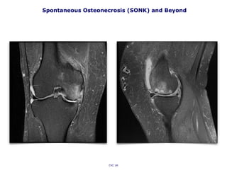
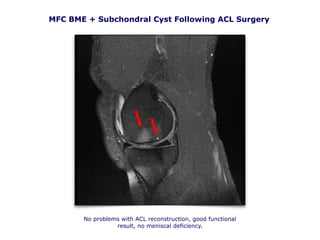
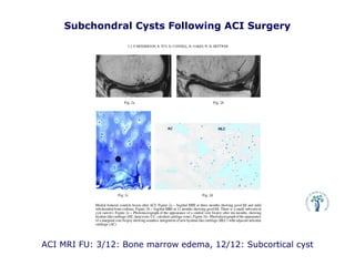
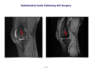
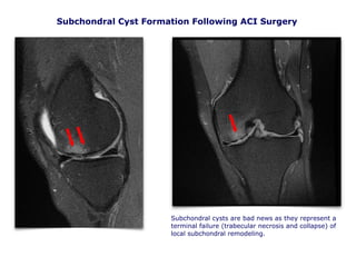
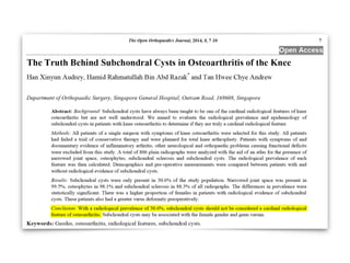
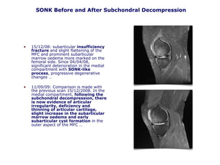
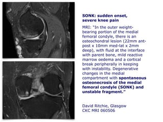
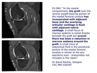
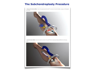
Articular Cartilage,
Subchondral Bone and
Osteochondral Unit
Prof. Vladimir Bobić, MD FRCS Ed
Chester Knee Clinic www.kneeclinic.info office@kneeclinic.info @ChesterKnee
The 4th BKS Annual Meeting
The Belfry Hotel and Resort, West Midlands
1st and 2nd February 2018
MARIARC MRI, UK (1997)
The orange pixels correspond to normal T2 values for bone. The blue and
purple pixels are anomalous: the T2 relaxation times are elevated because the
tissue is "wetter" than normal (the fluid interface between recipient and
donor bone).
OAT MRI analysis
“Chronic” BME and Cartilage Delamination
CKC UK
ICRS MR Cartilage
Imaging Protocol
January 2000
Osteochondral Functional Unit
Source: Dr Carl Winalski, Boston, USA, 2003
Well, not exactly a brand new concept:
Source: Francis Berenbaum, 3 November 2016
” … recent studies have shown that cartilage and subchondral bone act as a
single functional unit. This review highlights this novel concept.”
Osteoporosis Int. 2012
Cartilage + Subchondral Bone = a single functional unit
Nothing New!
Source: Francis Berenbaum, 3 November 2016
Radin et al. focused on subchondral role as an effective shock absorber. They found shear
stress in the articular cartilage always occurring whenever there is a discontinuity or
substantial gradient in stiffness of the subchondral plate. In former studies finite element
analysis showed increasing stress in the cartilage subsequent to subchondral plate stiffening. The
fact that these changes occurred without any evidence of metabolic or inflammatory
changes implied that the latter follow the mechanical changes, first in the bone and than
in the cartilage! December 1986.
The Role of Subchondral Bone (1986)
CKC UK
Chondrogenesis and OA
• The process of chondrogenesis is relevant to osteoarthritis (OA) in
two ways:
• 1st: evidence is beginning to emerge that osteoarthritic chondrocytes
are quite metabolically active and reinitiate synthesis of some
proteins that are characteristic of early developmental stages.
• 2nd: an understanding of cartilage differentiation and development will
provide guiding principles for tissue engineering of neo-cartilage, and
may, therefore, play a part in new therapies for this common disease.
• In the early phase of OA, the pathologic processes seem to
indicate that mechanisms of cartilage repair, rather than
degradation, are at work!
• There is substantial evidence to indicate that chondrocytes are
activated in OA and potentially could be stimulated to synthesize
appropriate cartilage ECM, or even recapitulate developmental
patterns.
14
OsteoArthritis or OsteoArthrosis?
or just wear and tear?
The Subchondral Unit: A New Frontier
re-drawn from Imhof et al. 1999
Henning Madry, Saarland University, Homburg/Saar, Germany
Imhof H, Breitenseher M, Kainberger F, Rand T, Trattnig S. (1999): Importance of subchondral
bone to articular cartilage in health and disease. Top Magn Reson Imaging 10:180–192
The Structure of Subchondral Bone
Redrawn from: Imhof H, Breitenseher M, Kainberger F, Rand T, Trattnig S. (1999): Importance of subchondral bone to
articular cartilage in health and disease. Top Magn Reson Imaging 10:180–192
A surprisingly high number of arterial and venous vessels, as well
as nerves, can be seen in the subchondral region sending tiny
branches into the calcified cartilage …
The Structure of Subchondral Bone
• This is extremely important for cartilage repair: the
tidemark is crossed by collagen fibrils extending
from the articular cartilage into the calcified
cartilage, while no collagen fibrils connect the
calcified cartilage to the subchondral bone plate.
• Blood vessels from the subchondral region can extend into
the overlying calcified cartilage through canals in the
subchondral bone plate.
• Therefore, nutrients can reach chondrocytes in the
calcified zone via these perforations.
• Unsurprisingly, the perforations are grouped
together in the regions of subchondral plate where
the stress is greatest.
CKC UK
… the structural integrity of articular cartilage
is dependent on normal subchondral bone
turnover, intact chondrocyte function and
ordinary biomechanics stresses …
The Structure of Subchondral Bone
The changes in the thickness of the subchondral bone plate depends on the
location and mechanical loads
Henning Madry, Saarland University, Homburg/Saar, Germany
• VB to DR email, 2006:
• Subject: SONK and all that jazz (re confusing MRI
appearance of different subchondral events):
• VB: “ There is something there and it seems it’s all
connected. We are probably looking at different stages of the
same thing:
• … it seems that subchondral repair and remodelling are a
common denominator, some of which is successful (traumatic
bone bruising, transient osteoporosis, SONK), partially
successful (persisting bone marrow oedema) or not at all
(progressive chronic bone marrow oedema, subchondral
cysts, AVN, osteonecrosis, and in the end secondary
osteoarthrosis). I don’t know, for some people most of this is
probably at various places on the same timeline, but I am not
sure if that makes sense.”
• DR: “The terminology is a bit confusing … “
• I would like to thank Dr David Ritchie and Dr Carl Winalski for
their unreserved help and patience over many years
(“Vladimir, stop staring at MR images and stick to your day
job.”)
Subchondral Events in a Nutshell:
The Terminology is a Bit Confusing …
• Disclaimer: Arthrex (OATS inventor), principal investigator for UK BioPoly clinical trial, former clinical advisor for TiGenix CCI.
• This is a non-EBM (orthopaedic surgeon’s) clinical overview of “subchondral events”, based on our clinical and MR imaging
experience since 1996.
• We have a problem at the outset: what are we actually talking about? How do
we treat a wide range of osteochondral problems which are still poorly defined
and understood?
• Bone Bruise (BB): a transient traumatic event = a series of trabecular
microfractures. No surgical treatment required.
• Bone Marrow Oedema (BME): MRI evidence of increased subchondral
metabolic activity. Remodelling or reparative process, or a failure of subchondral
remodelling or repair = a degenerative process? Initially a reparative process, but
if persistent it is probably a degenerative process (often associated with initial
formation of subchondral cysts, which are a consequence of failed local repair).
No surgical treatment required, but …
• Transient Osteoporosis (TOP): no history of trauma, the knee pain is
spontaneous and disabling, exacerbated on weight-bearing. Usually gets better
over many months, back to normal MRI and clinically. When multiple joints are
involved (in approx. 40% of patients) the condition is referred to as Transient
Regional Migratory Osteoporosis (TRMO). No surgical treatment required.
CKC UK
No Edema in Bone Marrow Edema!
• The correlation of bone marrow lesions with pain in knee OA has been
convincingly established. Here, another compelling association is established
between bone marrow (edema) lesions and risk for progression of knee OA.
What remains to be established is the cause-and-effect relationship between
the various variables.
• It is interesting that, histologically, the lesions that appear as bone
marrow edema on MRI contain very little edema at all. Rather, they
demonstrate fibrosis, osteonecrosis, and extensive bony REMODELLING
and are likely the result of contusions and focal microfractures.
• Also, it is not clear, whether an initial injury to articular cartilage
leads to mechanical malalignment and subsequent subchondral bone
destruction or rather subchondral bone damage leads to mechanical
malalignment and subsequent articular cartilage destruction.
Does bone marrow edema predict progression of knee arthritis?
A summary of Felson’s 2001 and 2003 AIM articles written by Jon Gilles, M.D., The John Hopkins Arthritis Center, 2003.
http://www.hopkins-arthritis.org/arthritis-news/2003/bone_edema_oa.html
Conclusion:
A majority of acutely
ACL injured knees
(92%) had a cortical
depression fracture,
which was associated
with larger BME
volumes.
This indicates strong
compressive forces
to the articular
cartilage at the time
of injury, which may
constitute an additional
risk factor for later
knee OA development.
CKC UK
ACL injury + extensive BB
CKC MRI 110206
7 months later
MFC ACI, 6/12: “In the medial
compartment, the ACI graft has
been placed over the central
weight-bearing portion of the
medial femoral condyle. Small
cartilage flap at the interface
peripherally in keeping with
minor delamination but
otherwise the graft appears good
with no cartilage overgrowth or
major defects. The
inhomogeneity of the implant
cartilage and mild marrow
oedema-like signal beneath
the graft are expected normal
findings 6 months after the
procedure.”
Unedited MRI report Dr
David Ritchie, Glasgow, UK
CKC MRI 260906
“Normal” Bone Marrow Oedema 6/12 after MFC ACI
Transient Regional Migratory Osteoporosis:
MRI report: “The diffuse bone marrow oedema pattern with development of subchondral linear fractures
would therefore suggest regional migratory osteoporosis rather than typical SONK lesions.”
Transient Osteoporosis – Extreme Bone Remodeling?
CKC UK
• The aetiology of TO and TRMO remains unclear:
• One of the likely explanations for the pathogenesis of TO is perhaps
that proposed by Frost and others.
• He stated that under noxious tissue stimuli, the ordinary biological
processes, including blood flow, cell metabolism and turnover
and also tissue modelling and remodelling, might be greatly
accelerated, called the Regional Acceleratory Phenomenon
(RAP). In his opinion a prolonged or exaggerated RAP in which a
large number of bone turnover foci are activated, is the cause of
TO.
• It has been hypothesized that symptoms may be related to bone
marrow edema demonstrated at MRI and to a transitory regional
arterial hyperflow observed at the early scintigraphic analysis.
Bone tissue micro damage is the most frequent noxious
stimulus that provokes RAP and bone tissue micro fracture
is the main consequence.
• Several elements support this hypothesis. The repeatedly observed
histological findings in patients with TO showing mild inflammatory
changes and osteoporosis, associated with an elevated bone
turnover with increased bone resorption and reactive bone
formation are a good description of ongoing TRMO.
The Terminology is Even More Confusing …
• Spontaneous Osteonecrosis (SONK): is the term used to describe a
subchondral insufficiency fracture that causes osteonecrosis. MRI
appearance: a thin linear hypointense subchondral focus on T1W and T2W
that blends in with overlying cortex and is typically surrounded by
diffuse BME. With or without subchondral fractures/deformity.
• Avascular Necrosis (AVN): an osteonecrotic lesion, low signal rim on
T1W and double line sign on T2W, with or without BME. The necrotic focus
often extends some distance away from the articular margin and may
contain fat, blood, fluid, fibrous tissue. With or without subchondral
fractures/deformity.
• Osteochondritis Dissecans (OCD): semidetached osteochondral
fragment (essentially a non-union) with fluid layer at osseous interface and
seemingly intact articulating surface. A traumatic or metabolic event, or
both?
• Secondary Osteoarthrosis (not –itis): if localized, this is perhaps the
end result of more extensive progressive failure of subchondral
remodelling. Increased but unsuccessful subchondral activity (progressive
BME + multiple cysts?) seems to be a primary event, with secondary loss
of articulating surface, which fails gradually as it is not supported by
normal elastic trabecular bone. May go as far back as injury-induced BB,
with or without visible initial chondral damage.
CKC UK
BME and Insufficiency Fracture
Recent localised incomplete subarticular fracture of the outer aspect of the MFC (15 x 5 x 3
mm) with slight depression of the overlying articular cortex and prominent surrounding
marrow and soft tissue oedema but no obvious disruption of the overlying articular
cartilage or unstable osteochondral fragment.
CKC UK
Insufficiency Fracture and BME
AJSM 2011
SONK (Spontaneous Osteonecrosis)
CKC UK
Spontaneous Osteonecrosis (SONK)
• Ahlback et al first described
spontaneous osteonecrosis of the
knee as a distinct clinical entity in
1968.
• Osteonecrosis of the knee has also
been described as a postsurgical
complication following
arthroscopic meniscectomy
(Muscolo et al., Prues-Latour et al.)
and following radiofrequency-
assisted arthroscopic treatments,
mainly in 50+ age groups.
• The pathophysiology of osteonecrosis
following these arthroscopic
procedures is not fully understood
(vascular isufficiency, trabecular
microfractures?), or, more likely, a
consequence of pre-arthrosopy
osteopoenia and altered focal
biomechanics (bone density
should be looked into).
Spontaneous Osteonecrosis of Both Femoral Condyles
Shifting Bone Marrow Oedema is a self-contained disorder involving both femoral condyles. On MRI it
exhibits vast marrow oedema and is most likely an event on the SONK timeline.
SONK Histology
In the early stages of the
condition a subchondral
fracture was noted in the
absence of any features of
osteonecrosis, whereas in
advanced stages, osteonecrotic
lesions were confined to the area
distal to the site of the fracture
which showed impaired healing.
In such cases, formation of
cartilage and fibrous tissue,
occurred indicating delayed
or non-union.
These findings strongly suggest
that the histopathology at each
stage of spontaneous
osteonecrosis is characterised by
different types of repair reaction
for subchondral fractures.
Spontaneous Osteonecrosis (SONK) and Beyond
CKC UK
MFC BME + Subchondral Cyst Following ACL Surgery
No problems with ACL reconstruction, good functional
result, no meniscal deficiency.
ACI MRI FU: 3/12: Bone marrow edema, 12/12: Subcortical cyst
Subchondral Cysts Following ACI Surgery
CKC UK
Subchondral Cyst Formation Following ACI Surgery
Subchondral cysts are bad news as they represent a
terminal failure (trabecular necrosis and collapse) of
local subchondral remodeling.
• Conclusions: Delivery of bone
marrow concentrate can result
in healing of acute full-thickness
cartilage defects that is superior
to that after microfracture
alone in an equine model.
• If this is the case, looking at
osteochondral defects, is this
combination working better
because microfracture (multiple
perforations and tunnelling) of
subchondral bone is making it less
stiff but also allows “biologic
fuel” (bone marrow, blood and
who knows what else) to reach
deeper areas, re-establish
nutrition and facilitate local
osteochondral repair?
ABMA: An Essential Ingredient for Octeochondral Repair?
JBJS A August 2010
Lateral Femoral Trochlea:
a reliable source of good cancellous bone and bone marrow, even in advanced OA
CKC UK
MFC AVN
An alternative approach to the treatment of
femoral and tibial Osteonecrosis, Chronic SONK
and Secondary OA:
• The knee is often not too bad (all 3 compartments) or it is too early
for a partial or a full knee replacement.
• Classic Microfracture and Core Decompression are probably not deep
enough.
• Looking at most MRIs it seems that we need to reach at least 15 to 20
mm deep into subchondral bone, which is where any cylindrical
osteochondral harvesters are very handy.
• Effectively, this is a combination of OAT and deep core (subchondral)
decompression, with a hand driven K-wire, through the bottom of the
recipient socket, with
• a mixture of autologous blood + bone marrow injected into the
recipient socket,
• and capped with 10 mm OATS plug, which was soaked in the same
mixture of bone marrow and blood.
• This “integrated” subchondral repair concept makes sense, it gives
most people quick and durable pain relief and better knee function,
but it is based on huge assumptions.
• The main question is weather unprocessed (and not concentrated)
autologous bone marrow, is powerful enough biologically?
CKC UK
“Pre-TKR” option: OATS combined with autologous bone marrow aspirate
Autologous Bone Marrow Aspirate
AANA Annual Meeting San Francisco 2011
Autologous Bone Marrow
• Red marrow has significant haematopoietic stem cell potential
and still persists in adults in certain areas such as the iliac
crests.
• The anterolateral trochlea (the usual OATS donor site) is often
spared even in advanced OA and seems to contain reasonably
good bone marrow, which can be aspirated through the donor
site.
• Pluripotent haematopoietic stem cells can differentiate into any and all
of the cells of circulating blood and the immune system.
• MRI studies have indicated that the conversion of red to fatty marrow
occurs prematurely in some patients with avascular necrosis.
• Osteonecrosis is associated with a decrease in progenitor cells in the
proximal femur. Bone marrow also contains osteogenic progenitors,
with a potential for effective bone regeneration.
• It seems sensible to use core decompression but also to deliver
better “biologic fuel” with pluripotent cells to the affected area.
• The quantity and quality of good autologous bone marrow is
questionable, but as it seems that stem cells do not to do the
actual work (they seem to go around and boss other cells and
tell them where to go and what to do) a few mils of bone
marrow aspirate may be good enough to kick-start the process.
CKC UK
JBJS B June 2006
SONK Before and After Subchondral Decompression
• 15/12/08: subarticular insufficiency
fracture and slight flattening of the
MFC and prominent subarticular
marrow oedema more marked on the
femoral side. Since 04/04/08,
significant deterioration in the medial
compartment with SONK-like
process, progressive degenerative
changes …
• 11/09/09: Comparison is made with
the previous scan 15/12/2008. In the
medial compartment, following the
subchondral decompression, there
is now evidence of articular
irregularity, deficiency and
thinning of articular cartilage,
slight increase in the subarticular
marrow oedema and early
subarticular cyst formation in the
outer aspect of the MFC …
SONK: sudden onset,
severe knee pain
MRI: “In the outer weight-
bearing portion of the medial
femoral condyle, there is an
osteochondral lesion (22mm ant-
post x 10mm med-lat x 2mm
deep), with fluid at the interface
with parent bone, mild reactive
marrow oedema and a cortical
break peripherally in keeping
with instability. Degenerative
changes in the medial
compartment with spontaneous
osteonecrosis of the medial
femoral condyle (SONK) and
unstable fragment.”
David Ritchie, Glasgow
CKC MRI 060506
FU MRI: “In the medial
compartment, the graft over the
central weight-bearing portion of
the medial femoral condyle has
incorporated with adjacent
bone and the overlying
articular cartilage is flush
with adjacent native
cartilage. A small focus of
marrow oedema is noted directly
beneath the graft but overall
there has been a reduction in
marrow oedema around the
graft. A small trace of
subcortical fluid in the peripheral
portion of the medial femoral
condyle is similar to the pre-
operative scan - presumably not
included in the repair.”
Dr David Ritchie, Glasgow
CKC MRI 030307
The Subchondroplasty Procedure
Great idea, but it seems that this entirely new concept (as it is) is based on huge
assumptions.
Arguably, subchondroplasty is indicated mainly for the treatment of subchondral
cysts and cavities, rather than various bone marrow oedema conditions. Add image
of ReproBone
Bone marrow oedema, as metabolic (vascular) “event” does not lack bone
(therefore injecting bone substitute is not the right ingredient). To the contrary,
injecting bone paste will clog many interconnected cellular spaces and will slow
down or prevent subchondral repair and remodelling. Not surprised to hear that
patients "should expect 3 days of severe pain" (!) as injected and cured bone paste
will increase intra-osseous pressure (which is already higher than normal and which
is why some conditions like SONK are very painful to start with) and block
metabolic (vascular) pathways!
However, the real (biologically) desirable ingredient is autologous bone marrow
aspirate, delivered to the area affected with bone marrow oedema.
This is where subchondroplasty becomes a bit more intelligent and gets entirely
new biological meaning and a lot more street cred.
I rest my case! Questions?
Frustration after Subchondroplasty presentation at Sports Knee 17:
@BioKneeSociety
Thank You
Articular Cartilage
Subchondral Bone and
Osteochondral Unit

Bobic Vladimir - Subchondral Activity - BKS Meeting UK 2 Feb 2018

  • 1.
    Articular Cartilage, Subchondral Boneand Osteochondral Unit Prof. Vladimir Bobić, MD FRCS Ed Chester Knee Clinic www.kneeclinic.info office@kneeclinic.info @ChesterKnee The 4th BKS Annual Meeting The Belfry Hotel and Resort, West Midlands 1st and 2nd February 2018
  • 3.
    MARIARC MRI, UK(1997) The orange pixels correspond to normal T2 values for bone. The blue and purple pixels are anomalous: the T2 relaxation times are elevated because the tissue is "wetter" than normal (the fluid interface between recipient and donor bone). OAT MRI analysis
  • 5.
    “Chronic” BME andCartilage Delamination CKC UK
  • 6.
    ICRS MR Cartilage ImagingProtocol January 2000
  • 8.
    Osteochondral Functional Unit Source:Dr Carl Winalski, Boston, USA, 2003
  • 9.
    Well, not exactlya brand new concept: Source: Francis Berenbaum, 3 November 2016
  • 10.
    ” … recentstudies have shown that cartilage and subchondral bone act as a single functional unit. This review highlights this novel concept.” Osteoporosis Int. 2012 Cartilage + Subchondral Bone = a single functional unit
  • 12.
    Nothing New! Source: FrancisBerenbaum, 3 November 2016
  • 13.
    Radin et al.focused on subchondral role as an effective shock absorber. They found shear stress in the articular cartilage always occurring whenever there is a discontinuity or substantial gradient in stiffness of the subchondral plate. In former studies finite element analysis showed increasing stress in the cartilage subsequent to subchondral plate stiffening. The fact that these changes occurred without any evidence of metabolic or inflammatory changes implied that the latter follow the mechanical changes, first in the bone and than in the cartilage! December 1986. The Role of Subchondral Bone (1986) CKC UK
  • 14.
    Chondrogenesis and OA •The process of chondrogenesis is relevant to osteoarthritis (OA) in two ways: • 1st: evidence is beginning to emerge that osteoarthritic chondrocytes are quite metabolically active and reinitiate synthesis of some proteins that are characteristic of early developmental stages. • 2nd: an understanding of cartilage differentiation and development will provide guiding principles for tissue engineering of neo-cartilage, and may, therefore, play a part in new therapies for this common disease. • In the early phase of OA, the pathologic processes seem to indicate that mechanisms of cartilage repair, rather than degradation, are at work! • There is substantial evidence to indicate that chondrocytes are activated in OA and potentially could be stimulated to synthesize appropriate cartilage ECM, or even recapitulate developmental patterns. 14
  • 15.
  • 17.
    The Subchondral Unit:A New Frontier re-drawn from Imhof et al. 1999 Henning Madry, Saarland University, Homburg/Saar, Germany Imhof H, Breitenseher M, Kainberger F, Rand T, Trattnig S. (1999): Importance of subchondral bone to articular cartilage in health and disease. Top Magn Reson Imaging 10:180–192
  • 18.
    The Structure ofSubchondral Bone Redrawn from: Imhof H, Breitenseher M, Kainberger F, Rand T, Trattnig S. (1999): Importance of subchondral bone to articular cartilage in health and disease. Top Magn Reson Imaging 10:180–192 A surprisingly high number of arterial and venous vessels, as well as nerves, can be seen in the subchondral region sending tiny branches into the calcified cartilage …
  • 19.
    The Structure ofSubchondral Bone • This is extremely important for cartilage repair: the tidemark is crossed by collagen fibrils extending from the articular cartilage into the calcified cartilage, while no collagen fibrils connect the calcified cartilage to the subchondral bone plate. • Blood vessels from the subchondral region can extend into the overlying calcified cartilage through canals in the subchondral bone plate. • Therefore, nutrients can reach chondrocytes in the calcified zone via these perforations. • Unsurprisingly, the perforations are grouped together in the regions of subchondral plate where the stress is greatest. CKC UK
  • 21.
    … the structuralintegrity of articular cartilage is dependent on normal subchondral bone turnover, intact chondrocyte function and ordinary biomechanics stresses …
  • 22.
    The Structure ofSubchondral Bone The changes in the thickness of the subchondral bone plate depends on the location and mechanical loads Henning Madry, Saarland University, Homburg/Saar, Germany
  • 23.
    • VB toDR email, 2006: • Subject: SONK and all that jazz (re confusing MRI appearance of different subchondral events): • VB: “ There is something there and it seems it’s all connected. We are probably looking at different stages of the same thing: • … it seems that subchondral repair and remodelling are a common denominator, some of which is successful (traumatic bone bruising, transient osteoporosis, SONK), partially successful (persisting bone marrow oedema) or not at all (progressive chronic bone marrow oedema, subchondral cysts, AVN, osteonecrosis, and in the end secondary osteoarthrosis). I don’t know, for some people most of this is probably at various places on the same timeline, but I am not sure if that makes sense.” • DR: “The terminology is a bit confusing … “ • I would like to thank Dr David Ritchie and Dr Carl Winalski for their unreserved help and patience over many years (“Vladimir, stop staring at MR images and stick to your day job.”) Subchondral Events in a Nutshell:
  • 24.
    The Terminology isa Bit Confusing … • Disclaimer: Arthrex (OATS inventor), principal investigator for UK BioPoly clinical trial, former clinical advisor for TiGenix CCI. • This is a non-EBM (orthopaedic surgeon’s) clinical overview of “subchondral events”, based on our clinical and MR imaging experience since 1996. • We have a problem at the outset: what are we actually talking about? How do we treat a wide range of osteochondral problems which are still poorly defined and understood? • Bone Bruise (BB): a transient traumatic event = a series of trabecular microfractures. No surgical treatment required. • Bone Marrow Oedema (BME): MRI evidence of increased subchondral metabolic activity. Remodelling or reparative process, or a failure of subchondral remodelling or repair = a degenerative process? Initially a reparative process, but if persistent it is probably a degenerative process (often associated with initial formation of subchondral cysts, which are a consequence of failed local repair). No surgical treatment required, but … • Transient Osteoporosis (TOP): no history of trauma, the knee pain is spontaneous and disabling, exacerbated on weight-bearing. Usually gets better over many months, back to normal MRI and clinically. When multiple joints are involved (in approx. 40% of patients) the condition is referred to as Transient Regional Migratory Osteoporosis (TRMO). No surgical treatment required. CKC UK
  • 25.
    No Edema inBone Marrow Edema! • The correlation of bone marrow lesions with pain in knee OA has been convincingly established. Here, another compelling association is established between bone marrow (edema) lesions and risk for progression of knee OA. What remains to be established is the cause-and-effect relationship between the various variables. • It is interesting that, histologically, the lesions that appear as bone marrow edema on MRI contain very little edema at all. Rather, they demonstrate fibrosis, osteonecrosis, and extensive bony REMODELLING and are likely the result of contusions and focal microfractures. • Also, it is not clear, whether an initial injury to articular cartilage leads to mechanical malalignment and subsequent subchondral bone destruction or rather subchondral bone damage leads to mechanical malalignment and subsequent articular cartilage destruction. Does bone marrow edema predict progression of knee arthritis? A summary of Felson’s 2001 and 2003 AIM articles written by Jon Gilles, M.D., The John Hopkins Arthritis Center, 2003. http://www.hopkins-arthritis.org/arthritis-news/2003/bone_edema_oa.html
  • 26.
    Conclusion: A majority ofacutely ACL injured knees (92%) had a cortical depression fracture, which was associated with larger BME volumes. This indicates strong compressive forces to the articular cartilage at the time of injury, which may constitute an additional risk factor for later knee OA development. CKC UK
  • 27.
    ACL injury +extensive BB CKC MRI 110206 7 months later
  • 28.
    MFC ACI, 6/12:“In the medial compartment, the ACI graft has been placed over the central weight-bearing portion of the medial femoral condyle. Small cartilage flap at the interface peripherally in keeping with minor delamination but otherwise the graft appears good with no cartilage overgrowth or major defects. The inhomogeneity of the implant cartilage and mild marrow oedema-like signal beneath the graft are expected normal findings 6 months after the procedure.” Unedited MRI report Dr David Ritchie, Glasgow, UK CKC MRI 260906 “Normal” Bone Marrow Oedema 6/12 after MFC ACI
  • 30.
    Transient Regional MigratoryOsteoporosis: MRI report: “The diffuse bone marrow oedema pattern with development of subchondral linear fractures would therefore suggest regional migratory osteoporosis rather than typical SONK lesions.”
  • 31.
    Transient Osteoporosis –Extreme Bone Remodeling? CKC UK • The aetiology of TO and TRMO remains unclear: • One of the likely explanations for the pathogenesis of TO is perhaps that proposed by Frost and others. • He stated that under noxious tissue stimuli, the ordinary biological processes, including blood flow, cell metabolism and turnover and also tissue modelling and remodelling, might be greatly accelerated, called the Regional Acceleratory Phenomenon (RAP). In his opinion a prolonged or exaggerated RAP in which a large number of bone turnover foci are activated, is the cause of TO. • It has been hypothesized that symptoms may be related to bone marrow edema demonstrated at MRI and to a transitory regional arterial hyperflow observed at the early scintigraphic analysis. Bone tissue micro damage is the most frequent noxious stimulus that provokes RAP and bone tissue micro fracture is the main consequence. • Several elements support this hypothesis. The repeatedly observed histological findings in patients with TO showing mild inflammatory changes and osteoporosis, associated with an elevated bone turnover with increased bone resorption and reactive bone formation are a good description of ongoing TRMO.
  • 32.
    The Terminology isEven More Confusing … • Spontaneous Osteonecrosis (SONK): is the term used to describe a subchondral insufficiency fracture that causes osteonecrosis. MRI appearance: a thin linear hypointense subchondral focus on T1W and T2W that blends in with overlying cortex and is typically surrounded by diffuse BME. With or without subchondral fractures/deformity. • Avascular Necrosis (AVN): an osteonecrotic lesion, low signal rim on T1W and double line sign on T2W, with or without BME. The necrotic focus often extends some distance away from the articular margin and may contain fat, blood, fluid, fibrous tissue. With or without subchondral fractures/deformity. • Osteochondritis Dissecans (OCD): semidetached osteochondral fragment (essentially a non-union) with fluid layer at osseous interface and seemingly intact articulating surface. A traumatic or metabolic event, or both? • Secondary Osteoarthrosis (not –itis): if localized, this is perhaps the end result of more extensive progressive failure of subchondral remodelling. Increased but unsuccessful subchondral activity (progressive BME + multiple cysts?) seems to be a primary event, with secondary loss of articulating surface, which fails gradually as it is not supported by normal elastic trabecular bone. May go as far back as injury-induced BB, with or without visible initial chondral damage. CKC UK
  • 33.
    BME and InsufficiencyFracture Recent localised incomplete subarticular fracture of the outer aspect of the MFC (15 x 5 x 3 mm) with slight depression of the overlying articular cortex and prominent surrounding marrow and soft tissue oedema but no obvious disruption of the overlying articular cartilage or unstable osteochondral fragment. CKC UK
  • 34.
  • 35.
  • 36.
  • 38.
  • 39.
  • 40.
    Spontaneous Osteonecrosis (SONK) •Ahlback et al first described spontaneous osteonecrosis of the knee as a distinct clinical entity in 1968. • Osteonecrosis of the knee has also been described as a postsurgical complication following arthroscopic meniscectomy (Muscolo et al., Prues-Latour et al.) and following radiofrequency- assisted arthroscopic treatments, mainly in 50+ age groups. • The pathophysiology of osteonecrosis following these arthroscopic procedures is not fully understood (vascular isufficiency, trabecular microfractures?), or, more likely, a consequence of pre-arthrosopy osteopoenia and altered focal biomechanics (bone density should be looked into).
  • 41.
    Spontaneous Osteonecrosis ofBoth Femoral Condyles Shifting Bone Marrow Oedema is a self-contained disorder involving both femoral condyles. On MRI it exhibits vast marrow oedema and is most likely an event on the SONK timeline.
  • 42.
    SONK Histology In theearly stages of the condition a subchondral fracture was noted in the absence of any features of osteonecrosis, whereas in advanced stages, osteonecrotic lesions were confined to the area distal to the site of the fracture which showed impaired healing. In such cases, formation of cartilage and fibrous tissue, occurred indicating delayed or non-union. These findings strongly suggest that the histopathology at each stage of spontaneous osteonecrosis is characterised by different types of repair reaction for subchondral fractures.
  • 43.
  • 44.
    MFC BME +Subchondral Cyst Following ACL Surgery No problems with ACL reconstruction, good functional result, no meniscal deficiency.
  • 45.
    ACI MRI FU:3/12: Bone marrow edema, 12/12: Subcortical cyst Subchondral Cysts Following ACI Surgery
  • 46.
    Subchondral Cysts FollowingACI Surgery CKC UK
  • 47.
    Subchondral Cyst FormationFollowing ACI Surgery Subchondral cysts are bad news as they represent a terminal failure (trabecular necrosis and collapse) of local subchondral remodeling.
  • 50.
    • Conclusions: Deliveryof bone marrow concentrate can result in healing of acute full-thickness cartilage defects that is superior to that after microfracture alone in an equine model. • If this is the case, looking at osteochondral defects, is this combination working better because microfracture (multiple perforations and tunnelling) of subchondral bone is making it less stiff but also allows “biologic fuel” (bone marrow, blood and who knows what else) to reach deeper areas, re-establish nutrition and facilitate local osteochondral repair? ABMA: An Essential Ingredient for Octeochondral Repair? JBJS A August 2010
  • 51.
    Lateral Femoral Trochlea: areliable source of good cancellous bone and bone marrow, even in advanced OA CKC UK MFC AVN
  • 52.
    An alternative approachto the treatment of femoral and tibial Osteonecrosis, Chronic SONK and Secondary OA: • The knee is often not too bad (all 3 compartments) or it is too early for a partial or a full knee replacement. • Classic Microfracture and Core Decompression are probably not deep enough. • Looking at most MRIs it seems that we need to reach at least 15 to 20 mm deep into subchondral bone, which is where any cylindrical osteochondral harvesters are very handy. • Effectively, this is a combination of OAT and deep core (subchondral) decompression, with a hand driven K-wire, through the bottom of the recipient socket, with • a mixture of autologous blood + bone marrow injected into the recipient socket, • and capped with 10 mm OATS plug, which was soaked in the same mixture of bone marrow and blood. • This “integrated” subchondral repair concept makes sense, it gives most people quick and durable pain relief and better knee function, but it is based on huge assumptions. • The main question is weather unprocessed (and not concentrated) autologous bone marrow, is powerful enough biologically? CKC UK
  • 53.
    “Pre-TKR” option: OATScombined with autologous bone marrow aspirate
  • 54.
  • 55.
    Autologous Bone MarrowAspirate AANA Annual Meeting San Francisco 2011
  • 56.
    Autologous Bone Marrow •Red marrow has significant haematopoietic stem cell potential and still persists in adults in certain areas such as the iliac crests. • The anterolateral trochlea (the usual OATS donor site) is often spared even in advanced OA and seems to contain reasonably good bone marrow, which can be aspirated through the donor site. • Pluripotent haematopoietic stem cells can differentiate into any and all of the cells of circulating blood and the immune system. • MRI studies have indicated that the conversion of red to fatty marrow occurs prematurely in some patients with avascular necrosis. • Osteonecrosis is associated with a decrease in progenitor cells in the proximal femur. Bone marrow also contains osteogenic progenitors, with a potential for effective bone regeneration. • It seems sensible to use core decompression but also to deliver better “biologic fuel” with pluripotent cells to the affected area. • The quantity and quality of good autologous bone marrow is questionable, but as it seems that stem cells do not to do the actual work (they seem to go around and boss other cells and tell them where to go and what to do) a few mils of bone marrow aspirate may be good enough to kick-start the process. CKC UK
  • 58.
  • 59.
    SONK Before andAfter Subchondral Decompression • 15/12/08: subarticular insufficiency fracture and slight flattening of the MFC and prominent subarticular marrow oedema more marked on the femoral side. Since 04/04/08, significant deterioration in the medial compartment with SONK-like process, progressive degenerative changes … • 11/09/09: Comparison is made with the previous scan 15/12/2008. In the medial compartment, following the subchondral decompression, there is now evidence of articular irregularity, deficiency and thinning of articular cartilage, slight increase in the subarticular marrow oedema and early subarticular cyst formation in the outer aspect of the MFC …
  • 60.
    SONK: sudden onset, severeknee pain MRI: “In the outer weight- bearing portion of the medial femoral condyle, there is an osteochondral lesion (22mm ant- post x 10mm med-lat x 2mm deep), with fluid at the interface with parent bone, mild reactive marrow oedema and a cortical break peripherally in keeping with instability. Degenerative changes in the medial compartment with spontaneous osteonecrosis of the medial femoral condyle (SONK) and unstable fragment.” David Ritchie, Glasgow CKC MRI 060506
  • 61.
    FU MRI: “Inthe medial compartment, the graft over the central weight-bearing portion of the medial femoral condyle has incorporated with adjacent bone and the overlying articular cartilage is flush with adjacent native cartilage. A small focus of marrow oedema is noted directly beneath the graft but overall there has been a reduction in marrow oedema around the graft. A small trace of subcortical fluid in the peripheral portion of the medial femoral condyle is similar to the pre- operative scan - presumably not included in the repair.” Dr David Ritchie, Glasgow CKC MRI 030307
  • 62.
  • 64.
    The Subchondroplasty Procedure Greatidea, but it seems that this entirely new concept (as it is) is based on huge assumptions. Arguably, subchondroplasty is indicated mainly for the treatment of subchondral cysts and cavities, rather than various bone marrow oedema conditions. Add image of ReproBone Bone marrow oedema, as metabolic (vascular) “event” does not lack bone (therefore injecting bone substitute is not the right ingredient). To the contrary, injecting bone paste will clog many interconnected cellular spaces and will slow down or prevent subchondral repair and remodelling. Not surprised to hear that patients "should expect 3 days of severe pain" (!) as injected and cured bone paste will increase intra-osseous pressure (which is already higher than normal and which is why some conditions like SONK are very painful to start with) and block metabolic (vascular) pathways! However, the real (biologically) desirable ingredient is autologous bone marrow aspirate, delivered to the area affected with bone marrow oedema. This is where subchondroplasty becomes a bit more intelligent and gets entirely new biological meaning and a lot more street cred. I rest my case! Questions?
  • 65.
    Frustration after Subchondroplastypresentation at Sports Knee 17:
  • 66.
  • 67.
    Thank You Articular Cartilage SubchondralBone and Osteochondral Unit