DBS PROTOCOL POSTER.pptx
•Download as PPTX, PDF•
0 likes•98 views

THIS POSTER INCLUSEDS MRI GUIDED DEEP BRAIN STIMULATION PROTOCOL, WHICH CONSITS OF ITS INDICATIONSCONTRAINDICATIONS,PREPARATION, PROCEDURES ,PROTOCAL , RISKS
Recommended

Recommended
More Related Content
Similar to DBS PROTOCOL POSTER.pptx
Similar to DBS PROTOCOL POSTER.pptx (20)
More from VANI PUSHPA MUDAVATH
More from VANI PUSHPA MUDAVATH (11)
Recently uploaded
Recently uploaded (20)
DBS PROTOCOL POSTER.pptx
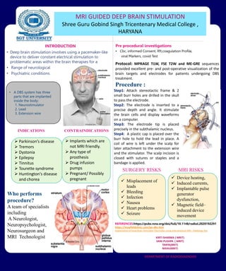
- 1. • Deep brain stimulation involves using a pacemaker-like device to deliver constant electrical stimulation to problematic areas within the brain therapies for a • Range of neurological • Psychiatric conditions. INTRODUCTION INDICATIONS CONTRAINDICATIONS Pre procedural investigations • Cbc, informed Consent, Rft,coagulation Profile, viral Markers, covid Test REFERENCES:https://pubs.rsna.org/doi/full/10.1148/radiol.2020192291 https://mayfieldclinic.com/pe-dbs.htm Implantation of Deep Brain Stimulator Electrodes Using Interventional MRI | Radiology Key KIRTI SHARMA ( MRIT) VANI PUSHPA ( MRIT) TANYA(BRIT) NISHU(BRIT) DEPARTMENT OF RADIODIAGNOSIS SURGERY RISKS MRI RISKS • A DBS system has three parts that are implanted inside the body: 1. Neurostimulator 2. Lead 3. Extension wire Who performs procedure? A team of specialists including A Neurologist, Neuropsychologist, Neurosurgeon and MRI Technologist MRI GUIDED DEEP BRAIN STIMULATION Shree Guru Gobind Singh Tricentenary Medical College , HARYANA Parkinson’s disease Tremors Dystonia Epilepsy Tinnitus Tourette syndrome Huntington's disease and chorea Implants which are not MRI friendly. Any type of prosthesis Drug infusion pumps Pregnant/ Possibly pregnant Protocol: MPRAGE T1W, FSE T2W and ME-GRE sequences provided excellent pre- and post-operative visualization of the brain targets and electrodes for patients undergoing DBS treatment. Procedure : Misplacement of leads Bleeding Infection Nausea Heart problems Seizure Device heating, Induced currents, Implantable pulse generator dysfunction, Magnetic field– induced device movement Step1: Attach stereotactic frame & 2 small burr holes are drilled in the skull to pass the electrode. Step2: The electrode is inserted to a precise depth and angle. It stimulate the brain cells and display waveforms on a computer. Step3: The electrode tip is placed precisely in the subthalamic nucleus. Step4: A plastic cap is placed over the burr hole to hold the lead in place. A coil of wire is left under the scalp for later attachment to the extension wire and the stimulator. The scalp incision is closed with sutures or staples and a bandage is applied.