
Recommended
More Related Content
Similar to 356 MHR • Calculus and Vectors • Chapter 6Eighth pages.docx
Similar to 356 MHR • Calculus and Vectors • Chapter 6Eighth pages.docx (20)
More from tamicawaysmith
More from tamicawaysmith (20)
Recently uploaded
Recently uploaded (20)
356 MHR • Calculus and Vectors • Chapter 6Eighth pages.docx
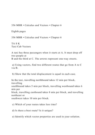
- 1. 356 MHR • Calculus and Vectors • Chapter 6 Eighth pages 356 MHR • Calculus and Vectors • Chapter 6 TA S K Taxi Cab Vectors A taxi has three passengers when it starts at A. It must drop off two people at B and the third at C. The arrows represent one-way streets. a) Using vectors, find two different routes that go from A to C via B. b) Show that the total displacement is equal in each case. In the taxi, travelling northbound takes 12 min per block, travelling southbound takes 5 min per block, travelling westbound takes 6 min per block, travelling eastbound takes 8 min per block, and travelling northeast or southwest takes 10 min per block. c) Which of your routes takes less time? d) Is there a best route? Is it unique? e) Identify which vector properties are used in your solution.
- 2. f) If the taxi charges for mileage are $0.50/rectangular block and the time charges are $0.10/minute, what is the cheapest route from A to C? How much should each passenger pay? 8.1 RNA Polymerases and Sigma Factors RNA polymerase is a complex enzyme that carries out transcription by making RNA copies (called transcripts) of a DNA template strand. In bacteria, the RNA pol holoenzyme is made up of: Core polymerase: a2, b, b′ Required for the elongation phase Sigma factor: s Required for the initiation phase ‹#› 1 Subunit Structure of RNA Polymerase ‹#› 2 FIGURE 8.3 ■ Subunit structure of RNA polymerase. Two views of RNA polymerase. The channel for the DNA template is shown by the yellow line. Subunits (αI, αII, β, β′, and ω) are color-coded dark green, medium green, light green, cyan, and gold, respectively. The function of the omega (ω) subunit is
- 3. currently unclear. Recent evidence suggests it may have a role in sigma factor competition for core polymerase. Sigma factor (red), which recognizes promoters on DNA, is shown separate from core polymerase in the left-hand panel. Different functional areas of sigma are labeled sigma 1 through sigma 4 (σ1–σ4). Sigma factor interacts with the alpha (α), beta (β), and beta-prime (β′) subunits. The molecule on the left is rotated 110° to give the image on the right. To view stereo images of RNA polymerase, locate code 1L9Z in the RCSB Protein Data Bank on the Internet. Source: Robert D. Finn et al. 2000. EMBO J. 19:6833–6844. Sigma Factors – 1 The sigma factor helps the core enzyme detect the promoter, which signals the beginning of the gene. Every cell has a “housekeeping” sigma factor. In Escherichia coli, it is sigma-70. Recognizes consensus sequences at the –10 and –35 positions, relative to the start of the RNA transcript (+1) A single bacterial species can make several different sigma factors. ‹#› 3 Sigma Factors – 2 ‹#› 4
- 4. FIGURE 8.4 ■ –10 and –35 sequences of E. coli promoters. A. Alignment of sigma-70 (σ70)-dependent promoters from different genes. Dots were added to help visualize alignments: biotin synthesis (bioB); cytochrome o (cyoA); galactose utilization (galE); lactose utilization (lacP); RNA polymerase (rpoD); small-subunit ribosome protein (rpsL); tryptophan synthesis (trp); glucose 6-phosphate dehydrogenase (zwf, zwischenferment). Yellow indicates conserved nucleotides; brown denotes transcript start sites (+1). B. The alignment in (A) generates a consensus sequence of σ70-dependent promoters (red-screened letters indicate nucleotide positions where different promoters show a high degree of variability). “N” indicates that any of the four standard nucleotides can occupy the position. Sigma Factors – 3 Mutations in the consensus sequence can affect the strength of the promoter. Some mutations can cause decreased transcription (called “down mutations”), while others cause increased transcription (“up mutations”). ‹#› 5 FIGURE 8.4 ■ –10 and –35 sequences of E. coli promoters. C. Mutations in the lacP promoter that affect promoter strength (lac genes encode proteins that are used to metabolize the carbohydrate lactose). Some mutations can cause decreased transcription (called “down mutations”), while others cause increased transcription (“up mutations”). Some E. coli Promoter Sequences Recognized by Different Sigma Factors - 2
- 5. ‹#› 6 FIGURE 8.5 ■ RNA polymerase holoenzyme bound to a promoter. A. The initial open complex forms when holoenzyme binds to a promoter. DNA –10 and –35 contacts with sigma factor are shown. Nontemplate strand is color-coded magenta; template strand, green. B. Blowup of (A), with the beta subunit removed to view the transcription bubble. Some bases in the nontemplate strand are flipped outward (yellow) to interact with sigma factor or the beta subunit after the transcription bubble is formed. The base at position +1 is the first base transcribed. 8.2 Transcription of DNA to RNA Transcription occurs in three phases: 1. Initiation: RNA pol holoenzyme binds to the promoter. This is followed by melting of the helix and synthesis of the first nucleotide of the RNA. 2. Elongation: the RNA chain is extended. 3. Termination: RNA pol detaches from the DNA, after the transcript is made. ‹#› 7 Transcription Initiation RNA polymerase holoenzyme forms a loosely bound, closed complex with DNA. Closed complex must become an open complex through the unwinding of one helical turn.
- 6. RNA polymerase in the open complex becomes tightly bound to DNA, and so begins transcription. The first ribonucleoside triphosphate (r N T P) of the new RNA chain is usually a purine (A or G). ‹#› 8 Initiation of Transcription ‹#› 9 FIGURE 8.6 ■ The initiation of transcription. Sigma factor helps RNA polymerase find promoters but is discarded after the first few RNA bases are polymerized. (Omega is not shown.) Transcription Elongation Elongation is the sequential addition of ribonucleotides from nucleoside triphosphates. The original RNA polymerase continues to move along the template, synthesizing RNA at ~ 45 bases/sec. The unwinding of DNA ahead of the moving complex forms a 17-bp transcription bubble. Positive supercoils ahead are removed by DNA topoisomerases. ‹#› 10
- 7. Transcription Termination All bacterial genes use one of two known transcription termination signals: 1. Rho-dependent Relies on a protein called Rho and a strong pause site at the 3′ end of the gen 2. Rho-independent Requires a GC-rich region of RNA, as well as 4–8 consecutive U residues ‹#› 11 The Termination of Transcription ‹#› 12 FIGURE 8.7 ■ The termination of transcription. A. Rho- dependent termination requires Rho factor but not NusA. B. Rho-independent termination requires NusA but not Rho. Antibiotics That Affect Transcription Antibiotics must meet two fundamental criteria: They must kill or retard the growth of a pathogen, and they must not harm the host. Rifamycin B Selectively binds to the bacterial RNA pol Inhibits transcription initiation Actinomycin D
- 8. Nonselectively binds to DNA Inhibits transcription elongation ‹#› 13 Structure and Mode of Action of Rifamycin ‹#› 14 FIGURE 8.8 ■ Structure and mode of action of rifamycin. A. Structure of rifamycin. The R groups indicated are added to alter the structure and pharmacology of the basic structure. B. Electron micrograph of Amycolatopsis. C. Contact points between rifamycin and residues in the beta subunit of RNA polymerase. Structure and Mode of Action of Actinomycin D ‹#› 15 FIGURE 8.9 ■ Structure and mode of action of actinomycin D. This antibiotic inserts its ring structure (A) between parallel DNA bases and wraps its side chains along the minor groove (B). (PDB code for B: 1DSC) Different Classes of RNA
- 9. There are several classes of RNA, each designed for a different purpose: Messenger RNA (mRNA): encodes proteins Ribosomal RNA (rRNA): forms ribosomes Transfer RNA (tRNA): shuttles amino acids Small RNA (sRNA): regulates transcription or translation tmRNA: frees ribosomes stuck on damaged mRNA Catalytic RNA: carries out enzymatic reactions ‹#› 16 RNA Stability RNA stability is measured in terms of half-life. The average half-life for mRNA is 1–3 minutes. The stabilities of the different kinds of RNAs differ drastically. The RNA degradosome is made up of an RNase, an RNA helicase, and two metabolic enzymes. Recent findings suggest that it is compartmentalized within the cell. ‹#› 17 8.3 Translation of RNA to Protein Once a gene has been copied into mRNA, the next stage is translation, the decoding of the RNA message to synthesize protein. An mRNA molecule can be thought of as a sentence in which triplets of nucleotides, called codons, represent individual
- 10. words, or amino acids. Ribosomes are the machines that read the language of mRNA and convert, or translate, it into protein. They do so via the genetic code. ‹#› 18 The Genetic Code – 1 Consists of nucleotide triplets called codons There are 64 possible codons: 61 specify amino acids Include the start codons 3 are stop codons The code is degenerate or redundant. Multiple codons can encode the same amino acid. The code operates universally across species. Remarkably, with very few exceptions ‹#› 19 The Genetic Code – 2 First baseSecond Base U- RNA codonSecond Base U- Amino acidSecond Base C-RNA codon Second Base C-Amino acidSecond Base A-RNA codonSecond Base A- Amino acidSecond Base G-RNA codonSecond Base g- Amino acidThird baseUUUUP h eU CUS e rU A UT y rU G UC y sUUUUCP h eUCCS e rU ACT y rUGCC y sCUU U AL e uU C AS e rU A AStopU G AStopAUU U GL e uU CGS e rU A GStopU GGT r pGCCU UL e uCCUP r oC A UH i sCG UA r
- 11. gUCCUCL e uCCCP r oC A CH i sCG CA r gCCCU AL e uCCAP r oC A AG l nCG AA r gACC U GL e uCCGP r oC A GG l nCGGA r gGAA U UI l eA C UT h rA A UA s nAG US e rUAAUGI l eA CCT h rA A CA s nAGCS e rCAA U AI l eA C AT h rAAAL y sAG AA r gAAAUGM e tA CGT h rA A GL y sAGGA r gGGG U UV a lGCUA l aG A UA s pG G UG l yUGG U CV a lGCCA l aG ACA s pGGCG l yCGG U AV a lG C AA l aG A AG l uG G AG l yAGG U GV a lG CGA l aGAGG l uGGGG l yG ‹#› 20 FIGURE 8.10 ■ The genetic code. Codons within a single box encode the same amino acid. Color-highlighted amino acids are encoded by codons in two boxes. Stop codons are highlighted red. Often, single-letter abbreviations for amino acids are used to convey protein sequences (see legend above). tRNA Molecules tRNAs are decoder molecules that convert the language of RNA into that of proteins. tRNAs are shaped like a clover leaf (in 2D) and a boomerang (in 3D). A tRNA molecule has two functional regions: Anticodon: hydrogen bonds with the mRNA codon specifying an amino acid 3′ (acceptor) end: binds the amino acid tRNAs contain a large number of unusual, modified bases. ‹#›
- 12. 21 Transfer RNA ‹#› 22 FIGURE 8.11 ■ Transfer RNA. A. Primary sequence. The letters D, M, Y, T, and Ψ stand for modified bases found in tRNA. B. Cloverleaf structure. DHU (or D) is dihydrouracil, which occurs only in this loop; TΨC consists of thymine, pseudouracil, and cytosine bases that occur as a triplet in this loop. The DHU and TΨC loops are named for the modified nucleotides that are characteristically found there. C. Three-dimensional structures. The anticodon loop binds to the codon, while the acceptor end binds to the amino acid. (PDB code: 1GIX) Codon-Anticodon Pairing ‹#› 23 FIGURE 8.12 ■ Codon-anticodon pairing. The tRNA anticodon consists of three nucleotides at the base of the anticodon loop. The anticodon hydrogen bonds with the mRNA codon in an antiparallel fashion. This tRNA is “charged” with an amino acid covalently attached to the 3′ end. Attaching Amino Acids to tRNA Each tRNA must be charged with the proper amino acid before it encounters the ribosome.
- 13. The charging of tRNAs is carried out by a set of enzymes called aminoacyl-tRNA synthetases. Each cell has generally 20 of these “match and attach” proteins, one for each amino acid. Each aminoacyl-tRNA synthetase must recognize its own tRNA but not bind to any other tRNA. So each tRNA has its own set of interaction sites that match only the proper synthetase. ‹#› 24 Charging of tRNA Molecules by Aminoacyl-tRNA Synthetases ‹#› 25 FIGURE 8.14 ■ Charging of tRNA molecules by aminoacyl- tRNA synthetases. At the end of this process, each amino acid is attached to the 3′ end of CCA on a specific tRNA molecule. Curved arrows indicate nucleophilic attack by electrons. The Ribosome, a Translation Machine – 1 The ribosome translates the language of the mRNA code into the amino acid sequences of proteins that conduct the activities of the cell. Ribosomes are composed of two subunits, each of which includes rRNA and proteins. In prokaryotes, the subunits are 30S and 50S and combine to form the 70S ribosome.
- 14. ‹#› 26 FIGURE 8.15 ■ Bacterial ribosome structure. As this schematic illustrates, note that a section of the 30S subunit (A) fits into the valley of the 50S subunit (B) when forming the 70S ribosome (C). The Ribosome, a Translation Machine – 2 The 70S ribosome harbors three binding sites for tRNA: A (acceptor) site: binds incoming aminoacyl-tRNA P (peptidyl-tRNA) site: harbors the tRNA with the growing polypeptide chain E (exit) site: binds a tRNA recently stripped of its polypeptide ‹#› 27 Binding of tRNA ‹#› 28 FIGURE 8.17 ■ Binding of tRNA. X-ray-crystallographic model of Thermus thermophilus ribosome with associated tRNAs. 50S is red, 30S is magenta, and tRNAs in the A, P, and E sites are blue, green, and yellow, respectively.) Inset: The formation of a peptide bond between the peptidyl-tRNA in the P site and aminoacyl-tRNA in the A site. The mRNA (light blue) travels
- 15. along the 30S subunit, and the growing peptide (yellow) exits from a channel formed in the 50S subunit. (PDB codes: 1GIX and 1GIY) The Ribosome Is a “Ribozyme” The ribosome makes the peptide bonds that stitch amino acids together using a remarkable enzymatic activity called peptidyltransferase. Peptidyltransferase is actually a ribozyme (an RNA molecule that carries out catalytic activity). Part of 23S r R N A of the large ribosomal subunit While highly conserved, there are differences in r R N A sequences that increase in relation to the evolutionary distance among species. So r R N A serves as a molecular clock. ‹#› 29 How Do Ribosomes Find the Right Reading Frame? Every m RNA DNA rRNA has three potential reading frames, so how does the ribosome find the right one? The upstream, untranslated leader RNA contains a purine-rich sequence with the consensus 5′-AGGAGGU-3′. Located 4–8 bases upstream of the start codon in Escherichia coli This Shine-Dalgarno sequence is complementary to a sequence at the 3′ end of 16S rRNA of the 30S subunit. ‹#›
- 16. 30 Polysomes Once a ribosome begins translating mRNA and moves off of the ribosome-binding site, another ribosome can immediately jump onto that site. The result is an RNA molecule with many ribosomes moving along its length at the same time. The multiribosome structure is known as a polysome. Ribosomes in a polysome are closely packed and arranged helically along the mRNA. Polysomes help protect the message from degrading RNases and enable the speedy production of protein from just a single mRNA molecule. ‹#› 31 Bacterial Transcription and Translation Are Coupled Different ribosomes can bind simultaneously to the start of each cistron within a polycistronic mRNA. Before RNA polymerase has even finished making an mRNA molecule, ribosomes will bind to the 5′ end of the mRNA and begin translating protein. This is called coupled transcription and translation. Eukaryotic microbes, on the other hand, use separate cellular compartments to carry out most of their transcription and translation. ‹#›
- 17. 32 Coupled Transcription and Translation in Bacteria ‹#› 33 FIGURE 8.18 ■ Coupled transcription and translation in bacteria. A. During coupled transcription and translation in prokaryotes, ribosomes attach at mRNA ribosome-binding sites and start synthesizing protein before transcription of the gene is complete. B. Model of E. coli polysome showing the nascent polypeptides (numbered) exiting from each ribosome. A representative ribosome is shown in the dashed circle. Note the helical arrangement of ribosomes along the chain, which are held together by mRNA (blue tracing). The closer the ribosome is to the 3′ end of the mRNA, the longer the synthesized protein molecules grow. Defining a Gene Before we discuss translation, it helps to illustrate the alignments between the DNA sequence of a structural gene, and the mRNA transcript containing translation signals and protein- coding sequences. ‹#› 34 FIGURE 8.20 ■ Alignment of structural genes in a bacterial operon, the mRNA transcript, and protein products. In this figure, the term “gene” refers to the region of DNA
- 18. corresponding to the entire mRNA transcript, including upstream and downstream untranslated areas. The Three Stages of Protein Synthesis Polypeptide synthesis occurs in three stages: Initiation: brings the two ribosomal subunits together, placing the first amino acid in position Elongation: sequentially adds amino acids as directed by mRNA transcript Termination: releases the completed protein and recycles ribosomal subunits Each phase requires a number of protein factors and energy in the form of GTP. ‹#› 35 Prof. Olave will show video in class Must know every detail of prokaryotic translation. I will only summarize once. ‹#› 36 FIGURE 8.23 ■ Termination of translation. The completed protein is released, and the ribosome subunits are recycled. Antibiotics That Affect Translation Streptomycin: inhibits 70S ribosome formation Tetracycline: inhibits aminoacyl-tRNA binding to the A site Chloramphenicol: inhibits peptidyltransferase
- 19. Puromycin: triggers peptidyltransferase prematurely Erythromycin: causes abortive translocation Fusidic acid: prevents translocation ‹#› 37 Antibiotics That Inhibit Protein Synthesis in Bacteria ‹#› 38 FIGURE 8.24 ■ Antibiotics that inhibit protein synthesis in bacteria. Streptomycin (A) and tetracycline (B) bind to the A site. Streptomycin causes mistranslation, tetracycline inhibits tRNA binding. Chloramphenicol (C) and erythromycin (D) bind to the peptidyltransferase site, thus inhibiting peptide bond formation. Unsticking Stuck Ribosomes The molecule tmRNA has properties of both tRNA and mRNA. It rescues ribosomes stuck on damaged mRNA that lacks a stop codon. ‹#› 39 FIGURE 8.25 ■ tmRNA and protein tagging. B. Mechanism of tmRNA tagging in E. coli.
- 20. 8.4 Protein Modification, Folding, and Degradation For many proteins, translation is not the last step in producing a functional molecule. Often a protein must be modified after translation either to achieve an appropriate 3D structure or to regulate its activity. Primary, secondary, and tertiary structures of proteins can be modified after the primary protein sequence has been assembled by the ribosomes. A healthy cell “cleans house” by degrading damaged or unneeded proteins. ‹#› 40 Protein Processing after Translation Protein structure may be modified after translation: N-formyl group may be removed by methionine deformylase. The entire methionine may be removed by methionyl aminopeptidase. Acetyl groups or AMP can be attached. Proteolytic cleavages may activate or inactivate a protein. ‹#› 41 Protein Folding Folding of many proteins requires assistance from other proteins called chaperones. GroEL and GroES chaperones
- 21. Form stacked ring with a hollow center The protein fits inside the open hole. DnaK chaperones Do not form rings Clamp down on a polypeptide to assist folding ‹#› 42 E. coli GroEL-GroES and DnaK Structures ‹#› 43 FIGURE 8.26 ■ E. coli GroEL-GroES and DnaK (HSP70) structures. A. Three-dimensional reconstructions of GroEL- ATP, GroEL GroES-ATP, and GroEL-GroES from cryo-EM. The first two panels are side views; the third panel is a top view. GroES is red. (PDB codes: 2C7E, 1PCQ) B. DnaK clamping down on a peptide (yellow). (PDB code: 1DKX) Protein Degradation: Cleaning House Many normal proteins contain degradation signals called degrons. The N-terminal rule suggests that the N-terminal amino acid of a protein directly correlates with its stability. Proteasomes are protein-degrading machines found in eukaryotes and archaea. Bacteria contain ATP-dependent proteases, such as L o n and C l p P.
- 22. ‹#› 44 Protein Degradation Machines ‹#› 45 FIGURE 8.27 ■ Protein degradation machines. A. Bacterial ClpY ATPase and ClpQ protease (Haemophilus influenzae). (PDB cod 1G3I) Two of the six subunits from each ring were removed to reveal the interior cavity. The active sites involved in peptide bond cleavage are indicated in pink. B. The 20S proteasome from the methanoarchaeon Methanosarcina thermophila. (PDB code: 1G0U) E. coli Protein Folding versus Degradation Triage Pathways Damaged proteins randomly enter chaperone-based refolding pathways or degradation pathways until the protein is repaired or destroyed. ‹#› 46 FIGURE 8.28 ■ E. coli protein folding versus degradation triage pathways. The diagram depicts what can happen to a newly synthesized protein. However, a protein that unfolds in response to environmental stress (for example, heat) will undergo the same triage process.
- 23. 8.5 Secretion: Protein Traffic Control Proteins destined for the bacterial cell membrane or envelope regions require special export systems. Proteins meant for the cell membrane are tagged with hydrophobic N-terminal signal sequences of 15–30 amino acids. These sequences are bound by the signal recognition particle (S R P). Proteins then undergo cotranslational export. ‹#› 47 SRP and Cotranslational Export in E. coli ‹#› 48 FIGURE 8.29 ■ SRP and cotranslational export in E. coli. A ribosome “paralyzed” by an SRP does not resume translating protein until encountering FtsY in the membrane. Translation can then recommence. Some proteins designated for integral membrane location are inserted directly (top). Other integral membrane proteins and proteins destined for the periplasm are inserted or secreted via the Sec system (bottom). Protein Export to the Periplasm Many periplasmic proteins, such as S O D and maltose-binding protein, are delivered to the periplasm by a common pathway called the S e c A-dependent general secretion pathway. The general secretion pathway has several steps, which can be
- 24. summarized as: The peptide is completely translated in the cytoplasm. The completed pre-secretion protein is then captured by a piloting protein called S e c B. S e c B unfolds and delivers the protein to S e c A, which is associated with the S e c Y E G translocon. ‹#› 49 The SecA-Dependent General Secretion Pathway ‹#› 50 FIGURE 8.30 ■ The SecA-dependent general secretion pathway. This pathway exports many proteins across the cell membranes of Gram-negative and Gram-positive bacteria. Protein Export in Gram-Positive Bacteria Gram-positive bacteria must also export proteins across the cell membrane and then fold and process them once they are secreted. Many streptococci cluster their secretion systems at the cell membrane in an anionic phospholipid microdomain called the ExPortal. The ExPortal is located near the cell septum and appears linked to peptidoglycan synthesis.
- 25. ‹#› 51 FIGURE 8.31 ■ Location of the ExPortal of Streptococcus pyogenes. HtrA was identified using immunofluorescence. Note that HtrA is located at the septum. Export of Prefolded Proteins to Periplasm The twin arginine translocase (T A T) can move a subset of already folded proteins across the inner membrane and into the periplasm. Powered by the proton motive force ‹#› 52 FIGURE 8.32 ■ The twin arginine translocase (TAT). Model for the Tat protein translocase, which includes proteins TatA, TatB, and TatC. Journeys to the Outer Membrane Gram-negative bacteria need to export proteins completely out of the cell. For example, digestive enzymes and toxins Seven elegant secretion systems have evolved: Labeled Type I–VII Some deliver the exported proteins to other dedicated transport proteins in the periplasm. Others provide nonstop service. ‹#› 53
- 26. Type I Protein Secretion ABC transporters are the simplest of the protein secretion systems and make up what is called the type I protein secretion. Type I protein secretion moves certain proteins directly from the cytoplasm to the environment. Type I systems all have three protein components: An outer membrane channel An ABC protein at the inner membrane A periplasmic protein lashed to the inner membrane ‹#› 54 Type I Secretion: The HlyABC Transporter ‹#› 55 FIGURE 8.33 ■ Type I secretion: the HlyABC transporter. A. Hemolysin (HlyA) is transported directly from the cytoplasm into the extracellular medium through a multicomponent ABC transport system. The HlyB and D proteins are dedicated to HlyA transport. TolC is shared with other transport systems. Not drawn to scale. B. Molecular model of TolC. The beta barrel channel spans the outer membrane, and the alpha helix tunnel extends into the periplasm. Three monomers (red, yellow, and blue) make up the channel. Source: Part A modified from Moat et al. 2002. Microbial Physiology, 4th ed. Wiley- Liss.
- 27. Name: _________________________ Biology 351- Homework Assignment #5 (10 points) This assignment is due on Tuesday February 20th in lab by 11:21AM. Give yourself enough time to print out your assignment in case you have printer problems. I will not accept electronic copies. Hardcopies only, and late assignments are not accepted in the biology department. 1. During transcriptional initiation RNA polymerase holoenzyme recognizes the consensus sequences within the promoter of E. coli. What part of the RNA polymerase holoenzyme recognizes the consensus sequence? 2. Does RNA polymerase holoenzyme recognize the sense, or antisense strand? The antisense strand is used for what purpose during transcription? 3. A single strand of bacterial DNA contains the base sequence -35 -10 +1 5’ CGTGTATTGACACTGGTGAGCCACTATCGTATATTCCCTA AGTGAGTATTGG 3’ a. What is the complementary sequence? Draw or type this sequence just below and indicate its polarity (directionality) in order to create a double-stranded DNA sequence. b. Under the double-stranded DNA sequence, draw or type the mRNA sequence that will be translated, and indicate its
- 28. polarity. c. Which strand of the DNA serves as the coding strand, and which serves as the template strand, for the synthesis of the RNA transcript for this hypothetical gene fragment. 4. If a stop codon is not included in the mRNA molecule, how would this affect the following: a. translocation on the mRNA by polyribosomes b. concentration of this specific polypeptide in the cell 5. How many different types of tRNA molecules exist in the cell? For what purpose (hint: why are there 20 different tRNA molecules)?