
Recommended
More Related Content
Similar to Special senses.ppsx
Similar to Special senses.ppsx (20)
More from lumaGhaziALzamel
More from lumaGhaziALzamel (15)
Recently uploaded
Recently uploaded (20)
Special senses.ppsx
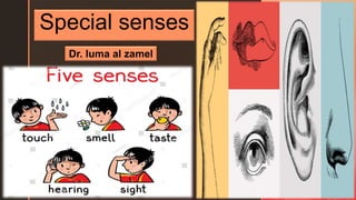
- 1. z Special senses Dr. luma al zamel
- 6. • Every sound produces sound waves or vibrations in the air, which travel at about 332 meters (1088 feet) per second. The auricle, because of its shape, concentrates the waves and directs them along the auditory meatus causing the tympanic membrane to vibrate. • Tympanic membrane vibrations are transmitted and amplified through the middle ear by movement of the ossicles. At their medial end the footplate of the stapes rocks to and from in the oval window, setting up fluid waves in the perilymph of the scala vestibuli. The fluid wave is finally expended into the middle ear by vibration of the membrane of the round window. • The semicircular canals have no auditory function although they are closely associated with the cochlea. They provide information about the position of the head in space, contributing to maintenance of posture and balance.
- 8. • The semicircular canals and the vestibule (utricle and saccule) are concerned with balance. Any change of position of the head causes movement in the perilymph and endolymph, which bends the hair cells and stimulates the sensory nerve endings in the utricle, saccule and ampullae. • The resultant nerve impulses are transmitted by the vestibular nerve which joins the cochlear nerve to form the vestibulocochlear nerve. The vestibular branch passes first to the vestibular nucleus, then to the cerebellum. • The cerebellum also receives nerve impulses from the eyes and proprioceptors (sensory receptors) in the skeletal muscles and joints. Impulses from these three sources are coordinated and efferent nerve impulses pass to the cerebrum and to skeletal muscles. • This results in awareness of body position, maintenance of upright posture and fixing of the eyes on the same point, independently of head movements.
- 11. • Before reaching the retina, light rays pass successively through the conjunctiva, cornea, aqueous fluid, lens and vitreous body. They are all denser than air and, apart from the lens, they have a constant refractory power, like that of water. • Light rays entering the eye need to be bent (refracted) to focus them on the retina. • The lens is a biconvex elastic transparent body suspended behind the iris from the ciliary body by the suspensory ligament. To increase the refractive power the ciliary muscle contracts, releasing its pull on the suspensory ligament and the anterior surface of the lens bulges forward, increasing its convexity. This focuses light rays from near objects on the retina. • Abnormal refraction within the eye is corrected using biconvex or biconcave lenses. • Pupil size influences accommodation by controlling the amount of light entering the eye. • The iris consists of one layer of circular and one of radiating smooth muscle fibres. Contraction of the circular fibres constricts the pupil, and contraction of the radiating fibres dilates it. The size of the pupil is controlled by the autonomic nervous system. Sympathetic stimulation dilates the pupils and parasympathetic stimulation causes constriction.
- 12. Extraocular muscles of the eye The eyeballs are moved by six extrinsic muscles, attached at one end to the eyeball and at the other to the walls of the orbital cavity. There are four straight (rectus) muscles and two oblique muscles. They are: • medial rectus • lateral rectus • superior rectus • inferior rectus • superior oblique • inferior oblique. Movement of the eyes to look in a particular direction is under voluntary control, but coordination of movement, needed for convergence and accommodation to near or distant vision, is under autonomic (involuntary) Control.
- 14. Accessory organs of the eye The eye is a delicate organ which is protected by several structures: • eyebrows : They protect the anterior aspect of the eyeball from sweat, dust and other foreign bodies. • eyelids and eyelashes: protect the eye from injury. • Reflex closure of the lids occurs when the conjunctiva or eyelashes are touched, when an object comes close to the eye or when a bright light shines into the eye — this is called the conjunctival or corneal reflex. • Blinking at about 3- to 7-second intervals spreads tears and oily secretions over the cornea, preventing drying.
- 15. • lacrimal apparatus: For each eye this consists of: • 1 lacrimal gland and its ducts • 2 lacrimal canaliculi • 1 lacrimal sac • 1 nasolacrimal duct. The fluid that fills the conjunctival sac consists of tears and the oily secretion of tarsal glands and is spread over the cornea by blinking. The functions of this mixture of fluids include: • washing away irritating materials, e.g. dust, grit • the bactericidal enzyme lysozyme prevents microbial infection. • its oiliness delays evaporation and prevents drying of the conjunctiva • nourishment of the cornea.
- 16. Physiology of olfactory The nasal cavity has a dual function: a passageway for respiration and sense of smell. Olfactory nerves (first cranial nerves). These are the sensory nerves of smell. They originate as specialized olfactory nerve endings (chemoreceptors) in the mucous membrane of the roof of the nasal cavity. All odorous materials give off volatile molecules, which are carried into the nose with the inhaled air and stimulate the olfactory chemoreceptors when dissolved in mucus.
- 17. • The air entering the nose is warmed and convection currents carry eddies of inspired air to the roof of the nasal cavity. 'Sniffing' concentrates volatile molecules in the roof of the nose. This increases the number of olfactory receptors stimulated and thus the perception of the smell. The sense of smell may affect the appetite. If the odors are pleasant the appetite may improve and vice versa. When accompanied by the sight of food, an appetizing smell increases salivation and stimulates the digestive system. The sense of smell may create long-lasting memories, especially to distinctive odors, e.g. hospital smells, favorite or least-liked foods. • Inflammation of the nasal mucosa prevents odorous substances from reaching the olfactory area of the nose, causing loss of the sense of smell (anosmia). The usual cause is the common cold.
- 18. Physiology of taste Taste buds contain sensory receptors (chemoreceptors) that are found in the papillae of the tongue and widely distributed in the epithelia of the tongue, soft palate, pharynx and epiglottis. They consist of small sensory nerve endings of the glossopharyngeal, facial and vagus nerves (cranial nerves VII, IX and X). Nerve impulses are generated and conducted along the glossopharyngeal, facial and vagus nerves before synapsing in the medulla and thalamus. Their destination is the taste area in the parietal lobe of the cerebral cortex where taste is perceived.
- 19. Four fundamental sensations of taste have been described — sweet, sour, bitter and salt. some tastes consistently stimulate taste buds in specific parts of the tongue: • sweet and salty, mainly at the tip • sour, at the sides • bitter, at the back. The sense of taste triggers salivation and the secretion of gastric juice. It also has a protective function, e.g., when foul- tasting food is eaten then reflex gagging or vomiting may be induced. The sense of taste is impaired when the mouth is dry because substances can be 'tasted' only if they are in solution.