Evaluation of Bowel and Mesenteric Blunt Trauma with Multidetector CT
•Download as PPTX, PDF•
12 likes•3,466 views

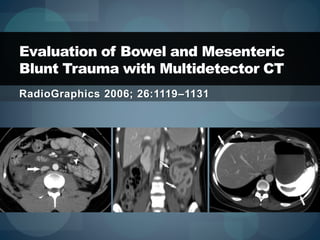
Evaluation of Bowel and Mesenteric Blunt Trauma with Multidetector CT RadioGraphics 2006; 26:1119–1131

Recommended
More Related Content
What's hot
What's hot (20)
Viewers also liked
Viewers also liked (20)
Similar to Evaluation of Bowel and Mesenteric Blunt Trauma with Multidetector CT
Similar to Evaluation of Bowel and Mesenteric Blunt Trauma with Multidetector CT (16)
More from Sun Yai-Cheng
More from Sun Yai-Cheng (20)
Recently uploaded
Recently uploaded (20)
Evaluation of Bowel and Mesenteric Blunt Trauma with Multidetector CT
- 1. RadioGraphics 2006; 26:1119–1131 Evaluation of Bowel and Mesenteric Blunt Trauma with Multidetector CT
- 2. Evaluation of Bowel and Mesenteric Blunt Trauma with Multidetector CT Findings Specific to Bowel Injury Bowel Wall Discontinuity Extraluminal Contrast Material Extraluminal Air Findings Less Specific to Bowel Injury Bowel Wall Thickening Abnormal Bowel Wall Enhancement Mesenteric Features
- 3. hypervascular thickened jejunum with a suspicious defect extraluminal air
- 4. free air free oral contrast material Traumatic devascularization of the left kidney
- 5. Traumatic devascularization of the left kidney thickened wall of the transverse colon with decreased enhancement
- 6. intraperitoneal areas of free contrast material free air intraperitoneal areas of free contrast material
- 7. retroperitoneal area of extraluminal contrast enhancement
- 8. thickening of the wall of the ascending colon with adjacent fat stranding free air hematoma of the right oblique internus muscle
- 9. retroperitoneal air pericecal fat stranding and thickening of lateroconal fascia
- 10. air in SMV contrast enhancement of the duodenal diverticulum
- 11. decreased enhancement of small bowel free fluid
- 12. mesenteric fat stranding subcutaneous emphysema extending to the extraperitoneal space producing pseudopneumoperitoneum
- 13. Evaluation of Bowel and Mesenteric Blunt Trauma with Multidetector CT Findings Specific to Mesenteric Injury Mesenteric Extravasation Mesenteric Vascular Beading Termination of Mesenteric Vessels Less Specific Findings Mesenteric Infiltration Mesenteric Hematoma Bowel Features
- 14. thickening of the intestinal wall
- 15. thickened cecal wall posterior abdominal wall tear
- 17. shock bowel without flattening of IVC
- 18. high-attenuation foci of intraperitoneal fluid
- 20. shock bowel
- 21. hypervascular thickened segments of small bowel abdominal wall tear
- 22. mesenteric fat stranding hypervascular thickened segments of small bowel
- 23. unenhanced small-bowel loops hypervascular thickened segments of small bowel
- 24. defect in the proximal jejunum
- 26. bowel infarct
- 27. mesenteric extravasation thickening and hypervascularity of proximal jejunum
- 28. mesenteric hematoma thickening and hypervascularity of proximal jejunum
- 31. lesser sac stranding abrupt termination of left gastric artery
- 33. abrupt termination of SMV
- 34. hematoma
- 35. Evaluation of Bowel and Mesenteric Blunt Trauma with Multidetector CT Common Features in Bowel and Mesenteric Injuries Intraperitoneal and Retroperitoneal Fluid Abdominal Wall Injury
- 38. abdominal wall tear mesenteric hematoma normal appearance of the sigmoid colon
Editor's Notes
- Figure 1. Jejunal perforation in a 66-year-old woman after a motor vehicle accident. Axial CT image shows hypervascular thickened jejunum with a suspicious defect (curved arrow) and with focal fluid, fat stranding, and extraluminal air (straight arrow) adjacent to jejunal loops. The patient later underwent resection of a 20-cm segment of the small bowel. No mesenteric injury was found at surgery.
- Figure 2a. Bowel injuries in a 37-year-old male pedestrian struck by a motor vehicle. (a) Axial CT image shows free air (curved arrow) and free oral contrast material in the left upper quadrant, adjacent to the jejunum (arrow). (b) Axial CT image at a level lower than a shows a thickened wall of the transverse colon with decreased enhancement (arrows). Traumatic devascularization of the left kidney (*) also is visible on both images. The patient underwent a partial small-bowel resection and primary surgical repair for jejunal rupture and necrosis, a segmental resection for transverse colon necrosis, and repair of a colonic serosal tear distal to the necrotic transverse colon. No mesenteric injury was seen at surgery.
- Figure 3a. Ruptured bladder in a 43-year-old female pedestrian who was struck by a car. (a) Axial abdominal CT image shows intraperitoneal areas of free contrast material (straight arrows) and free air (curved arrow). (b) Axial CT image shows a retroperitoneal area of extraluminal contrast enhancement (arrow). These features mimic those found in bowel injury but, instead, are secondary to a bladder rupture, which was found at surgery.
- Figure 4. False-positive findings of bowel injury in a 22-year-old woman who was a front-seat passenger and was wearing a seat belt during a motor vehicle accident. Axial CT image shows thickening of the wall of the ascending colon, with adjacent fat stranding (straight arrow), free air (curved arrows), and hematoma of the right oblique internus muscle (*). Although the free air was initially attributed to bowel injury, no bowel or gastric injuries were found at diagnostic laparoscopy. The free intraperitoneal air may instead be attributable to traumatic pneumothorax.
- Figure 5a. Bowel and mesenteric injuries in a 70-year-old male pedestrian struck by a car. (a) Axial CT image shows a trace of fluid that extends from the cecum, with pericecal fat stranding and thickening of the lateroconal fascia (curved arrow) and focal retroperitoneal air (straight arrow). (b) Axial CT image shows air in the superior mesenteric vein (arrow) as well as contrast enhancement of the duodenal diverticulum (arrowhead), a finding that simulates duodenal injury. At surgery, full-thickness cecal and mesenteric tears were identified and repaired.
- Figure 6a. Bowel and mesenteric injuries in a 56-year-old woman after a motor vehicle accident. (a) Axial CT image at the level of the lower abdomen shows a segment of the small bowel with decreased enhancement (straight arrow) and (curved arrow). (b) Axial CT image at the level of the middle abdomen shows mesenteric fat stranding (curved arrow), subcutaneous emphysema extending to the extraperitoneal space and producing pseudopneumoperitoneum (arrowheads), and hyperattenuating mesenteric nodes (straight arrows) that might be mistaken for foci of extravasated contrast material. At surgery, a 30-cm segment of the distal ileum that had been separated from its mesentery by shearing force, with resultant devascularization, was resected.
- Figure 7. Splenic laceration with active bleeding in a patient after a fall from a height of 6 feet onto a pole. Axial CT image shows apparent thickening of the intestinal wall in several small-bowel loops (arrows), an appearance caused by insufficient bowel distention. At surgery, no small-bowel injury was identified, but splenic laceration with active bleeding was seen.
- Figure 8a. False-positive findings of bowel injury in a 52-year-old woman after a jump from a height of four stories. (a) Axial CT image shows a thickened cecal wall (straight arrow) and a posterior abdominal wall tear on the right side (curved arrow), findings suggestive of bowel injury. (b) Axial CT image shows the thickened cecal wall (arrows) with attenuation similar to that of water, a finding suggestive of shock bowel. No bowel injuries were found at emergent laparotomy, and the cecal wall thickening was probably related to hypoperfusion complex.
- Figure 9. Axial CT image in a 25-year-old man after a motor vehicle accident shows evidence of shock bowel (arrowheads) likely related to fluid resuscitation, without flattening of the inferior vena cava (arrow). At surgery, hepatic laceration with active bleeding, renal laceration, and aortic dissection were noted. No small-bowel injury was identified.
- Figure 10a. Bowel and mesenteric injuries in a 32-year-old woman after a motor vehicle accident. Axial (a) and coronal (b) CT images show abnormal hypervascular thickened jejunal loops (arrows in b) and high-attenuation foci of intraperitoneal fluid (arrowheads in a) consistent with blood. No free or focal air was visible on CT images. At surgery, mesenteric tears in a middle segment of the jejunum and a distal segment of the ileum were found, with bleeding mesenteric vessels and multiple areas of perforation in the middle segment of the jejunum and the proximal and middle segments of the ileum. The affected segments of small bowel were resected.
- Figure 11. Bowel injury in an 18-year-old man with severe head trauma from a motor vehicle accident. Axial CT image shows hypervascular thickened jejunal loops with mucosal feathering (arrows), features characteristic of shock bowel. The patient died 1 day after hospital admission.
- Figure 12a. Bowel and mesenteric injuries in a 37-year-old woman after a motor vehicle accident in which she was a rear-seat passenger. CT images show hypervascular thickened segments of small bowel, especially evident in the left upper quadrant (*). Axial source images also show a tear in the abdominal wall on the right side (arrow in a) and mesenteric fat stranding (arrow in b). Oblique coronal image also shows unenhanced small-bowel loops in the lower abdomen (arrow in c). At surgery, shearing injury to the small-bowel mesentery was found, with active bleeding and with complete devascularization and necrosis of a 90-inch (229-cm) segment of the distal jejunum and ileum and a perforation of the middle jejunum.
- Figure 13a. Bowel injury in a 77-year-old man after a motor vehicle accident. Axial CT images show a defect in the proximal jejunum (arrow in a) and a mesenteric hematoma in the left upper quadrant (arrow in b). Although no free air was seen on CT images, a blowout perforation in the antimesenteric aspect of the proximal jejunum was found at surgery. No mesenteric injury was described in the surgical report.
- Figure 14a. Bowel and mesenteric injuries in a 52-year-old woman who was a front-seat passenger during a motor vehicle accident. (a) Coronal CT image shows an unenhanced segment of small bowel, a feature consistent with a bowel infarct (arrow). (b, c) Axial images show mesenteric extravasation (arrow in b), mesenteric hematoma (arrow in c), and thickening and hypervascularity of the proximal jejunum (*). At surgery, an extensive small-bowel mesenteric tear was found, with active bleeding from a jejunal branch of the superior mesenteric artery and with ischemia in a 20-cm-long segment of jejunum; the segment was resected. A small serosal tear also was found in the jejunum proximal to the margin of resection and in the cecum and was repaired.
- Figure 15a. Mesenteric injuries due to a motor vehicle accident. (a) Axial CT image shows a change in caliber, or beading, of some mesenteric vessels in the area of injury (arrows). At surgery, a tear was found in the ileocecal mesentery that warranted resection of the terminal ileum, cecum, and ascending colon. (b) Axial CT image from another patient shows beading of a mesenteric vessel in the area of injury (arrows). At surgery, a hematoma was found in the jejunal mesentery.
- Figure 16. Multiple injuries in a 58-year-old man after a 25-foot fall from a roof. Sagittal reformatted image from abdominal CT shows lesser sac stranding (arrow) and abrupt termination of the left gastric artery (arrowhead) at the level of stranding.
- Figure 17a. Bowel and mesenteric injuries in a 23-year-old man who was ejected from a car during a motor vehicle accident. At surgery, three small-bowel mesenteric tears were found. The most proximal tear was repaired, but the two more distal ones were larger and warranted resection of the affected small-bowel segment. No primary injury to the small bowel was found. Three subserosal tears in the colon also were found, one in the rectosigmoid segment and two in the ascending colon, and were repaired. (a) Coronal CT image at the level of the middle abdomen shows a mesenteric hematoma that extends to the right upper quadrant (arrows). (b) Oblique coronal CT image shows abrupt termination of the left-sided tributaries (arrowhead) of the superior mesenteric vein (SMV).
- Figure 19. Mesenteric injury in a 57-year-old man after a fall from a scaffold. Axial CT image shows a hematoma surrounded by fat stranding (arrow) in the splenic flexure mesocolon, with no evidence of active bleeding. At surgery, a nonexpanding mesenteric hematoma was found that did not require repair.
- Figure 20a. Mesenteric lacerations in a 45-year-old man after a head-on automobile collision. Axial CT images show a retroperitoneal hematoma near the cecum (arrow in a) and omental fat stranding on the left side of the abdomen (arrowhead in b). At surgery, a mesenteric tear with active bleeding was found in the midjejunal area. Despite the absence of obvious jejunal injury, this finding resulted in resection of a short (15-cm) segment of the small bowel. A second mesenteric tear was found near the cecum, with a retroperitoneal hematoma (repaired) and omental bleeding. No bowel injuries were found.
- Figure 21. Mesenteric injuries in a 19-year-old man after a motor vehicle accident. Axial CT image shows a sigmoid mesenteric hematoma (arrow) and a normal appearance of the sigmoid colon (arrowhead). A complete tear of the abdominal wall (*) is visible in the right lower quadrant. Avulsion of the sigmoid colon mesentery associated with an ischemic sigmoid colon segment (subsequently resected) was found at surgery.