1. Topic: Implant therapy outcomes, surgical aspectsTopic: Implant therapy outcomes, surgical aspects
Presented at the 20th Annual Scientific Meeting of the European Association of Osseointegration
13-15 October 2011, Athens, Greece
Presented at the 20th Annual Scientific Meeting of the European Association of Osseointegration
13-15 October 2011, Athens, Greece
ReferencesConclusions
Background and Aim
POST-EXTRACTION SOCKETS AUGMENTATION WITH ACELLULAR DERMAL MATRIX
AND ALLOGENIC BONE SUBSTITUTE IN THE AESTHETIC AREA. A CASE SERIES.
N.Maslova1 2 , A.Puisys1 2, T.Linkevicius1 2 3, E.Vindasiute1 2
1 Vilnius Implantology Center 2 Vilnius Research Group 3 Vilnius University, Institute of Odontology
Vilnius, Lithuania
171171
1.Araujo MG, Lindhe J. Dimensional ridge alterations followinng tooth extraction. An
experimental study in the dog. J Clin Periodontol 2005;32:212-218.
2.Fickl S, Zuhr O, Wachtel H, Stappert CF, Stein JM, Hurzeler M. Dimensional
changes of the alveolar ridge contour after different socket preservation
techniques. J Clin Periodontol 2008;35:906-913.
3.Fowler EB, Breault LG, Rebitski G. Ridge preservation utilizing an acellular dermal
allograft and demineralized freeze-dried bone allograft. Part I. A Report of 2
cases. J Periodontol 2000;71:1353-1359.
4. Khalid Al-Hezaimi, Paul Levi, Robert Rudy, Badar Al-Jandan, Abdulaziz Al-
Rasheed. An extraction socket classification developed using analysis og bone
type and blood suply to the buccal bone in monkeys. Int J Periodontics
Restorative Dent 2011;31:421-427
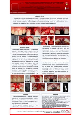
Study included 6 patients (aged from 24 to 37 y) who needed
to extract 6 central upper incisors. All patients were non
smokers. In all the cases the thin biotype (thickness less than
2 mm) of a gingiva with extremely thin buccal bone (thickness
was 0-0.5 mm) was observed. In 2 cases the anatomic
situation was even worse due to previous infections. After
tooth extraction flap with buccal and palatal sites was splitted
without a vertical incision. Thus the envelope from both sides
was generated. Alveolar post extraction socket was
augmented with allogenic bone substitute (maxgraft®, Botiss)
and 2 mm thickness xenogenic membrane (mucoderm®,
Botiss) was positioned on the internal part of an envelope.
Buccal and a palatal flap edges were sutured without a
tension. Patients received 500 mg of amoxicillin (1,5 g per day
for 7 days) and rinced with 0.12% chlorhexidine twice a day
for 7 to 10 days.
It is very important to keep favorable anatomical situation in the esthetic zone after tooth extraction. Many studies confirm loss
of a buccal bone wall after tooth being extracted, especially in the clinical cases with thin soft tissue biotype , bone destruction
due to periodontal pathology or as a result of fractured tooth root. Therefore the need of two surgical procedures occurs:
implantation with bone and soft tissue augmentation and healing abutments placement after 6 months.
In all six clinical cases after 4 months after socket
augmentation (despite previous extremely thin buccal bone)
there was enough bone to place implants without any
additional augmentation. All implants had excellent primary
stability of 35N, therefore one stage surgery could be chosen.
The procedure became time saving, less uncomfortable for
the patient and cost effective.
Results
Methods and Materials
After four months of healing the computer tomography has
been executed and evaluated. The width of bone was
calculated to be from 5,5 to 7 mm. A minimally invasive flap
without vertical incision was raised, all 6 placed implants
were positioned in the correct 3D positions with stability more
than 35N, healing abutments were connected the same day.
All implants were restored with cement-retained metal
ceramic restorations after 4-5 months after implantation.
Reduction of time and volume of the surgery conducted to
less traumatic and cost effective procedure for a patient.
Because of a good initial anatomic situation operation
occurred without a need of a vertical incision that resulted in a
better esthetics of the mucosal tissues.
This technique could be especially valuable in the cases
with a very thin buccal bone or resorbed buccal bone.