1. Topic: Implant therapy outcomes, surgical aspectsTopic: Implant therapy outcomes, surgical aspects
Presented at the 20th Annual Scientific Meeting of the European Association of Osseointegration
13-15 October 2011, Athens, Greece
Presented at the 20th Annual Scientific Meeting of the European Association of Osseointegration
13-15 October 2011, Athens, Greece
References
Conclusions
Background and Aim
The influence of mucosal tissue thickening on crestal
bone stability around bone level implants.
A.Puisys1 2, T.Linkevicius1 2 3, N.Maslova1 2 E.Vindasiute1 2
1 Vilnius Implantology Center 2 Vilnius Research Group 3 Vilnius University, Institute of Odontology
Vilnius, Lithuania
The influence of mucosal tissue thickening on crestal
bone stability around bone level implants.
A.Puisys1 2, T.Linkevicius1 2 3, N.Maslova1 2 E.Vindasiute1 2
1 Vilnius Implantology Center 2 Vilnius Research Group 3 Vilnius University, Institute of Odontology
Vilnius, Lithuania
188188
1. Tomas linkevicius, Peteris Apse, Simonas Grybauskas, Algirdas Puisys, The Influence of Soft Tissue Thickness on
Crestal Bone Changes Around Implants:A 1-Year Prospective Controlled Clinical Trial. Int J Oral Maxillofac Implants
2009;24:712-719
2. Hermann JS, Buser D, Schenk RK, Cochran DL. Crestal Bone Changes Around Titanium Implants. A Histometric
Evaluation of Unloaded non-submerged and submerged implants in the canine mandible. J Periodontol 200;71:1412-
1424
3. Barboza EP, Caula AL, Carvalho WR. Crestal Bone Loss Around Submerged and exposed unloaded dental implants: A
radiographic and microbiological descriptive study. Implant Dent 2002;11:162-169
Bone loss (mesially and distally) in two months in each group is
presented table below.
No statistical difference was observed between the groups:
Mucosal tissue thickness has been shown as an important
factor in etiology of early crestal bone loss around dental
implants. Few animal studies have shown that if we have thin
tissues during implant placement, crestal bone loss may occur
during the formation of the biologic width. Recently, prospective
controlled clinical study reported that if tissue thickness at the
crest was 2 mm or less, all implants, irrespective to their position
to the bone level, developed crestal bone loss within 1-year of
follow-up. On the contrary, in thick tissue pattern, supracrestally
placed implants had significantly less bone loss, compared to
crestally positioned implants.
Mucosal tissue thickness becomes very important also in short
implant placement. In regions with very limited bone height, when
only 6 mm or less implant could be installed, even the smallest
inch of the bone support is important for the survival and stability
of the implant. Sometimes limited bone height is accompanied by
thin tissue biotype, thus the risk to loose crestal bone around
short implants is very high.
Results
Methods and Materials
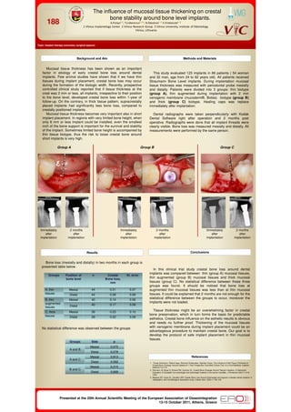
Group A Group B Group C
Immediately
after
implantation
Immediately
after
implantation
Immediately
after
implantation
2 months
after
implantation
2 months
after
implantation
2 months
after
implantation
This study evaluated 123 implants in 86 patients ( 54 woman
and 32 man, age from 24 to 62 years old). All patients received
Straumann Bone Level implants. During implantation mucosal
tissue thickness was measured with periodontal probe mesially
and distally. Patients were divided into 3 groups: thin biotype
(group A), thin augmented during implantation with 2 mm
xenogenic membrane (mucoderm®, Botiss) biotype (group B)
and thick (group C) biotype. Healing caps was replace
immediately after implantation.
Dental radiographs were taken perpendicularly with Kodak
Dental Software right after operation and 2 months post
operative. Radiographs were done that all implant threads were
clearly visible. Bone loss was measured mesially and distally. All
measurements were performed by the same person.
In this clinical trial study crestal bone loss around dental
implants was compared between thin (group A) mucosal tissues,
thin augmented (group B) mucosal tissues and thick mucosal
tissues (group C). No statistical difference between these three
groups was found. It should be noticed that bone loss at
augmented thin mucosal tissues was less than at thin mucosal
tissues. It could be explained that 2 months are not enough for the
statistical difference between the groups to occur, moreover the
implants were not loaded.
Tissue thickness might be an overwhelming factor in crestal
bone preservation, which in turn forms the basis for predictable
esthetics. Crestal bone influence on the esthetic results is obvious
and needs no further proof. Thickening of the mucosal tissues
with xenogenic membrane during implant placement could be an
advantageous procedure to maintain crestal bone. Our goal is to
develop the protocol of safe implant placement in thin mucosal
tissues.
Groups Position of
bone level
n Crestal
Bone loss,
mm
St. error
A, thin
tissues
Mesial 44 0.31 0.07
Distal 44 0.28 0.08
B, thin
augmented
tissues
Mesial 40 0.14 0.06
Distal 40 0.17 0.08
C, thick
tissues
Mesial 39 0.23 0.10
Distal 39 0.22 0.08
Groups Side p
A and B
Mesial 0,075
Distal 0,078
A and C
Mesial 0,913
Distal 0,582
B and C
Mesial 0,215
Distal 0,669