
Recommended
More Related Content
What's hot
What's hot (20)
Similar to Periodontal flaps
Similar to Periodontal flaps (20)
More from mikitha p
Recently uploaded
Recently uploaded (20)
Periodontal flaps
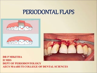
- 1. PERIODONTAL FLAPS DR P MIKITHA II MDS DEPT OF PERIODONTOLOGY AECS MAARUTI COLLEGE OF DENTAL SCIENCES
- 2. Contents • Introduction • Definition • Historical background • Objectives of flap surgery • Indications and contraindications • Advantages and disadvantages • Principle of flap design • Classification of flap • Properties of ideal flap • Types of incisions • Different flap techniques • Healing after flap surgery • Factors affecting the outcome of flap surgery • Conclusion • references
- 3. INTRODUCTION • The ultimate aim of periodontal therapy is to establish a healthy dentition with sound attachment appartus resulting in proper frrm, function and esthetics. • To achieve this goal many non-surgical and surgical techniques have been proposed to treat a variety of periodontal conditions most commonly the periodontal pocket. • Periodontal therapy comprises of initial non-surgical debridement followed by a re-evaluation at which stage the need for further treatment, usually surgical in nature is established.
- 4. DEFINITIONS • Periodontal flap is defined as a section of gingiva and/or mucosa surgically separated from the underlying tissues to provide visibility of and access to the bone and root surface. -(Carranza 10th edition). • Flap is defined as the separation of a section of tissue from the surrounding tissue except at its base, -(Glossary of periodontal terms) • A flap is defined as a mass of tissue, usually including skin, only partially removed from one part of the body so that it retains its own blood supply during transfer to another site. –(Dorland’s medical dictionary). Takei H, Carranza FA, Shin K. the flap technique for pocket therapy In: carranza’s clinical periodontolgy, Elsevier, 12, 2012; 593- 603.
- 5. Carl Partch- 19th century -1907 Partch incison Robert Neumann- 1912 1st introduced mucoperiosteal flap –”Neumann flap” Leonard Widman – 1918 Modified the Neumann flap – “Widmann flap” Cieszynski- 1918 Reverse bevel incision Kirkland – 1931 Modified flap procedure Carranza – 1939 Surgical treatment of periodontitis Nabers – 1954 Introduced “repositioning of attached gingiva” Ariaudo and Tyrrell – 1957 Modified Nabers procedure Friedman- 1962 Apically positioned flap Oschenbein and Bohannan – 1964 Palatal flap Morris – 1965 Unrepositioned mucoperiosteal flap Ramjford and Nissle – 1974 Modified Widmann flap Takei et al- 1985 Pappila preservation flap Trombelli et al – 2007 Single flap approach (SFA) Bianchi and Bassetti - 2009 Whales technique Takei H, Carranza FA, Shin K. the flap technique for pocket therapy In: carranza’s clinical periodontolgy, Elsevier, 12, 2012; 593- 603.
- 6. Objectives of flap surgery MAIN OBJECTIVE of periodontal surgery is to contribute to the long- term preservation of the periodontium by facilitating plaque removal and plaque control. -Jan Lindhe To enable visual instrumentation of root surfaces To re-establish the healthy, clinical status of periodontium with long term maintenance To restore the periodontal apparatus when attachment loss has occurred Cohen SE. fundamental of surgical therapy. In: Atlas of cosmetic and reconstructive periodontal surgery, BC Decker Inc, 3, 2007; 56-72.
- 7. 1. Access to roots and alveolar bone 2. Modification of osseous defects: • estabilish physiologic architecture of hard tissues through regeneration or resection • Augment alveolar ridge defects 3. Repair or regeneration of the periodontium 4. Pocket reduction: • Enhance maintenance by patient and therapist • Improves long term stability 5. Provide acceptable soft tissue contours • Enhances plaque control measures • Improve esthetics Cohen SE. fundamental of surgical therapy. In: Atlas of cosmetic and reconstructive periodontal surgery, BC Decker Inc, 3, 2007; 56-72.
- 8. indications • Irregular bony contours • Deep craters • Pockets on teeth in which a complete removal of root irritants is not clinically possible • Grade II or III furcation involvemnet • Root resection/ hemisection • Intrabony pockets on distal areas of last molars Takei H, Carranza FA, Shin K. the flap technique for pocket therapy In: carranza’s clinical periodontolgy, Elsevier, 12, 2012; 593- 603.
- 9. • Persistent inflammation in aresas with moderate to deep pockets • Unaccesible areas like root concavities, furctaion areas, etc • Deep periodontal pockets: Waerhaug stated that pocket depth greater tha 5mm demonstrated onlu an 11% efficacy in removal of plaque and calculus. • Osseous defect: morphology of osseous defects can limit the effectiveness of non-surgical therapy Takei H, Carranza FA, Shin K. the flap technique for pocket therapy In: carranza’s clinical periodontolgy, Elsevier, 12, 2012; 593- 603.
- 10. contraindications A. Patient non co-operation B. Poor plaque control C. High caries rate D. Systemic conditions: Cardiovascular diseases: Arterial HTN: patients consent should be taken and LA with adrenaline or without adrenaline must be used Angina pectoris: premedication with sedatives and LA, low in adrenaline is recommended. Takei H, Carranza FA, Shin K. the flap technique for pocket therapy In: carranza’s clinical periodontolgy, Elsevier, 12, 2012; 593- 603.
- 11. Myocardial infarction Anticoagulant treatment: the range in which SRP and surgical procedures can be safely performed is one and half to 2 times the avarage normal prothrombin time (12-14 sec) Aspirin and other NSAID drugs should not be used for post-op pain control Rheumatic endicarditis, congenital heart lesions and heart/vascukakr implants involve risk of transient bacteremia that follows manipulation of infection periodontal pockets, ADA- recommended antibiotic prophylaxis and antiseptic mouthrinsing 0.2% CHX prior to surgery Cohen SE. fundamental of surgical therapy. In: Atlas of cosmetic and reconstructive periodontal surgery, BC Decker Inc, 3, 2007; 56-72.
- 12. Organ transplantation: Prophylactic antibiotics are recommended in transplant patient taking immunosuppressive drugs Blood disorders: Patients suffering from acute leukemia, agranulocytosis and lymphogranulomatosis must not be subjected to periodontal surgery Diabetes Neurological disorders: Multiple sclerosis and parkinsons disease make periodontal surgery impossible Epilepsy Drugs used to treat epilepsy may cause gingival enlargements. Theese patients may withspecial restrictions be subjected to periodontal surgery. Cohen SE. fundamental of surgical therapy. In: Atlas of cosmetic and reconstructive periodontal surgery, BC Decker Inc, 3, 2007; 56-72.
- 13. advantages • Pocket epithelium is removed by the inverse bevel incision • The inter dental bone or infrabony defects can be covered by the flaps • No open wound persists postoperatively • Rapid healing. • Less post operative discomfort and fewer complications. • Less post operative gingival recession, therefore esthetic • Less dentin exposure. • Short surgical time • Direct healing disadvantages • When flaps are repositioned apically, the cervical areas of the teeth are often exposed, long and sensitive, due also to shrinkage of the tissues. • Possibility of deep periodontal pockets remaining after surgery. • Possibility of formation of post- operative gingival craters in proximal surface areas (especially in molars). • New attachment is unpredictable. • Less regeneration achieved compared to other regenerative procedures. Takei H, Carranza FA, Shin K. the flap technique for pocket therapy In: carranza’s clinical periodontolgy, Elsevier, 12, 2012; 593- 603.
- 14. Principles of flap design • According to HUPP 1933 following principles should be followed: Prevention of flap necrosis: The apex of flap should never be wider than the base Flap should either run parallel to each other or preferably converge from the base of the flap to its apex Flap length to base ratio should be no greater than 2:1 The major blood supply to a flap was found to exist at its base and travels in an apical to coronal direction. So, it was also determined that the greater the ratio of flap length to flap base, the greater the vascular compromise at the flap margins. Cohen SE. fundamental of surgical therapy. In: Atlas of cosmetic and reconstructive periodontal surgery, BC Decker Inc, 3, 2007; 56-72.
- 15. Whenever possible axial blood supply should be included in the base of the flap The base of the flap should not be excessively twisted or stretched (as either of these will compromise the supplying vessels) • Prevention of flap tearing: The access of the flap should be enough to avoid tearing If an envelope flap doesn’t provide sufficient access, another incision should be made Cohen SE. fundamental of surgical therapy. In: Atlas of cosmetic and reconstructive periodontal surgery, BC Decker Inc, 3, 2007; 56-72.
- 16. Vertical releasing incisions should be placed one full tooth anterior to the area of any anticipated bone removal The incision should be started at the line angle of the tooth and carried obliquely apically into the unattached gingiva. Takei H, Carranza FA, Shin K. the flap technique for pocket therapy In: carranza’s clinical periodontolgy, Elsevier, 12, 2012; 593- 603.
- 17. Properties of ideal flap Ideal flap/section of soft tissue: • Is outlined by a surgical incision • Carries its own blood supply • Allows surgical access to the underlying tissues • Can be placed in the original position • Can be maintained with sutures in a particular desired position • Expected to heal • Sharp incisions heal rapidly • Flap extension- 2 teeth anterior and 1 tooth posterior to the area of surgery • Incisions- over intact bone/ 6-8mm away from the diseased bone (Peterson) Takei H, Carranza FA, Shin K. the flap technique for pocket therapy In: carranza’s clinical periodontolgy, Elsevier, 12, 2012; 593- 603.
- 18. Types of incision Principles governing incision placement According to LASKIN 1980: • The incision should not be made over the operative site but in the adjacent, undisturbed areas so that the flap will be supported by normal tissue and the potential for rapid revascularization is preserved • The incision should be placed do that major nerves are not transected unless necessary • An adequate blood supply should be maintained by incising parallel to the major vessels • Incisions should not be made in areas of thinned mucosa like that found over an exostosis because the blood supply is reduced, suturing is difficult and rate of dehiscence is very high. Cohen SE. fundamental of surgical therapy. In: Atlas of cosmetic and reconstructive periodontal surgery, BC Decker Inc, 3, 2007; 56-72.
- 19. • When developing flaps around teeth, incisions should be made in gingival crevice. • Important to maintain the integrity of the interdental papillae. • If access is inadequate, the surgeon may extend the length of the incision or make a releasing incision. The releasing incision is usually made at about an angle of 45 degrees from the direction of the parent incision • If the flap is to include both mucosa and the periosteum the incision should be made directly to the bone with one cut and it should be elevated in one piece without tearing the periosteum • After the necessary surgery, the clotted blood should be removed from beneath the flap to lessen the possibility of infection and permits tissue fluid to penetrate more readily. Cohen SE. fundamental of surgical therapy. In: Atlas of cosmetic and reconstructive periodontal surgery, BC Decker Inc, 3, 2007; 56-72.
- 20. Seven main incision types are commonly used in periodontal surgery • External bevel or gingivectomy incision • Horizontal incision: a. internal bevel incision b. Crevicular incision/ sulcular incision c. Interdental incision • Vertical/ oblique releasing incision • Cutback incisions • Thinning incisions • Distal wedge incisions • Periosteal releasing incisions Cohen SE. fundamental of surgical therapy. In: Atlas of cosmetic and reconstructive periodontal surgery, BC Decker Inc, 3, 2007; 56-72.
- 21. THE EXTERNAL BEVEL OR GINGIVECTOMY INCISIONS • It is contained in the gingiva and coronally directed with the surgical objectives of pocket elimination, access to roots and improved gingival contours. • Indications: 1. To treat gingival enlargement and to perform esthetic crow lengthening 2. Used in conjunction with flap surgery when there is need to thin the tissues externally before flap reflection. Eg: severe gingival enlargement with lobulated gingiva and highly irregular gingival margins Takei H, Carranza FA, Shin K. the flap technique for pocket therapy In: carranza’s clinical periodontolgy, Elsevier, 12, 2012; 593- 603.
- 22. Typesof horizontalincisons a. The internal bevel incision- starts at a distance from the gngival margin and is aimed at the bone crest. b. The crevicular incision- starts at the bottom of the pocket and is directed to the bone margins c. The interdental incision- performed after the flap is elevated Takei H, Carranza FA, Shin K. the flap technique for pocket therapy In: carranza’s clinical periodontolgy, Elsevier, 12, 2012; 593- 603.
- 23. Internalbevelincision • First incision- it is initial incision in the reflection of the flap • Reverse bevel incision- its bevel is in reverse direct from the gingivectomy imcison • #11 or #15 surgical scalpel is used mostly Objectives of internal bevel incision: 1. Removes pocket lining and areas of tissue invaded by microorganisms. 2. Chief advantage- eliminates the part of the gingival margin which has been penetrated by the pathogens 3. Conserves the relatively less involved outter surface of gingiva 4. Produces sharp, thin flap margin for adaptation to the bone tooth junction Takei H, Carranza FA, Shin K. the flap technique for pocket therapy In: carranza’s clinical periodontolgy, Elsevier, 12, 2012; 593- 603.
- 24. Indications: • Primary incision of the flap surgery if there is sufficient band of attavhed gingiva • Desire to correct bone morphology • Thick gingiva • Deep periodontal pockets and bone defect • Desire to lengthen clinical crown Incision design: The placement of primary incision is determined by the following factors: 1. Band of attached gingiva 2. Method of periodontal surgery 3. Periodontal pocket depth Takei H, Carranza FA, Shin K. the flap technique for pocket therapy In: carranza’s clinical periodontolgy, Elsevier, 12, 2012; 593- 603.
- 25. 4. Whether osteoplasty and ostectomy are necessary 5. Esthetics 6. Whether restorative treatment is necessary after periodontal surgery 7. Clinical crown length needed for abutment Takei H, Carranza FA, Shin K. the flap technique for pocket therapy In: carranza’s clinical periodontolgy, Elsevier, 12, 2012; 593- 603.
- 26. Variations in the type of internal bevel incision for different types of flaps: Modified widmann flap doesn’t intend to remove the pocket wall, but eliminates pocket lining. Starts close, no more than 1-2mm apically to the gingival margin and follows the normal scalloping of the gingival margin. Apically displaced flap, pocket wall is to be preserved; so, incision is to be made as close to the tooth as possible 0.5- 1mm Undisplaced flap, incision is initiated at or near a point just coronal to the projection of the bottom of the pocket on the outer surface of gingiva Takei H, Carranza FA, Shin K. the flap technique for pocket therapy In: carranza’s clinical periodontolgy, Elsevier, 12, 2012; 593- 603.
- 27. Studies in favor of the benefits of removal of pocket epithelium by internal bevel incision Morris 1949 •Removal of pocket epithelium is necessary for new CT attachment Stone 1966 •Any residual epithelium on the wound edge could serve as a seed area and result in rapid proliferation of the JE along the root surface Yukna 1976 •Successfully removed all epithelium with internal bevel incision as described by ENAP Caffesse at al 1968 •Observed that all pocket epithelium was removed with the reverse bevel incision as described in the modified widman flap procedure carranza •Stated that placement of the scalloped internal bevel incision 1mm subcrestally will remove most of the granulation tissue contained in the lateral wall of pocket
- 28. Sulcularor crevicularincision • It is selected if preservation of all existing keratinized tissue is desirable • Scalpel blade is inserted into the gingival crevice, aligned parallel to the long axis of the tooth, and angled towards the alveolar crest. Interproximally the incision is extended into the embrasure space to include as much papilla as possible Takei H, Carranza FA, Shin K. the flap technique for pocket therapy In: carranza’s clinical periodontolgy, Elsevier, 12, 2012; 593- 603.
- 29. Indications • Narrow band of attached gingica • Thin gingiva and alveolar process • Shallow periodontal pocket • Esthetic reason • As a secondary incison of usual flap surgery • Bone graft or GTR • Facilitate the removal of inflammatory granulation tissue surrounding the cervical area and the secondary flap of soft tissue walls of the periodontal pocket Takei H, Carranza FA, Shin K. the flap technique for pocket therapy In: carranza’s clinical periodontolgy, Elsevier, 12, 2012; 593- 603.
- 30. Interdentalincison • After first 2 incisions have been placed, periosteal elevator is inserted into the initial internal bevel incision, and the flap is separated from the bone. With this access the interdental incision is placed to separate the collar of gingiva (around facial, lingual and interdental areas that is left around the tooth) • Orban knife is used Takei H, Carranza FA, Shin K. the flap technique for pocket therapy In: carranza’s clinical periodontolgy, Elsevier, 12, 2012; 593- 603.
- 31. Verticalreleasingincisions • They are normally perpendicular to the gingival margins and placed at the line angles of the tooth Advantages: • Increase access • Decrease tension on retracted flap • Allow apical and coronal positioning of flaps • Vertical incisions in the lingual and palatal areas are avoided • Facial vertical incision shouls always be placed at the line angles of the teeth and never over the height of contour of the root Takei H, Carranza FA, Shin K. the flap technique for pocket therapy In: carranza’s clinical periodontolgy, Elsevier, 12, 2012; 593- 603.
- 32. • As a rule, when trying to decide on what side of the interproximal space to place the releasing incision, it is best to include papilla with the flap to enhance the blood supply to the flap and to allow for ease of suturing • Suture vertical incisions before horizontal portion of flap Takei H, Carranza FA, Shin K. the flap technique for pocket therapy In: carranza’s clinical periodontolgy, Elsevier, 12, 2012; 593- 603.
- 33. Cutbackincisions • Vertical incisions may be used to move the flap lateraaly as in pedicle flap • Vertical incision is made at an acute angle to the horizontal incision, in the direction toward which flap is moved, placing the base of the pedicle at the recipient site. This is termed as cutback incision. • Care must be taken not to extended cutback incisions more than 2-3mm to minimize disruption of the remaining blood supply to the flap. Indicated to prevent tension in tissues during healing, and to prevent the displacement of laterally displaced flap Takei H, Carranza FA, Shin K. the flap technique for pocket therapy In: carranza’s clinical periodontolgy, Elsevier, 12, 2012; 593- 603.
- 34. Thinningincisions • Reduces the bulk of CT from the underside of the flap and are used to reduce the thickness of flaps before reflection • Such incisions are used as a part of distal wedge procedures and to thin the bulky papillae. Cohen SE. fundamental of surgical therapy. In: Atlas of cosmetic and reconstructive periodontal surgery, BC Decker Inc, 3, 2007; 56-72.
- 35. Distalwedgeincisions • Triangular: placed creating the apex of the triangel close to the hammular notch and the base of the triangle next to the distal surface of terminal tooth. • The thinning or undermining incisions are accomplished before full reflection of tissue and are extended 2-3mm apical to the crestal aspect of tuberosiy Takei H, Carranza FA, Shin K. the flap technique for pocket therapy In: carranza’s clinical periodontolgy, Elsevier, 12, 2012; 593- 603.
- 36. • The linear distal wedge- 2 parallel incisions over the crest of the tuberosity that extend from the proximal surface of the terminal molar to hammular notch area. • The distance between 2 incisions is determined by the thickness of the tissues, with wider separation of the incisions in thicker tissues. Takei H, Carranza FA, Shin K. the flap technique for pocket therapy In: carranza’s clinical periodontolgy, Elsevier, 12, 2012; 593- 603.
- 37. Periostealreleasingincisions • Used when coronal or lateral advamcement of a flap onto the root or crown of the tooth is indicated. • The periosteum on the underside of the flap is scored with a scalpel blade to increase flap mobility allowing passive coronal advancement of the flap. • This incision which severs the underlying periosteum at the base of full-thickness flap, allows tension free coronal positioning of the flap to cover exposed root surfaces and to provide primary closure over barrier membranes used in GTR and GBR procedures. Cohen SE. fundamental of surgical therapy. In: Atlas of cosmetic and reconstructive periodontal surgery, BC Decker Inc, 3, 2007; 56-72.
- 38. Incisions Description Indication External bevel Coronally directed Gingivectomy, crown lengthening, gingivoplasty Internal bevel Apically directed placed at crest of the gingival margin or stepped back from the margin 0.5-2mm ENAP, Modified widman flap, falp and curettage, crow lengthening Sulcular / crevicular Apically directed placed in gingival crevice and directed towards the alveolar crest When preservation of gingiva is critical, esthetic areas, GTR procedures Releasing Perpendicular to the gingical margin at the line angles of teeth Increase access, to allow apical or coronal positioning of flap Thinning Internal or undermining incison extemding from gingival margin towards the base of flap to decrease the bulk of CT underside of the flap Palatal flaps, distal wedge procedures, internal bevel gingivectomy, bulky paipillae periosteal Incisions at the base of flap severing the underlying periosteum To release flap tension allowing coronal advancement of flap Cohen SE. fundamental of surgical therapy. In: Atlas of cosmetic and reconstructive periodontal surgery, BC Decker Inc, 3, 2007; 56-72.
- 39. Classification of flaps • Based on bone exposure after flap reflection (carranza 1979) • Based on flap placement after surgery (carranza 1990) • Based on management of papillae • Based o presence/ absence of releasing incisions • Based on the main purpose of procedure (Ramfjord 1979) • Based on the anatomic type of mucosa Cohen SE. fundamental of surgical therapy. In: Atlas of cosmetic and reconstructive periodontal surgery, BC Decker Inc, 3, 2007; 56-72.
- 40. • Mucoperiosteal or full thickness flap • Partial thickness or mucosal flap • Combination flap Based on bone exposure after flap reflection • Non displaced flap • Displaced flap/ positioned flap: • apical displaced flap • Coronal displaced flap • Lateral displaced flap Based on placement of flap after surgery • Conventional flap • Pappila preservation flap Based on management of papillae Based on presence / absence of releasing incisions •Flap with releasing incisions •Flaps without releasing incision Takei H, Carranza FA, Shin K. the flap technique for pocket therapy In: carranza’s clinical periodontolgy, Elsevier, 12, 2012; 593- 603.
- 41. • Pocket elimination flap • Reattachment flap surgery • Mucogingival repair Acc. To main purpose of the procedure (Ramfjord 1979) • Gingival flap • Mucogingival flap: extends beyond the mucogingival junction to include alveolar mucosa Based on the anatomic type of mucosa Cohen SE. fundamental of surgical therapy. In: Atlas of cosmetic and reconstructive periodontal surgery, BC Decker Inc, 3, 2007; 56-72.
- 43. FullthicknessFlap • In this flap all the soft tissue along with the periosteum is reflected to expose the underlying bone. Advantages: Improved visibility Associated with less bleeding and post-op pain Most common type of flap used when access to the bone is indicated for resective or regenerative procedure Can be used to reduce or eliminate periodontal pockets, but there must be a sufficient band of attached gingiva and sufficient alveolar crest width to achieve this Cohen SE. fundamental of surgical therapy. In: Atlas of cosmetic and reconstructive periodontal surgery, BC Decker Inc, 3, 2007; 56-72.
- 44. Contraindications: • Thin periodontal tissues with probable osseous dehiscence and osseous fenestration • Area where alveolar bone Is thin Cohen SE. fundamental of surgical therapy. In: Atlas of cosmetic and reconstructive periodontal surgery, BC Decker Inc, 3, 2007; 56-72.
- 45. Partial/splitthicknessflap • In this only the epithelium and a layer of the underlying CT are included. The bone remains covered by a layer of CT, including the periosteum. Indications: • When the flap is to be positioned apically or when the operator doesn’t want to expose the bone. • Indicated on buccal surfaces. Palatal and lingual surfaces with their wide zones of attached gingiva and thick alveolar bone do not require split thickness flap Cohen SE. fundamental of surgical therapy. In: Atlas of cosmetic and reconstructive periodontal surgery, BC Decker Inc, 3, 2007; 56-72.
- 46. Contraindications: • Thin areas of gingiva • Posterior areas of the mandible Advantages: • Favorable in augmentation of attached gingiva with thin bone (done by positioning flap apically or laterally) Disadvantages: • The biggest problem- thickness of remaining periosteum-connective tissue bed on bone. • If it is less than 0.5-1mm the remaining periosteum-connective tissue may become necrotic. Cohen SE. fundamental of surgical therapy. In: Atlas of cosmetic and reconstructive periodontal surgery, BC Decker Inc, 3, 2007; 56-72.
- 48. ComparisonbETwEEnfull thicknessand partial thickness Cohen SE. fundamental of surgical therapy. In: Atlas of cosmetic and reconstructive periodontal surgery, BC Decker Inc, 3, 2007; 56-72.
- 49. Combination flap • A useful variation of these 2 flaps is the combination or split-full-split flap. • 1st a crevicular incision is made lateral to the periodontal pocket and down to the crest of ther alveolar bone (split) • 2nd periodontal elevator is used to bluntly dissect the flap down to the approximate level of the mucogingival junction (full) • 3rd scalpel is again used to split the alveolar mucosa apically beyond the mucogingival junction (split) Cohen SE. fundamental of surgical therapy. In: Atlas of cosmetic and reconstructive periodontal surgery, BC Decker Inc, 3, 2007; 56-72.
- 50. The original widman flap • One of the first detailed descriptions of the use of a flap procedure for pocket elimination was published in 1918 by Leonard Widman. • Widman described a mucoperiosteal flap design aimed at removing the pocket epithelium and the inflamed connective tissue, thereby facilitating optimal cleaning of the root surfaces. Cohen SE. fundamental of surgical therapy. In: Atlas of cosmetic and reconstructive periodontal surgery, BC Decker Inc, 3, 2007; 56-72.
- 51. Advantages • Less discomfort for the oatient since healing was by primary intention • Re-establish a proper contour of the alveolar bone in sites with angular bony defects Disadvantages • Exposure of root surfaces • Vertical incisions Cohen SE. fundamental of surgical therapy. In: Atlas of cosmetic and reconstructive periodontal surgery, BC Decker Inc, 3, 2007; 56-72.
- 52. PROCEDURE • 2 releasing incisions demarcate the area scheduled for surgical therapy. A scalloped reverse bevel incision is made in the gingival margin to connect the 2 releasing incisions. • The collar of inflamed gingival tissue is removed following the elevation of a mucoperiosteal flap • By bone recontouring a physiologic contour of the alveolar bone may be reestablished • The coronall ends of the buccal and lingual flaps are placed at the alveolar bone crest and secured in this position by interdentally placed sutures. Cohen SE. fundamental of surgical therapy. In: Atlas of cosmetic and reconstructive periodontal surgery, BC Decker Inc, 3, 2007; 56-72.
- 53. The Neumann Flap • Neumann (1920, 26) suggested the use of a flaps procedure which in some respects was different from the originally described by Widman. Technique • An intracrevicular incision was made through the base of the gingival pockets, and the entire gingiva (and a part of the alveolar mucosa) was elevated in a mucoperiosteal flap. • Sectional releasing incisions well made to demarcate the area of surgery. • Following flap elevation, the inside of the flap was curetted to remove the pocket epithelium and the granulation tissue. Cohen SE. fundamental of surgical therapy. In: Atlas of cosmetic and reconstructive periodontal surgery, BC Decker Inc, 3, 2007; 56-72.
- 54. • The root surfaces were subsequently carefully “cleaned”. Any irregularities of the alveolar bone were corrected to give the bone crest a horizontal outline. • The flaps were trimmed to allow both an optimal adaptation to the teeth and a proper coverage. Cohen SE. fundamental of surgical therapy. In: Atlas of cosmetic and reconstructive periodontal surgery, BC Decker Inc, 3, 2007; 56-72.
- 55. Vertical incisonsi Exposed root surfaces subjected to mechanical debridement Suturing Intra crevicular incision Retracted gingiva to expose the diseased root surface
- 56. The modified flap or Kirkland Flap • In a publication from 1931 Kirkland described a surgical procedure to be used in the treatment of “Periodontal Pus Pockets”. The procedure was called the modified flap operation, and is basically an access flap for proper root debridement. Advantages: • Less extensive procedure • Less postop pain and swelling • More esthetic results • No apical displacement of the gingival margins • More chances of bone regeneration Cohen SE. fundamental of surgical therapy. In: Atlas of cosmetic and reconstructive periodontal surgery, BC Decker Inc, 3, 2007; 56-72.
- 57. Technique: • Intracrevicular incision • The gingiva is retracted to expose the diseased root surfaces • Exposed root surfaces are subjected to mechanical debridement • Flaps are replced to their original position and sutured. Cohen SE. fundamental of surgical therapy. In: Atlas of cosmetic and reconstructive periodontal surgery, BC Decker Inc, 3, 2007; 56-72.
- 58. Modified widman flap • Ramfjord and Nissle 1974 and it’s a open flap curettage technique • Original widman flap= apical displacement+ osseous recontouring • Modified widman flap= doesn’t meet above objectives Indications : • Whenever reattachment with minimal gingival recession is desired • Especially effective with pocket depths of 5-7mm • Moderate furcation involvement • Patient with a high caries rate and root senstivity problem Takei H, Carranza FA, Shin K. the flap technique for pocket therapy In: carranza’s clinical periodontolgy, Elsevier, 12, 2012; 593- 603.
- 59. Contraindications : • Very thin and narrow attached gingiva • Osseous surgical procedures with very deep osseous defects and irregular bone loss. Advantages : • Tissue friendly • Reparative with healing byb primary intention • Minimal crestal bone resorption • Lack of post-op discomfort Disadvantages: • Unfavourable proximal architecture immediately following surgery • Pockets are not completely eliminated • Cant be used for regenerative purposes Takei H, Carranza FA, Shin K. the flap technique for pocket therapy In: carranza’s clinical periodontolgy, Elsevier, 12, 2012; 593- 603.
- 60. PRINCIPLES 1. Initial incision- continuous, scalloping, paramarginal (intragingival) incisions; no vertical releasing incision 2. 2nd incision- sulcular incision 3. 3rd incision- horizontal incision, also interdentally; removal of the delineated tissue and all granulation tissue 4. Root cleaning and planing with direct vision 5. Flap adaptation complete coverage interdentally Takei H, Carranza FA, Shin K. the flap technique for pocket therapy In: carranza’s clinical periodontolgy, Elsevier, 12, 2012; 593- 603.
- 61. 1. Initial incision is an internal bevel incision to the alveolar crest starting 0.5-1mm away from the gingival margin 2. Gingiva is reflected 3. Crevicular incision is made from the bottom of the pocket to the bone 4. 3rd incision is mae in interdental spaces coronal to the bone with a curette or an interproximal kife, and gingival collar is removed 5. Tissue tags and granulation tissue are removed 6. Adapt the facial and lingual interproximal tissue adjacent to each other in such a way that no interproximal bone remains exposed at the time of suturing. Interrupted direct sutures are placed Takei H, Carranza FA, Shin K. the flap technique for pocket therapy In: carranza’s clinical periodontolgy, Elsevier, 12, 2012; 593- 603.
- 62. Takei H, Carranza FA, Shin K. the flap technique for pocket therapy In: carranza’s clinical periodontolgy, Elsevier, 12, 2012; 593- 603.
- 63. Differencesbetween modified and original widman flap Takei H, Carranza FA, Shin K. the flap technique for pocket therapy In: carranza’s clinical periodontolgy, Elsevier, 12, 2012; 593- 603.
- 64. Laser assisted MWF • Current alterations to the Modified Widman Flap include the use of diode laser to aid in the removal of the epithelial lining of pockets and improve clinical outcomes of flap surgery. • Treatment protocols include the steps of incision and reflection of modified widman flap • In addition, the application of an 810- nm diode laser to all surfaces of the flap, the exposed bone, and the tooth surface takes place • While this study included only a small sample size over a rela tively short follow-up period, they show promising results in decreasing postopera tive pain while improving clinical measurements of probing depth and attachment level
- 65. Post op PD CAL at baseline Post debridement in modified widman flap A. using laser b. alone
- 66. Removal of pocket lining using diode laser - Sanjeev et al 2010
- 67. Apically repositioned flap • In 1950s and 1960s new surgical techniques for the removal of soft tissue were described • Importance of maintaining an adequate zone of attached gingiva after surgery was emphasized • Apically positioned flap surgery, in which flaps are reflected with an internal bevel incision and sutured apical to pre-op position • Norberg (1926)- advocated this technique for mucogingival problems in periodontal disease • Nabers(1954)- described this technique for the preservation of the gingiva following surgery Cohen SE. fundamental of surgical therapy. In: Atlas of cosmetic and reconstructive periodontal surgery, BC Decker Inc, 3, 2007; 56-72.
- 68. ADVANTAGES • Minimum pocket depth post operatively. • If optimal soft tissue coverage of the alveolar bone is obtained, the post surgical bone loss is minimal. • Preserves attached gingiva and increases its width. • Establishes gingival morphology facilitating good hygiene • Ensures healthy root surface necessary for the biologic width on alveolar margin and lengthened clinical crown. DISADVANTAGES • May cause esthetic problems due to root exposure • May cause attachment loss due to surgery. • May cause hypersensitivity • May increase risk of root caries. • Unsuitable for treatment of deep periodontal pockets. • Possibility of exposure of furcations and roots, which complicates post operative supragingival plaque control.
- 69. INDICATIONS • To eliminate periodontal pockets. • To increase the width of attached gingiva. • To lengthen the clinical crown for prosthetic treatment. • To improve gingival and gingival alveolar bone morphology. CONTRAINDICATIONS • Periodontal pockets in severe periodontal disease. • Periodontal pockets in areas where esthetics is critical • Deep intrabony defects • Patient at high risk for caries. • Severe hypersensitivity. • Tooth with marked mobility and severe attachment loss • Tooth with extremely unfavorable clinical crown / root ratio. Cohen SE. fundamental of surgical therapy. In: Atlas of cosmetic and reconstructive periodontal surgery, BC Decker Inc, 3, 2007; 56-72.
- 70. TECHNIQUE • Following a vertical releasing incision the reverse bevel incision is made through the gingiva and the periosteum to separate the inflamed tissue adjacent to the tooth from the flap • A mucoperiosteal flap is raised and the tissue collar remaining around the teeth including the pocket epithelium and inflamed CT is removed with curette • Osseous surgery is performed with the use of a rotating bur Cohen SE. fundamental of surgical therapy. In: Atlas of cosmetic and reconstructive periodontal surgery, BC Decker Inc, 3, 2007; 56-72.
- 71. • The flaps are repositioned in an apical direction to the level of the recontoured alveolar bone crest and retained in this position by sutures • A periodontal dressing is placed over the surgical area to ensure that the flaps remain in the correct position during healing. Cohen SE. fundamental of surgical therapy. In: Atlas of cosmetic and reconstructive periodontal surgery, BC Decker Inc, 3, 2007; 56-72.
- 72. Pappila preservation flap • Proposed by Takei et al 1985, cortellini et al 1995, 1999; described modifications of flap design to be used in combination with the regenerative procedures • For esthetic reasons, papillae preservation technique is often utilized in the surgical treatment of anterior tooth regions 2 types: Modified papilla preservation (cortellini et al 1995) Simplified papilla preservation (cortellini et al 1999) Cohen SE. fundamental of surgical therapy. In: Atlas of cosmetic and reconstructive periodontal surgery, BC Decker Inc, 3, 2007; 56-72.
- 73. TECHNIQUE a. An intrasulcular incision is made along the lingual/palatal aspects of the teeth with a semi-lunar incision made across each interdental area b. Curette or interproximal knife is used to carefully free the interdental papilla from the underlying hard tissue C-d. the detached interdental tissue is pushed through the embrasure with a blunt instrument to be included in the facial flap e. The flap is replaced and sutures are placed on the palatal aspect of the interdental areas. Cohen SE. fundamental of surgical therapy. In: Atlas of cosmetic and reconstructive periodontal surgery, BC Decker Inc, 3, 2007; 56-72.
- 74. Modified papilla preservation (Cortellini et al 1995) • Access to the interdental defects consists of a horizontal incision buccal keratinized gingiva at the base of the papilla • Connected with mesio-distal buccal intrasulcular incisions for elevation of full-thickness buccal flap • Residual interdental tissues are dissected from neighboring teeth and the underlying bone and elevated towards the palatal aspect • Elevation of full thickness palatal flap, including the interdental papilla, interdental defect exposure Cohen SE. fundamental of surgical therapy. In: Atlas of cosmetic and reconstructive periodontal surgery, BC Decker Inc, 3, 2007; 56-72.
- 75. Cohen SE. fundamental of surgical therapy. In: Atlas of cosmetic and reconstructive periodontal surgery, BC Decker Inc, 3, 2007; 56-72.
- 76. Simplified papillapreservation (Cortellini et al 1999) • To overcome the technical problems encountered with MPPT: • Difficult application in narrow interdental spaces and In posterior areas • Suturing technique not appropriate for use with non supportive barriers • Modified papilla preservation is used in wide interdental spaces(>2mm) especially in anterior dentition. Cohen SE. fundamental of surgical therapy. In: Atlas of cosmetic and reconstructive periodontal surgery, BC Decker Inc, 3, 2007; 56-72.
- 77. Distal molar surgery • Described by Robinson and Braden and modified by several other investigators. Objectives of wedge procedure: • Eliminate periodontal pockets • Maintain and preserve the attached gingiva • Make area accessible to instruments • Lengthen clinical crown • Create easily clearable gingival- alveolar form Cohen SE. fundamental of surgical therapy. In: Atlas of cosmetic and reconstructive periodontal surgery, BC Decker Inc, 3, 2007; 56-72.
- 78. Maxillary molars: Usually simpler than mandibular molars because: • Tuberosity presents a greater amount of fibrous attached gingiva • Anatomy of tuberosity extending distally is more adaptable to pocket elimination than is that of mandibular molars Cohen SE. fundamental of surgical therapy. In: Atlas of cosmetic and reconstructive periodontal surgery, BC Decker Inc, 3, 2007; 56-72.
- 79. Mandibular molars: Differences from the treatment in the maxillary tuberosity region due to: • Retromolar pad area doent usually present as much fibrous attached gingiva • Keratinized gingiva if present may not be directly to the molar • The greatest amount may be distolingual or distofacial and may be over the bony crest • The ascending ramus of the mandible may also create a short horizontal area distal to the terminal molar. Cohen SE. fundamental of surgical therapy. In: Atlas of cosmetic and reconstructive periodontal surgery, BC Decker Inc, 3, 2007; 56-72.
- 80. Modified distal wedge procedure • Buccal and palatal flaps are elevated a. Rectandular wedge is released from the tooth and underlying bone by sharp dissection and removed. b. Following bone recontouring and root debridement, the flaps are trimmed and shortened to avoid overlappoing wound margins and sutured. c. A close soft tisuue adaption should be accomplished to the distal surface of the molar. The remaining fibrous tissue pad distal to the buccolingual incision line is leveled by the use of gingivectomy incision Cohen SE. fundamental of surgical therapy. In: Atlas of cosmetic and reconstructive periodontal surgery, BC Decker Inc, 3, 2007; 56-72.
- 82. The palatal flap • The surgical approach to the palatal area differs from that for the other areas because of the character of the palatal tissue and the anatomy of the area. • The palatal tissue is all attached, keratinized tissue and has none of the elastic properties associated with other other gingival tissues. Therefore the palatal tissues cant be apically displaced and a partial- thickness flap cant be accomplished 2 methods for eliminating palatal flap: • One incision is an internal bevel • The other procedure uses a gingivectomy incision, which is followed by internal bevel incision Cohen SE. fundamental of surgical therapy. In: Atlas of cosmetic and reconstructive periodontal surgery, BC Decker Inc, 3, 2007; 56-72.
- 83. Procedure: • Primary incison is made intracrevicularly through the bottom of the periodontal pocket • The palatal flap is replaced and osseous recontouring is performed in the surgucal area • A secondary, scalloped reverse bevel incision is made to adjust the length of the flap to the height of the remianing alveolar bone • The shortened and thinned flap is replaced over the alveolar bone and in close contact with root surfaces Cohen SE. fundamental of surgical therapy. In: Atlas of cosmetic and reconstructive periodontal surgery, BC Decker Inc, 3, 2007; 56-72.
- 84. The single flap approach (SFA) • SFA is a simplified, minimally invasive surgical approach to access intra-osseous periodontal defect. – Trombelli et al 2007 Advantages: • Facilitate flap repositioning and suturing; flap can easily be stabilized to the undetached papilla, thus optimizing wound closure for primary intention healing. • By limiting surgical trauma on the vascular supply of the interproximal supracrestal soft tissues due to a limited flap elevation, a faster wound-healing process, particular;y at the level of the incision line • Wound stabilization and preservation of an intact interdental papilla may laso minimize the post-surgery shrinkage of gingival tissues and therefore limit the esthetic improvement of the patient. Trombelli L, Farina R et al. single flap approach with buccal access in periodontal reconstructive procedures. J Periodntol 2009; 80 (2): 353-60.
- 85. Trombelli L, Farina R et al. single flap approach with buccal access in periodontal reconstructive procedures. J Periodntol 2009; 80 (2): 353-60.
- 86. Buccal SFA and rh-pdgf-bband b-tcp • When combined with rhPDGF-BB and b-TCP, the SFA ma result in similar clinical outcomes, better quality of early wound healing and lower pain and consumption of analegsics during the first post-op days compared to the DFA . -Schincaglia GP, 2015 Trombelli L, Simonelli A et al. Single-flap approach for surgical debridement of deep intraosseous defects: A randomized controlled trial. J Periodontol. 2012; 83: 27-35.
- 87. CTG+SFA • The adjunctive use of a CTG in the regenerative treatment of intraosseous defects associated with buccal bone dehiscence accessed by buccal SFA may support the stability of the gingival profile. –Leonardo Trombelli 2016 Trombelli L, Farina R et al. single flap approach with and without GTR and a hydroxyapatite biomaterial in the management of intraosseous periodontal defects. J Periodontal. 2010; 81: 1256- 1263.
- 88. Whales technique • Bianchi and Bassetti (2009) described a new surgical technique- the whale’s tail technique, which was designed for the treatment of wide intrabony defects in the esthetic zone. • This technique involves the elevation of large flap from the buccal to the palatal side to facilitate access and visualization of the intrabony defects and was created especially to perform regeneration while maintaining interdental tissue over grafting materials Vijay DM, Deepika P C, Sharma H M. Whale's tail technique. J Int Clin Dent Res Organ 2019; 11: 110-3.
- 89. Vijay DM, Deepika P C, Sharma H M. Whale's tail technique. J Int Clin Dent Res Organ 2019; 11: 110-3.
- 90. advantages • Good access to defect area • Handling of interdental papilla is easier and more convienient indications • Surgical treatment of anterior teeth with diastema present • Therapies aimed at regeneration of periodontal defects such as bone grafts, membrane or both Contraindication • High frenal attachments • recession • Diastema <2mm Vijay DM, Deepika P C, Sharma H M. Whale's tail technique. J Int Clin Dent Res Organ 2019; 11: 110-3.
- 91. Healingafter flap surgery Homlay W, Greenwell H. Periodontal surgery. J Periodontol 2000, 2001, 25: 89-105.
- 92. Factors affecting the outcome of flap surgery • Pre therapeutic causes • Therapeutic causes • Post therapeutic cases 1. pre-threapeutic causes: Incorrect patient selection Improper diagnosis Inappropriate dental restorations Morphology of root surfaces Habits Occlusal trauma Homlay W, Greenwell H. Periodontal surgery. J Periodontol 2000, 2001, 25: 89-105.
- 93. 2. Therapeutic causes: Improper selection of surgical technique: Width of attached gingiva Height of remaining bone Pocket depth Mobility Co-operation of patient Patient systemic background Improper asepsis of surgical field and patient, improper sterilization of the imstruments Homlay W, Greenwell H. Periodontal surgery. J Periodontol 2000, 2001, 25: 89-105.
- 94. • Improper flap design: A properly designed flap will anatomically fall into correct position on its bony base following surgery If a mucoperiosteal flap is not designed correctly it may Rise too high coronally- redundant tissue with subsequent repocketing Fall for short of the osseous margin- resorption or sequestra formation Inadequately cover the bone graft- minimizes the opportunity for ideal healing Incomplete debridement Improper suturing Homlay W, Greenwell H. Periodontal surgery. J Periodontol 2000, 2001, 25: 89-105.
- 95. • Improper incision: the rationale of periodontal flap surgery is to gain access to underlying root and bone surfaces • If incisions are not made upto the bone/root surfaces and a mucosal flap is elevated which hinders in gaining proper access to the underlying root surface, can cause increased amount of bone resorption • Reflection of flap: elevation of flap shoud be such that only around 1 mm of marginal bone is exposed • Over reflection- bone resoprtion • Under reflection- limited access to the underlying rot/bone surface Homlay W, Greenwell H. Periodontal surgery. J Periodontol 2000, 2001, 25: 89-105.
- 96. • Debridement of the root surfaces and bone: complete debridement with removal of plaque and calculus from the root surface • Suturing of the flaps should be done to closely adapt the flap to tooth margins • Failure to properly place suture- gaping of the wound and hence recurrence of the disease Homlay W, Greenwell H. Periodontal surgery. J Periodontol 2000, 2001, 25: 89-105.
- 97. 3. post-therapeutic causes: • Unsupervised healing • Inadequate restorations post- surgically: • Failure to replce the missing teeth • Correction of overhanging restorations • Correct carious lesions Homlay W, Greenwell H. Periodontal surgery. J Periodontol 2000, 2001, 25: 89-105.
- 98. conclusion • The periodontal flap is one of the most commonly employed procedures, particularly for moderate and deep pockets in posterior areas. • The design of the flap is dictated by the surgical judgement of the operator and may depend on the objectives of the operation. The degree of access to the underlying bone and root surfaces necessary and the final position of the flap must be considered in designing the flap. Preservation of good blood supply to the flap is an important consideration. • The entire surgical procedure should be planned is every detail before the intervention is begun.
- 99. References • Takei H, Carranza FA, Shin K. the flap technique for pocket therapy In: carranza’s clinical periodontology, Elsevier, 12, 2012; 593- 603. • Cohen SE. fundamental of surgical therapy. In: Atlas of cosmetic and reconstructive periodontal surgery, BC Decker Inc, 3, 2007; 56-72. • Lindhe J, Karring T, Lang NP. Periodontal surgery: Access therapy In: Clinical periodontology and implant dentistry, Wiley Blackwell, 6, 2015; 767- 803. • Homlay W, Greenwell H. Periodontal surgery. J Periodontol 2000, 2001, 25: 89-105. • Sato N. Periodontal surgery In: A clinical atlas of periodontology, 3, 2003; 353- 392.
- 100. • Vijay DM, Deepika P C, Sharma H M. Whale's tail technique. J Int Clin Dent Res Organ 2019; 11: 110-3. • Trombelli L, Farina R et al. single flap approach with buccal access in periodontal reconstructive procedures. J Periodntol 2009; 80 (2): 353-60. • Trombelli L, Simonelli A et al. Single-flap approach for surgical debridement of deep intraosseous defects: A randomized controlled trial. J Periodontol. 2012; 83: 27-35. • Trombelli L, Farina R et al. single flap approach with and without GTR and a hydroxyapatite biomaterial in the management of intraosseous periodontal defects. J Periodontal. 2010; 81: 1256-1263.