
Recommended
More Related Content
Similar to TBR ACLS.pptx
Similar to TBR ACLS.pptx (20)
Recently uploaded
Recently uploaded (20)
TBR ACLS.pptx
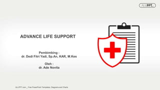
- 1. Pembimbing : dr. Dedi Fitri Yadi, Sp.An, KAR, M.Kes ADVANCE LIFE SUPPORT ALLPPT.com _ Free PowerPoint Templates, Diagrams and Charts Oleh : dr. Ade Novita
- 2. 2020 American Heart Association Chains of Survival for IHCA and OHCA
- 3. Adult Cardiac Arrest Algorithm • High Quality CPR • Shock Energy for Defibrillation • Drug Therapy • Advanced Airway • Reversible Causes
- 4. HIGH QUALITY CPR Push hard and fast Allow complete chest recoil Minimize interruptions Avoid excessive ventilation Change compressor every 2 minutes If no advance airway, 30:2 compression-ventilation ratio Quatitative waveform capnography, if PETCO2 is low or decreasing, reassess CPR quality Chest Compression Fraction >80%
- 5. ASISTOL and PEA TRUE ASISTOL •Check lead and cable connections •Monitor power on? •Monitor gain up? •Verify asystole in another lead? Asystole • complete absence of demonstrable electrical & mechanical cardiac activity. PEA • any one of a heterogeneous group of organized electrocardiographic rhythms without sufficient mechanical contraction of the heart to produce a palpable pulse or measurable blood pressure.
- 6. Ventricular tachycardia Monomorphic V T Polymorphic VT Ventricular tachycardia refers to a wide QRS complex heart rhythm, QRS duration beyond 120 milliseconds Type : monomorphic and polymorphic duration : sustained or non-sustained
- 7. Ventricular Fibrilation •Fibrillation waves of varying amplitude and shape. •No identifiable P waves, QRS complexes, or T waves •Heart rate anywhere between 150 to 500 per minute
- 8. Defibrilasi Defibrilasi merupakan proses pemberian sejumlah arus listrik untuk kejut jantung melalui alat defibrillator yang diharapkan dapat mengembalikan irama menjadi normal. Keberhasilan defibrilasi akan menurun jika dilakukan semakin lama dan VF cenderung berubah menjadi asistol dalam beberapa menit. Angka kematian meningkat 7-10% setiap menit yang terlewati tanpa dilakukan resusitasi.
- 9. Defibrilasi Buka pakaian pasien bagian dada, siapkan are apex jantung dan bagian sternum Lakukan kompresi dada dan ventilasi hingga alat defibrillator siap Nyalakan alat defibrillator, gunakan dosis energi maksimum (bifasik 200j, mon ofasik 360j) Siapkan gel pada paddles paddle pada posisi sternal-apical. Lempeng dada kanan (sternal) diletakkan pada dada bagian supero-anterior bagian kanan dan lempeng apical (kiri) dilet akkan pada dada bagian infero-lateral kiri. Lakukan charging, Ketika alat defibrillator sudah charge hingga penuh, beri aba apa kepada tim supaya tidak menyentuh pasien, hentikan kompresi dan v entilasis Pastikan irama masih menunjukkan VT/VF, tekan kedua tombol di paddle defi brillator untuk melepas energi shock, berikan tekanan 12,5 kg Ketika akan mel akukan defibrilasis Setelah selesai defibrilasis, segera lanjutkan kompresi dan ventilasi selama 5 siklus atau 2 menit.
- 11. Defibrilator The main difference between monophasic and biphasic shock delivery is that a monophasic electrical current moves in a single direction while a biphasic current is bidirectional (moving in a straight line and then reversing its direction). 1.Biphasic Defibrillators Are More Effective 2.Biphasic Defibrillation Is Less Likely to Burn the Patient 3.Biphasic Waveforms Take a Smaller Toll on Battery Life
- 12. Drug Therapy • Epinephrine Epinephrine α-Adrenergic effects increase myocardial and cerebral blood flow. β-Adrenergic effects may increase myocardial work and decrease subendocardial perfusion and cerebral blood flow Initially, 1 mg (10 mL of a 1:10,000 solution), may repeat as often as every 3-5 dosage. For endotracheal doses: 2-2.5 mg every 3-5 minutes
- 13. Drug Therapy • Amiodaron Amiodarone is a class III antiarrhythmic agent. Its main effect is to lengthen the action potential and refractory period in the myocardial tissue. It blocks the Na, K, and Ca channels Initially, 300 mg (or 5 mg/kg) via rapid inj. Additional 150 mg (or 2.5 mg/kg)
- 14. Drug Therapy • Atropin Initially, 0.5 mg repeated every 3-5 minutes. Max total: 3 mg. Atropine is an anticholinergic agent which competitively blocks the binding of acetylcholine to muscarinic receptors at the parasympathetic sites in the CNS and peripheral tissues such as the heart, intestines, bronchial muscles, iris and secretory glands.
- 15. Drug Therapy • Lidocaine 1-1.5 mg/kg repeated as necessary. Max: 3 mg/kg Lidocaine is classified as a class Ib antiarrhythmic that d ecreases the permeability of the neuron membrane to sod ium, which causes inhibition of depolarization, resulting in blocked conduction
- 16. Advanced Airway Tracheal intubation •Tracheal intubation should be attempted as soon as practical. Do not interrupt ventilation for more than 10 s. After intubation, the patient can be ventilated with a selfinflating bag capable of delivering high oxygen concentrations. A ratio of 8–10 breaths/min in a secure airway should be maintained, as high respiratory rates can impede cardiac output in a cardiac arrest situation. Wavefrom Capnography or capnometry to confirm and monitor ETT tube placement
- 17. Reversible Causes 5 H • Hypovolemia • Hypoxia • Hydrogen ion • Hyper / Hypokalemia • Hypothermia 5 T • Toxins • Tamponade Cardia • Tension pneumothorax • Thrombosis coronary (ACS) • Thrombosis pulmonary (embolism)
- 18. Reversible Causes Hypovolemia Hypoxia Hydrogen Ion (acidosis) Hypo/Hyperkalemia Hypotermia Loss of fluid volume in circulatory system most important interv ention is to obtain IV access and adminis ter IV Fluid Use fluid challenge to determine if the ar ret is related to hypov olemia An adequate oxygen supply Ensure that the airway is open Ensure adequare venti lation, and bilateral br eath sounds Obtain an arterial blo od gas to determine r espiratory acidosis Provide adequate ventilations Use sodium bicarbona te to prevent metabol ic acidosis if necessar y Sign of hiperkalemia : Taller, peaked T- wav es, and widening of t he QRS complex Sign of hipokalemia : flattened T-wave, pro minent U-waves, wide ned QRS complex If patients has been e xposed to the cold, w arming measures sho uld be taken. Core tempt should be above 30 C as soo n as possible
- 19. Reversible Causes Toxins Tamponade Tension Pneumoth orax Thrombosis Coronary Thrombosis Pulmo nal Most common : tricyclics, digoxin, beta blockers and CCB Cocaine Physical sign : bradyca rdia, pupil symptoms, other neurological cha nges Fluid buil-up in the pericardium result in ineffective pumping of the blood which can le ad to pulseless arrest ECG : narrow QRS complex and rapid hea rt rate physical sign : jugular vein distention, no pul se, muffled heart soun f ECG sign : narrow QRS complexes and rapid h eart rate physical sign: JVD, tracheal deviation, unequal breath sound, difficulty with ventilati on ECG signs : 12 lead ECG with ST segment changes, T inversions, and or Q waves Elevated cardiac markers ECG sign : narrow QRS complex and rapid hea rt rate Positive D-dimer test, positive test for DVT or PE
- 20. THANK YOU