
Recommended
More Related Content
What's hot
What's hot (20)
Similar to Aortic dissection .pptx
Similar to Aortic dissection .pptx (20)
Recently uploaded
Recently uploaded (20)
Aortic dissection .pptx
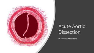
- 1. Acute Aortic Dissection Dr Mubarik Ahmed Jan
- 2. Definition • The Aortic Dissection is a condition that occurs due to tear in tunica intima, which is the innermost layer of the aorta. • The blood flowing in the wall dissects the layers of the aorta, that may lead to aneurysm, bleeding, end- organ damage and even death.
- 3. Epidemiology: The Aortic dissection is a rare diagnosis with an incidence of around 3 - 4 per 100,000 person - years. The Aortic dissection is more common in men than women with a ratio of ~2:1. The average age of diagnosis is in the 60’s, although patients with connective tissue disorder have a much lower age of presentation often in their 30’s.
- 4. Predisposing Factors: History of previous dissection Hemodynamic Stressors like Hypertension and Cocaine use. Connective Tissue Disorders (Marfan Syndrome, Ehlers-Danlos Syndrome) Anatomic Abnormalities that cause abnormal flow (Bicuspid aortic valve) Questionable predisposing factors: PCOS, Pregnancy, Family History
- 5. Marfan’s syndrome: Famous People With Marfan Syndrome Look - The patient doesn’t always know they have Marfan’s so need to look for the signs. Arachnodactyly - Elongated fingers. Pectus excavatum : sternal excavation Lanky limbs
- 6. Classification: Stanford and Debakey’s Type A ● Involves ascending aorta. ● Can extend distally ad infinitum. ● Surgery is usually indicated. Type B ● Involves aorta beyond left subclavian artery only. ● Often managed medically with BP control. Stanford’s - More commonly used
- 7. Classification: • DeBakey’s • Type 1: Involves ascending aorta, aortic arch, and descending aorta • Type 2: Ascending aorta only • Type 3: Descending aorta only
- 8. Aortic Dissection Variants Intramural thrombus • An infarction in the aortic media, most often due to an injury to the vaso vasorum, that results in a thrombus formation within the aortic wall, which may extend or resolve spontaneously. • Often a precursor to dissection
- 9. AD Variants Penetrating ulcer • Ulcer formation due to atherosclerosis which can lead to intramural thrombus, dissection or aortic perforation
- 11. Signs and Symptoms • Although cardiac tamponade is a relatively rare presentation of AD (~4%), it is the most common cause of death in AD
- 12. History + Physical Examination: • Classic presentation: • Sudden onset of tearing chest pain radiating to the back. • However, dissection may occur anywhere along the aorta and thus the presentation may be broad and mimic other common disorders • Variant presentations include: (Due to associated end organ damage) • Chest pain or back pain + vomiting • Chest pain or back pain with neurologic findings (may be due to dissection into the carotid arteries) • Chest pain or back pain + limb ischemia • Cardiac tamponade • Only 51% of AD patients have the classic tearing chest pain
- 13. History + Physical Examination: • Presenting blood pressure • Hypertension: 49% • Normotension: 33% • Hypotension: 18% • Classic Risk Factors (Hagan 2000) • 9% of patient’s have Marfan syndrome, these patients are often young. • 72% had a history of HTN. • 9% had prior cardiac surgery. • Physical Examination (Hagan 2000) • Pulse deficit: Present in only 28%. Defined as >20 SBP point difference between arms
- 14. Work-Up • CBC - Leukocytosis • INR/PTT • Renal function - Cr elevation with renal artery involvement. • Troponin elevated if dissection causes myocardial ischaemia. • D-dimer – If negative dissection is very unlikely, but not sufficient to rule out • Cross-match - (Possible surgery and need for blood products). • Various biomarkers are being investigated (e.g. elastin fragments, smooth muscle myosin heavy-chain protein).
- 15. ECG: Normal - >30% of patients have no ECG changes (Hagan 2000). 40% will show non-specific ST-T wave changes. Inferior ST elevation (right coronary dissection) but can also be any STEMI (0.1% of STEMIs are dissections) Pericarditis changes, electrical alternans (tamponade).
- 16. CXR ~ 60% will have a wide mediastinum on CXR, while ~16% will have a completely normal CXR. Large dilated tortuous aortic arch and descending aorta with mass effect on the trachea (displacing it to the right and mildly narrowing it).
- 17. CXR • Loss of the aortic knob/aortic- pulmonary window
- 18. • Look for a white line of calcium within the aortic knob. Then measure the distance from there to the outer edge of the aortic knob. • A distance > 0.5cm is considered a positive calcium sign and a distance > 1.0cm is considered highly suspicious for aortic dissection.
- 19. Trans Thoracic ECHO: • May be helpful in identifying cardiac tamponade in an unstable patient. • Tamponade is the common cause of hypotensive presentation of AD • ACEP Level B guideline: • Do not rely on abnormal bedside TTE result to establish diagnosis of thoracic aortic dissection.
- 20. Trans Esophageal ECHO: • Excellent option in patients with CKD or where CTA may not be an available. • It has a great sensitivity 98%.
- 21. The ADD Risk Score: Grading the pretest probability • The Aortic Dissection Detection Risk Score (ADD-RS) is a clinical decision tool that aids in grading the pretest probability of an acute aortic dissection. • Scores range from 0-3, • where 0 is classed as low risk, • 1 is moderate risk and • 2-3 is high risk
- 22. CT Angio Modality of choice with high specificity and sensitivity. Can identify a false lumen, location of dissection flap, extension into the great vessels, signs of aortic rupture and end-organ damage CTA of Chest/Abdomen/Pelvis should be done in patients with high suspicion to visualise the entire length of dissection
- 24. • Type B Aortic Dissection
- 25. MRA: ● This is the best imaging study for AD but is limited by availability and time. ● Sensitivity and specificity 98%.
- 28. Treatment depends on the type of dissection whether Type A or Type B. Type A dissections almost always require open surgical repair. Mobilize consultants as early as possible (Cardiothoracic surgery, interventional radiology). Mortality increases by 1-2% for every hour from symptom onset to definitive treatment. Type A Dissection:
- 29. Management • Type B dissections can often be managed medically, if uncomplicated, or with endovascular repair. • Thoracic Endo Vascular Aortic Repair (TEVAR) is now favored compared to open repair of Type B dissections as this has been shown to reduce morbidity and mortality.
- 30. Stepwise treatment : • The initial treatment is the same for both type A and type B and includes a stepwise fashion of: • Treating Pain, • Then Heart Rate, • Then Blood Pressure, • With consultation to Cardiothoracic surgery or vascular surgery depending on the site.
- 31. Treat Pain: One of the best ways to control BP in these patients is to aggressively treat pain. The pain is severe, and to effectively treat BP, first need to control the pain. Treat anything that can increase Blood Pressure or cause Valsalva Nausea can also be an issue and should be dealt with antiemetics.
- 32. Treat HR: • Treat HR first to avoid the shear force caused by the stroke volume of each beat. • Start with rate control because antihypertensive agent may cause reflex tachycardia which can worsen shear force. • Esmolol is the preferred agent for controlling heart rate with a goal HR<60. • Esmolol is given as a 500 mcg/kg bolus over 1 min, started at 50 mcg/kg/min infusion and increased gradually. • Max infusion for esmolol is 200 mcg/kg/min.
- 33. Treat HR: Labetalol IV 10 or 20 mg is also an option if esmolol infusion is not readily available and need HR control fast. Also consider Labetalol in cases with cocaine as provides both alpha and beta-blockade. Labetalol is also available as an infusion with rates from 30 to 120 mg / hour.
- 34. Treat Blood Pressure: • The goal SBP is <110. If unable to achieve the goal SBP after maxing esmolol to goal HR<60, then nicardipine or nitroprusside can be added. • Nicardipine is typically started as an infusion of 5 mg/hr and increased gradually until max of 30 mg/hr. • Nitroprusside: is a pure vasodilator. • Infusion rate is 0.3-0.5 mcg/kg/min to start. • Typical dose for BP control is 3-4 mcg/kg/min infusion.
- 35. Consult surgeon: Type A: Immediately to be pushed to Operation Theatre. Type B: Surgery may be needed. Patient will be admitted to ICU for • Conservative management if uncomplicated, • Analgesia, • Optimizing hemodynamics and • Managing post operative complications.
- 36. Post-operative complications: • The most common post-operative complications following endovascular repair of type B dissection includes: • Stent Graft migration, • Stent Graft fracture, • Endoleak, • Retrograde dissection, • Stroke, • Paraplegia and • Lower limb ischemia.
- 37. ACC/ AHA- Aortic- Dissection- guideline Acute AoD Management Pathway
- 38. Historical Fact: Dr. Michael Debakey This is Dr. Michael Debakey of Debakey Classification fame, born in 1908. He pioneered the first aortic repairs of aortic dissection which bears his name. At age 97, he actually suffered from an aortic dissection himself. Initially, he opted for medical management, but after becoming unresponsive, it was decided to proceed with surgical intervention. After a complicated post-op course and 8 months in the hospital, he returned to good health and continued to practice medicine until his death at age 99.
- 39. References: ● https://www.ahajournals.org/doi/full/10.1161/cir.0b013e3181d4739e ● https://emupdates.com/accaha-aortic-dissection-guideline/ ● https://litfl.com/acute-aortic-dissection/ ● https://rushemergencymedicine.org/2020/01/21/aortic-dissection/ ● http://www.emdocs.net/core-em-aortic-dissection/