Cell division essentials
•Download as PPTX, PDF•
7 likes•7,404 views

Cell division is essential but must be controlled. There are two phases of cell division - interphase and mitosis. Interphase is the non-dividing phase where the cell grows and carries out normal functions. Mitosis is the dividing phase where the nucleus divides into two identical daughter nuclei through the stages of prophase, metaphase, anaphase and telophase. Cytokinesis then divides the cytoplasm. Chromosomes condense through supercoiling during mitosis. Cyclins control progression through the cell cycle. Mutations from mutagens can lead to cancer development if they occur in oncogenes and are not repaired. Smoking strongly correlates with increased lung cancer rates, with a lag time between smoking and cancer development

Recommended
More Related Content
What's hot
What's hot (20)
Similar to Cell division essentials
Similar to Cell division essentials (20)
More from Bob Smullen
More from Bob Smullen (20)
Recently uploaded
Recently uploaded (20)
Cell division essentials
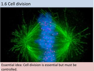
- 1. 1.6 Cell division Essential idea: Cell division is essential but must be controlled.
- 2. Understandings Statement Guidance 1.6 U.1 Mitosis is division of the nucleus into two genetically identical daughter nuclei. The sequence of events in the four phases of mitosis should be known. To avoid confusion in terminology, teachers are encouraged to refer to the two parts of a chromosome as sister chromatids, while they are attached to each other by a centromere in the early stages of mitosis. From anaphase onwards, when sister chromatids have separated to form individual structures, they should be referred to as chromosomes. 1.6 U.2 Chromosomes condense by supercoiling during mitosis. 1.6 U.3 Cytokinesis occurs after mitosis and is different in plant and animal cells. 1.6 U.4 Interphase is a very active phase of the cell cycle with many processes occurring in the nucleus and cytoplasm. 1.6 U.5 Cyclins are involved in the control of the cell cycle. 1.6 U.6 Mutagens, oncogenes and metastasis are involved in the development of primary and secondary tumours.
- 3. Applications and Skills Statement Guidance 1.6 A.1 The correlation between smoking and incidence of cancers. 1.6 S.1 Identification of phases of mitosis in cells viewed with a microscope or in a micrograph. Preparation of temporary mounts of root squashes is recommended but phases in mitosis can also be viewed using permanent slides. 1.6 S.2 Determination of a mitotic index from a micrograph.
- 4. Why do cells divide: • Growth: Multicellular organisms increase their size by increasing their number of cells through mitosis • Asexual reproduction: Certain eukaryotic organisms may reproduce asexually by mitosis (e.g. vegetative reproduction) • Tissue Repair: Damaged tissue can recover by replacing dead or damaged cells • Embryonic development: A fertilized egg (zygote) will undergo mitosis and differentiation in order to develop into an embryo
- 5. • Cellular division in eukaryotic cells. • Chromatin is arranged into chromosomes. • Chromosomes double. • Cell grows in size. • Cells divide. • Is cellular cloning. Cell division
- 6. 2 phases: 1. Interphase 2. M phase (mitotic phase) a. Prophase b. Metaphase c. Anaphase d. Telophase & cytokinesis Figure 12.4 The cell cycle Phases of the Cell Cycle (life cycle of a cell)
- 7. Interphase • The non-dividing phase in a cell • Lasts about ~ 90% of the cell cycle. • The cell grows and replicates DNA preparing for Mitosis. • There are three periods: 3 periods of Interphase 1. Go – a cell functioning as normal 2. G1 phase – first growth phase 3. S phase- synthesis of DNA 4. G2 phase- 2nd growth phase Mitosis is a reliable process. Only one error occurs per 100,000 cell divisions. 1.6 U.4 Interphase is a very active phase of the cell cycle with many processes occurring in the nucleus and cytoplasm.
- 8. 1.6 U.5 Cyclins are involved in the control of the cell cycle. Cyclinsare a family of proteins that control the progression of cells through the cell cycle Cells cannot progress to the next stage of the cell cycle unless the specific cyclin reaches it threshold. http://upload.wikimedia.org/wikipedia/commons/thumb/9/99/Protein_CCNE1_PDB_1w98.png/800px-Protein_CCNE1_PDB_1w98.png Cyclins bind to enzymes called cyclin-dependent kinases These kinases then become active and attach phosphate groups to other proteins in the cell. The attachment of phosphate triggers the other proteins to become active and carry out tasks (specific to one of the phases of the cell cycle). 4 3 2 1
- 9. http://upload.wikimedia.org/wikipedia/commons/thumb/9/99/Protein_CCNE1_PDB_1w98.png/800px-Protein_CCNE1_PDB_1w98.png Triggers cells to move from G0 to G1 and from G1 into S phase. prepares the cell for DNA replication in S phase. activates DNA replication inside the nucleus in S phase. promotes the assembly of the mitotic spindle and other tasks in the cytoplasm to prepare for mitosis. Progression through parts of the cell cycle are affected in various ways by specific cyclins
- 10. 1.6 U.4 Interphase is a very active phase of the cell cycle with many processes occurring in the nucleus and cytoplasm. Interphase This when the cell carries out it’s normal functions Metabolic reactions (e.g. respiration to produce ATP) are necessary for the life of the cell Protein synthesis - proteins and enzymes are necessary to allow cell grow Organelles numbers are increased to first support the enlarged cell DNA is replicated to ensure a second copy is available to enable mitosis Cells spend the majority of their time in interphase. It is a very active phase of the cycle. Mr P O D http://botit.botany.wisc.edu/Resources/Botany/Mitosis/Allium/Various%20views/Interphase%20prophase.JPG
- 11. 1.6 U.1 Mitosis is division of the nucleus into two genetically identical daughter nuclei. http://commons.wikimedia.org/wiki/File:Chromosome.svg centromere is the part of a chromosome that links sister chromatids Sister chromatids are duplicated chromosomes attached by a centromere Get the terminology right centrioles organise spindle microtubules Spindle microtubules (also referred to as spindle fibres) In animal cells two centrioles are held by a protein mass referred to as a centrosome After anaphase when the sister chromatids separate they should then be referred to as chromosomes It is easy to misuse the terms chromatid and chromosome. It is even easier to confuse the terms centromere, centriole and centrosome due to their similar spelling. Keep the terms clear in your mind to avoid losing marks. http://commons.wikimedia.org/wiki/Mitosis#mediaviewer/File:Mitosis_cells_sequence.svg
- 12. http://highered.mheducation.com/sites/0072495 855/student_view0/chapter2/animation__mitosis _and_cytokinesis.html Use the animated tutorials to learn about mitosis http://www.johnkyrk.com/mitosis.html http://www.sumanasinc.com/webcontent/animations/content /mitosis.html http://outreach.mcb.harvard.edu/animations/cellcycle. swf
- 13. 1.6 U.2 Chromosomes condense by supercoiling during mitosis. Why supercoil chromosomes? Human cells are on average 10μm in diameter and the nucelus within each is less than 5 μm in diameter. Human chromosomes are 15mm to 85mm (15,000μm to 85,000 μm) in length. Chromosomes need to be stored compactly to fit within the nuclei of cells. This problem becomes more acute during mitosis when chromosomes need to be short and compact enough that they can be separated and moved to each end of the cell. http://www.genome.gov/dmd/img.cfm?node=Photos/Graphics&id=85282 Chromatin fibres
- 14. 1.6 U.2 Chromosomes condense by supercoiling during mitosis. How are chromosomes supercoiled? Strain is placed on a DNA helix by over winding or under winding of the helix This causes the DNA molecule to coil back on itself becoming shorter and wider n.b. in eukaryotes proteins called histones aid the process http://www.maths.uq.edu.au/~infinity/Infinity7/images/supercoiling.gifhttp://vanat.cvm.umn.edu/mMeiosis/images/chromosome-X.jpg
- 15. Prophase • The nucleolus disappears. • Chromatin condenses into visible chromosomes. • There are two sister chromatids held together by a centromere. • The mitotic spindle forms in the cytoplasm. . 1.6 S.1 Identification of phases of mitosis in cells viewed with a microscope or in a micrograph
- 16. Metaphase • The nuclear envelope disappears. • Spindle fibers extend from each pole to the cell’s equator. • Spindle fibers attach to the centromeres.
- 17. Figure 12.3 Chromosome duplication and distribution during mitosis
- 18. Anaphase • Characterized by movement. It begins when pairs of sister chromatids pull apart. • Sister chromatids move to opposite poles of the cell. • Chromosomes look like a “V” as they are pulled. • At the end of anaphase, the two poles have identical number and types of chromosomes.
- 19. Telophase • Microtubules elongate the cell. • Daughter nuclei begin to form at the two poles. • Nuclear envelopes re-form. • Nucleolus reappears. • Chromatin uncoils. • The cells cytoplasm begins to pinch. • It is basically the opposite of prophase.
- 20. 1.6 U.3 Cytokinesis occurs after mitosis and is different in plant and animal cells. mitosis is the division of the nucleus, cytokinesis is the division of the cytoplasm to create two cells Though mitosis is similar for animal and plant cells cytokinesis is very different. http://wwwprod.biochem.wisc.edu/biochem/faculty/bednarek/images/figure_color.gif http://glencoe.mheducation.com/sites/983 4092339/student_view0/chapter10/animati on_-_cytokinesis.html http://www.haroldsmithlab.com/images/pg_HeLa_cell_division.jpg
- 21. Figure 12.8 Cytokinesis in animal and plant cells
- 22. 1.6 S.1 Identification of phases of mitosis in cells viewed with a microscope or in a micrograph. 1.6 S.2 Determination of a mitotic index from a micrograph. http://www.nuffieldfoundation.org/practical-biology/investigating-mitosis-allium-root-tip-squash A very good, well explained lab outline for creating slides and calculating the mitotic index. http://www.biology.arizona.edu/cell_bio/activities/cell_cycle/cell_cycle.html An excellent online alternative if resources don’t permit students to create and view their own slides
- 23. 1.6 U.6 Mutagens, oncogenes and metastasis are involved in the development of primary and secondary tumors. Tumors are abnormal growth of tissue that develop at any stage of life in any part of the body. A cancer is a malignant tumour and is named after the part of the body where the cancer (primary tumour) first develops. Use the links to find out: • most common types of cancer • what causes cancer and associated risk factors • how cancer can be treated http://www.cancer.gov/cancertopics/types/commoncancers http://youtu.be/8BJ8_5Gyhg8 http://www.cancerresearchuk.org/cancer- info/cancerandresearch/all-about-cancer/what-is-cancer/ What causes cancer? http://www.e-learningforkids.org/health/lesson/cancer/
- 24. 1.6 U.6 Mutagens, oncogenes and metastasis are involved in the development of primary and secondary tumors. Mutagens are agents that cause gene mutations. Not all mutations result in cancers, but anything that causes a mutation has the potential to cause a cancer. Mutagens can be: • chemicals that cause mutations are referred to as carcinogens • high energy radiation such as X-rays • short-wave ultraviolet light • Some viruses A mutation is a change in an organisms genetic code. A mutation/change in the base sequence of a certain genes can result in cancer. http://en.wikipedia.org/wiki/Oncogene#mediaviewer/File:Oncogenes_illustration.jpg
- 25. 1.6 U.6 Mutagens, oncogenes and metastasis are involved in the development of primary and secondary tumors. mutation in a oncogene If a mutation occurs in an oncogenes it can become cancerous. In normal cells oncogenes control of the cell cycle and cell division. http://en.wikipedia.org/wiki/Oncogene#mediaviewer/File:Oncogenes_illustration.jpg uncontrolled cell division tumour formation malfunction in the control of the cell cycle
- 26. 1.6 U.6 Mutagens, oncogenes and metastasis are involved in the development of primary and secondary tumours. Factors (other than exposure to mutagens) that increase the probability of tumour development include: • The vast number of cells in a human body – the greater the number of cells the greater the chance of a mutation. • The longer a life span the greater the chance of a mutation. Several mutations must occur in the same cell for it to become a tumour causing cell. The probability of this happening in a single cell is extremely small. http://en.wikipedia.org/wiki/Oncogene#mediaviewer/File:Oncogenes_illustration.jpg
- 27. There are two major types of tumor: 1. Benign Tumors this is a mass of cancerous cells that do not invade other areas of the body. These are not as dangerous to health but may still require removing to prevent effects on neighboring tissue 1.6 U.6 Mutagens, oncogenes and metastasis are involved in the development of primary and secondary tumors.
- 28. 2. Malignant Tumors is a mass of cancer cells that may invade surrounding tissues or spread to distant areas of the body. Cancer cells replace normal functioning cells in distant sites: e.g. replacing blood forming cells in the bone marrow, replacing bones leading to increased calcium levels in the blood, or in the heart muscles so that the heart fails. 1. Image is a normal CT. Images 2, 3 & 4 Are PET scans, Light green/blue areas show cancer cells
- 29. 1.6 A.1 The correlation between smoking and incidence of cancers. http://en.wikipedia.org/wiki/File:Smoking_lung_cancer.png There are many other similar surveys in different countries, with different demographics that show similar results. Along with lung cancer, cancers of mouth and throat are very common as these areas are in direct contact with the smoke too. It might surprise you that the following cancers are also more common in smokers: • Head and neck • Bladder • Kidneys • Breast • Pancreas • Colon
- 30. a. Describe the relationship shown. b. What type of correlation is shown c. How strong is the correlation? Justify your answer by discussing the evidence. d. The correlation shown here is lagged. A lag is a time gap between the factors. Estimate the size of the lag between cigarette consumption and lung cancer death.
- 31. http://en.wikipedia.org/wiki/File:Smoking_lung_cancer.png a. Describe the relationship shown. b. What type of correlation is shown c. How strong is the correlation? Justify your answer by discussing the evidence. d. The correlation shown here is lagged. A lag is a time gap between the factors. Estimate the size of the lag between cigarette consumption and lung cancer death. There are many other similar surveys in different countries, with different demographics that show similar results. Along with lung cancer, cancers of mouth and throat are very common as these areas are in direct contact with the smoke too. It might surprise you that the following cancers are also more common in smokers: • Head and neck • Bladder • Kidneys • Breast • Pancreas • Colon
- 32. Bibliography / Acknowledgments Jason de Nys