1. The document discusses wound healing, defining it as the normal physiological process that results in the repair of injured tissue.
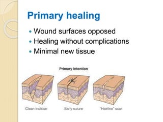
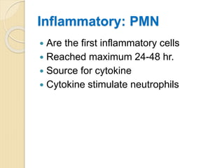
2. It describes the three phases of wound healing: inflammatory phase, proliferative phase, and remodeling phase. The inflammatory phase involves hemostasis and inflammation in the first 4-6 days. The proliferative phase involves re-epithelialization, angiogenesis, and matrix formation from days 4-14. The remodeling phase involves collagen remodeling from days 8 onwards.
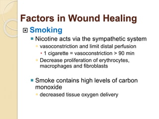
3. It also discusses factors that affect wound healing such as oxygen, nutrition, infection, diabetes, radiation therapy, medications, and smoking. Syndromes associated with abnormal wound healing such as cutis laxa and Ehlers