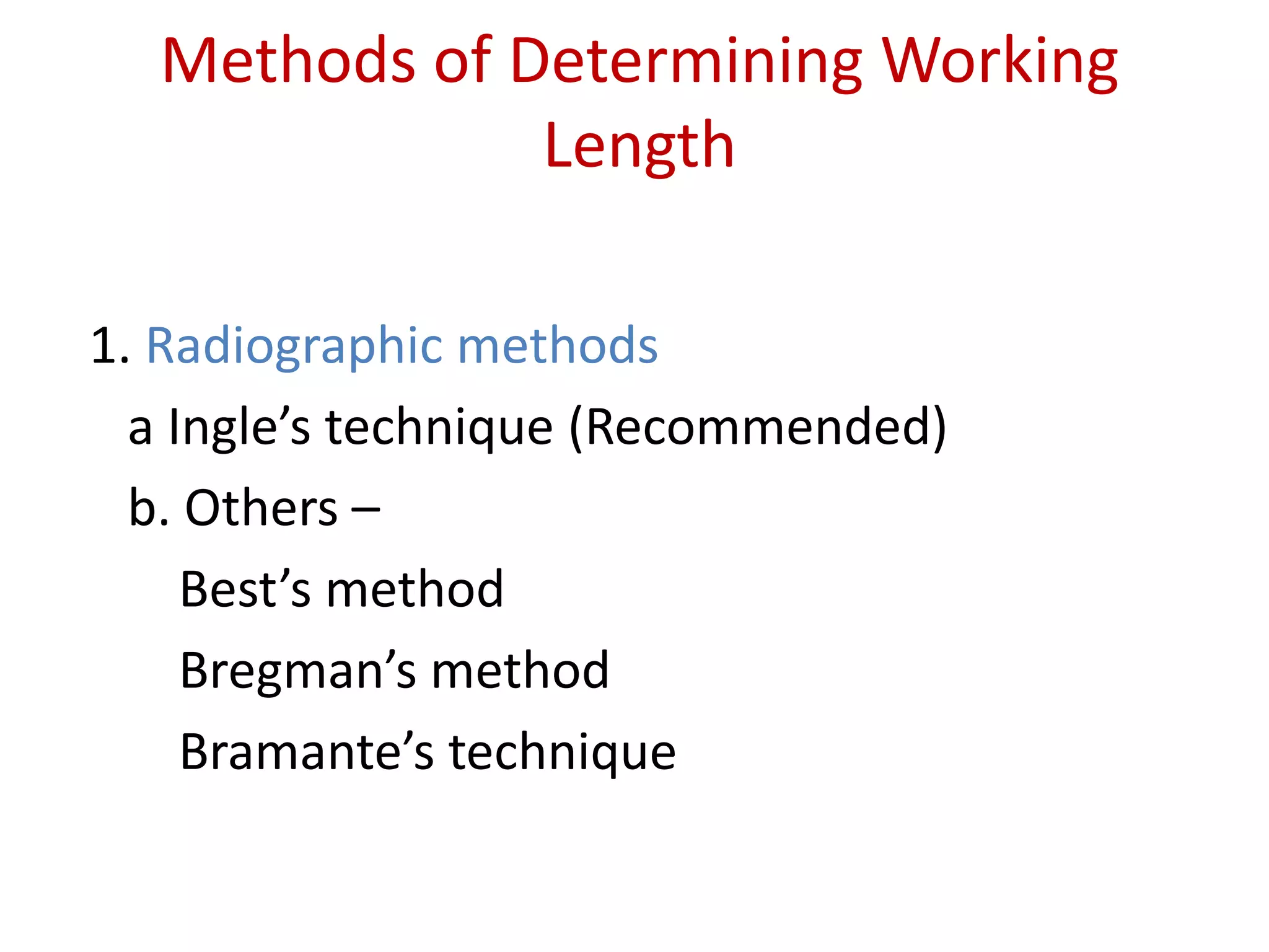
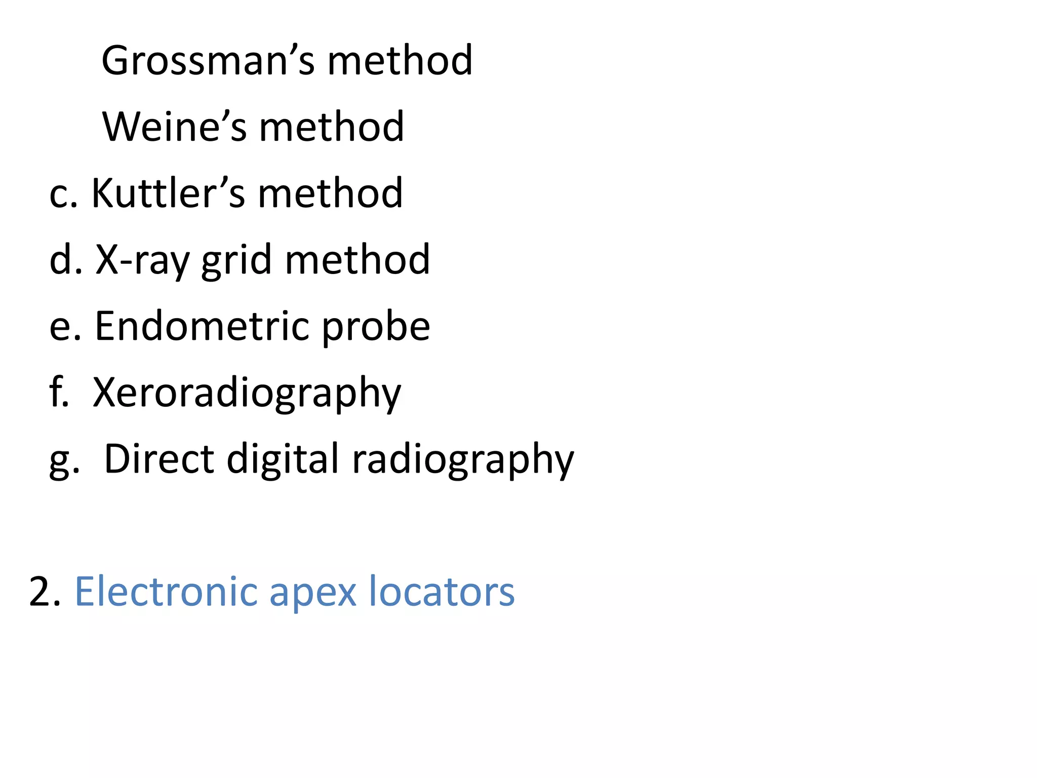
Working length determination
Presented by: Shrisha Pant
Methods of Determining Working
Length
1. Radiographic methods
a Ingle’s technique (Recommended)
b. Others –
Best’s method
Bregman’s method
Bramante’s technique
Grossman’s method
Weine’s method
c. Kuttler’s method
d. X-ray grid method
e. Endometric probe
f. Xeroradiography
g. Direct digital radiography
2. Electronic apex locators
3.Nonradiographic methods (not recommended)
a. Digital tactile sense method
b. Apical periodontal sensitivity
c. Paper point method
d. Average tooth length
Weine’s Modification method
Modification in Ingle’s Method
where final working length under following condition
obtained by:
No resorption cases : subtract1mm
Periapical bone resorption : subtract1.5mm
Periapical bone and root apex resorption : subtract
2mm , for compensation of loss of apical constriction.
Grossman method
It is based on simple mathematical formulation to calculate the working
length:
An instrument is inserted into the canal
Stopper is fixed to the reference point
Radiograph is taken
The formula used for calculation of actual length is as follows:
Actual length of tooth = Apparent length of tooth in radiograph
Actual length of instrument Apparent length of instrument in radiograph
Actual length of the tooth= actual length of the instrument X apparent length of the
t tooth in radiography
Apparent length of instrument in radiograph
Kuttler’s method
• According to Kuttler’s canal preparation should terminate
at apical constriction(minor diameter).
locate the minor and major diameter on preoperative
radiograph
Estimate length of roots from preoperative radiograph
Estimate canal width and insert no.10 or 15 file in canal up
to estimated length
take a radiography
If file is too long or short by more than 1mm from the minor
diameter , readjust the file and take another radiograph
If file reach the major diameter , subtract 0.5 mm from it for
younger patients and 0.6mm for older patient
Advantage
1.Allows rapid development of apical dentinal
matrix
2.Minimal error
3.Has shown many successful result.
Disadvantage
1.Time consuming
2.Calculation is complicated
3.Radiograph of excellent quality and
magnification is required.
Radiographic grid method
• Designed by Everett & Fixott in1963
• It is a simple method in which a millimeter grid is
superimposed on the radiograph.
• This over comes the need for calculation.
• But it is not good method if radiograph is bent during
exposure
ADVANTAGES :
Simple method
No need for calculations
DISADVANTAGES :
Cannot be used if radiograph is bent during
exposure
Endometric Probe
• In this method, one uses the graduations on
diagnostic file which are visible on radiograph.
• But its main disadvantage is that the smallest
file size to be used is number 25.
Xero-radiography
• The term Xero-radiography is derived from the Greek
word XEROS which literally means dry which
differentiates this from the conventional photochemical
system.
• Images are recorded on an aluminum plate coated with
selenium particles.
• Image are sharper.
• Edge enhancement improves the visualization of files and
various anatomical structure such as apical foramen and
periodontal ligament.
The aluminum plate is removed from the cassette
subjected to relaxation which removes old images
These are electrostatically charged
Inserted into cassette
Projection of radiation
Forms latent image and is converted to real images by
process called ‘development’ in the processor unit.
Direct digital radiography
• In this digital image is formed which is
represented by spatially distributed set of
discrete sensors and pixels.
• Two types of digital radiography:
a. Radiovisiography
b. Phosphor imaging system
ADVANTAGES of radiographic method
1.Can see anatomy of tooth
2.Can see curvature in roots
3.Can see relationship b/w adjacent teeth & anatomic
structures
DISADVANTAGES of radiographic method
1.Varies with different observers
2.Superimposition of anatomic structures
3.2D view of a 3D object
4.Radiation exposure
5.Cannot interpret if apical foramen has buccal or lingual
exit
6.Limited accuracy
7.Time consuming
Non radiographic method
1. Digital Tactile method
• There is increase in
resistance as the film
approaches apical 2-3mm.
• In this region the canal
frequently constricts (minor
diameter).
• Adjunct to radiograph or
apex locator.
ADVANTAGES :
• Time saving
• No radiation exposure
DISADVANTAGES :
• Does not always provide the accurate readings.
• In case of narrow canals, one may feel increased
resistance as file approaches apical 2-3 mm.
• In case of teeth with immature apex instrument can
go periapically
2.Paper point measurement
• Most reliable in cases of open apex where apical constriction
is lost because of perforation or resorption.
• Blunt end of paper point is passed gently in canal after LA.
• Moisture or blood present on apical part of paper point
indicates paper point has passed beyond estimated working
length.
This method, however, may
give unreliable data :
1. If the pulp not
completely removed.
2. If the tooth is pulp less
but a periapical lesion
rich in blood supply is
present.
3. If paper point is left in
canal for a long time
3.Periodontal sensitivity test
Based on patient’s response to pain
• This method does not provide accurate readings,
for example in case of narrow canals, instrument
may feel increased response in apical 2-3mm,
immature apex, file goes beyond apex.
• In case of canals with necrotic pulp, instrument can
pass beyond apical constriction, and in case of vital
or inflamed pulp, pain may occur several mm
before periapex is crossed by instrument
4.Average tooth length
It can be taken as a guideline.
Conclusion
• There are several method to determine working
length.
• Combination of electronic apex locator and ingle’s
radiographic technique is most appropriate
technique.
• Non radiographic method is not recommended.
Reference:
• Grossman’s ENDODONTIC PRACTICE , 13TH
EDITION
• Textbook of Endodontics , 2ND EDITION pg no
204-205
Working length determination

Working length determination

  • 1.
  • 2.
    Methods of DeterminingWorking Length 1. Radiographic methods a Ingle’s technique (Recommended) b. Others – Best’s method Bregman’s method Bramante’s technique
  • 3.
    Grossman’s method Weine’s method c.Kuttler’s method d. X-ray grid method e. Endometric probe f. Xeroradiography g. Direct digital radiography 2. Electronic apex locators
  • 4.
    3.Nonradiographic methods (notrecommended) a. Digital tactile sense method b. Apical periodontal sensitivity c. Paper point method d. Average tooth length
  • 5.
    Weine’s Modification method Modificationin Ingle’s Method where final working length under following condition obtained by: No resorption cases : subtract1mm Periapical bone resorption : subtract1.5mm Periapical bone and root apex resorption : subtract 2mm , for compensation of loss of apical constriction.
  • 7.
    Grossman method It isbased on simple mathematical formulation to calculate the working length: An instrument is inserted into the canal Stopper is fixed to the reference point Radiograph is taken The formula used for calculation of actual length is as follows:
  • 8.
    Actual length oftooth = Apparent length of tooth in radiograph Actual length of instrument Apparent length of instrument in radiograph Actual length of the tooth= actual length of the instrument X apparent length of the t tooth in radiography Apparent length of instrument in radiograph
  • 9.
    Kuttler’s method • Accordingto Kuttler’s canal preparation should terminate at apical constriction(minor diameter). locate the minor and major diameter on preoperative radiograph Estimate length of roots from preoperative radiograph Estimate canal width and insert no.10 or 15 file in canal up to estimated length
  • 10.
    take a radiography Iffile is too long or short by more than 1mm from the minor diameter , readjust the file and take another radiograph If file reach the major diameter , subtract 0.5 mm from it for younger patients and 0.6mm for older patient
  • 11.
    Advantage 1.Allows rapid developmentof apical dentinal matrix 2.Minimal error 3.Has shown many successful result. Disadvantage 1.Time consuming 2.Calculation is complicated 3.Radiograph of excellent quality and magnification is required.
  • 12.
    Radiographic grid method •Designed by Everett & Fixott in1963 • It is a simple method in which a millimeter grid is superimposed on the radiograph. • This over comes the need for calculation. • But it is not good method if radiograph is bent during exposure
  • 13.
    ADVANTAGES : Simple method Noneed for calculations DISADVANTAGES : Cannot be used if radiograph is bent during exposure
  • 14.
    Endometric Probe • Inthis method, one uses the graduations on diagnostic file which are visible on radiograph. • But its main disadvantage is that the smallest file size to be used is number 25.
  • 15.
    Xero-radiography • The termXero-radiography is derived from the Greek word XEROS which literally means dry which differentiates this from the conventional photochemical system. • Images are recorded on an aluminum plate coated with selenium particles. • Image are sharper. • Edge enhancement improves the visualization of files and various anatomical structure such as apical foramen and periodontal ligament.
  • 16.
    The aluminum plateis removed from the cassette subjected to relaxation which removes old images These are electrostatically charged Inserted into cassette Projection of radiation Forms latent image and is converted to real images by process called ‘development’ in the processor unit.
  • 17.
    Direct digital radiography •In this digital image is formed which is represented by spatially distributed set of discrete sensors and pixels. • Two types of digital radiography: a. Radiovisiography b. Phosphor imaging system
  • 18.
    ADVANTAGES of radiographicmethod 1.Can see anatomy of tooth 2.Can see curvature in roots 3.Can see relationship b/w adjacent teeth & anatomic structures DISADVANTAGES of radiographic method 1.Varies with different observers 2.Superimposition of anatomic structures 3.2D view of a 3D object 4.Radiation exposure 5.Cannot interpret if apical foramen has buccal or lingual exit 6.Limited accuracy 7.Time consuming
  • 19.
    Non radiographic method 1.Digital Tactile method • There is increase in resistance as the film approaches apical 2-3mm. • In this region the canal frequently constricts (minor diameter). • Adjunct to radiograph or apex locator.
  • 20.
    ADVANTAGES : • Timesaving • No radiation exposure DISADVANTAGES : • Does not always provide the accurate readings. • In case of narrow canals, one may feel increased resistance as file approaches apical 2-3 mm. • In case of teeth with immature apex instrument can go periapically
  • 21.
    2.Paper point measurement •Most reliable in cases of open apex where apical constriction is lost because of perforation or resorption. • Blunt end of paper point is passed gently in canal after LA. • Moisture or blood present on apical part of paper point indicates paper point has passed beyond estimated working length.
  • 22.
    This method, however,may give unreliable data : 1. If the pulp not completely removed. 2. If the tooth is pulp less but a periapical lesion rich in blood supply is present. 3. If paper point is left in canal for a long time
  • 23.
    3.Periodontal sensitivity test Basedon patient’s response to pain • This method does not provide accurate readings, for example in case of narrow canals, instrument may feel increased response in apical 2-3mm, immature apex, file goes beyond apex. • In case of canals with necrotic pulp, instrument can pass beyond apical constriction, and in case of vital or inflamed pulp, pain may occur several mm before periapex is crossed by instrument
  • 24.
    4.Average tooth length Itcan be taken as a guideline.
  • 25.
    Conclusion • There areseveral method to determine working length. • Combination of electronic apex locator and ingle’s radiographic technique is most appropriate technique. • Non radiographic method is not recommended.
  • 26.
    Reference: • Grossman’s ENDODONTICPRACTICE , 13TH EDITION • Textbook of Endodontics , 2ND EDITION pg no 204-205