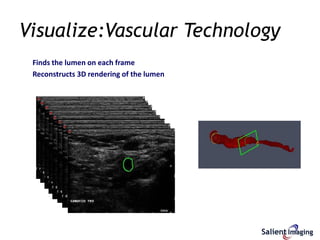
Document #:600-03-00021 Rev AA
Salient Imaging
pioneer in the development of medical imaging software algorithms
 Private U.S. Corporation
 Patented Technology in Imaging Algorithms
 U.S. FDA certified product: Visualize:Vascular™
 1st 3D rendering from 2D ultrasound focusing on the true lumen
Company
Salient Imaging’s technology is based on proven pattern recognition principles
Pattern Recognition is a well established and widely used method of identifying
and classifying complex data sets
 Mammography
 Lung
 Colon
 Liver
 Brain Tumor
Salient Imaging applies these proven practices to medical imaging to specifically
focus on ultrasound
Technology
Visualize:Vascular
Current technology The crucial diagnostic The Salient Image
shows the outside information is inside of the residual lumen
of the carotid artery the carotid artery in the ICA
 Find the overriding pattern
 Identify the anomaly
 Create focused 3D rendering
 Automatically measure and calculate luminal reduction / Stenosis
 Provide clinicians with focus on the salient diagnostic detail
Technology
Works on a transverse cine clip of the artery
Technology
Finds the lumen on each frame
Visualize:Vascular Technology
Finds the lumen on each frame
Reconstructs 3D rendering of the lumen
Visualize:Vascular Technology
Clinical Value
Comparison of CDE to Visualize:Vascular to MRA
• 100 consecutive cases
• Any case where Visualize disagreed with Doppler or >70% on either also had MRA
Key Findings:
Correlation between CDE to Visualize 75%
Correlation between MRA and Visualize 99%
Gurell, et. al. ACR/SRU 2012
Key Findings:
Correlation between CDE to Visualize 75%
Correlation between MRA and Visualize 99%
The images to the left are from a study comparing the
results of 2D Ultrasound to 3D Visualize:Vascular over
the same section of the carotid artery
Top Image: Ultrasound color flow can mis-lead
 Measured Moderate Stenosis of 50 to 69%
 Measurement is made in color block at left which is beyond the
Stenosis (black)
 Ultrasound data indicates patient call back in 1 yr
Bottom Image: Visualize:Vascular is more accurate
 Measured Severe Stenosis (85%)
 Visualize data indicates immediate surgery
Visualize:Vascular is Correct
 Pathology confirmed that Visualize:Vascular is correct. Patient has
severe amount of plaque in vessel.
Clinical Significance – Shadowing
Ultrasound
Some plaque
The complete outline and
extent of the plaque
Visualize:Vascular
Magnetic Resonance Angiography (MRA) Break
Clinical Significance – Poor Fidelity
The images to the right are from a case study
comparing the results of 2D Ultrasound and
MRA technologies to Visualize:Vascular the
over the same section of artery
Top Image: Ultrasound
shows the outer arterial walls with limited detail of the
lumen pathway, no significant plaque
Middle Image: Visualize:Vascular
Significant vulnerable plaque
provides the most clinical detail about the region of
interest
Bottom Image: MRA
shows that there is an occlusion by the break, but does not
give specific detail
Pathology confirmed results from Visualize
Ultrasound
Velocities show 80-89%
Visualize:Vascular
▪No significant concern, 52%
MRA
No significant concern, no break
MRA confirmed results from Visualize
Clinical Significance – Tortuosity
Ultrasound
Velocities show 70-79%
Visualize:Vasclar
Over 80%
Clinical Significance –Turbulence
Ultrasound
Velocities show 70-79%
Visualize:Vascular
Over 80%
Clinical Significance –Turbulence
Ultrasound
Velocities show 70-79%
Visualize:Vascular
Over 80%
Clinical Significance –Turbulence
Ultrasound
Velocities show 50-69%
Visualize:Vascular
Significant concern, 92%
CTA
Significant concern, 90%
CTA confirmed results from Visualize
Clinical Significance – Artifact?
The procedure should be performed on patients who demonstrate medical
necessity observed during the primary exam:
Noted Presence of Plaque
Noted Presence of Tortuosity
Noted Presence of Artifacts: Shadowing, Speckling, Fidelity Concerns
Noted Presence of Turbulent Flow Conditions
Follow-up to Surgery or Interventional Procedure
Medical Necessity
Document #:600-02-00003 Rev AA

Visualize vascular (slides)

  • 1.
  • 2.
    Salient Imaging pioneer inthe development of medical imaging software algorithms  Private U.S. Corporation  Patented Technology in Imaging Algorithms  U.S. FDA certified product: Visualize:Vascular™  1st 3D rendering from 2D ultrasound focusing on the true lumen Company
  • 3.
    Salient Imaging’s technologyis based on proven pattern recognition principles Pattern Recognition is a well established and widely used method of identifying and classifying complex data sets  Mammography  Lung  Colon  Liver  Brain Tumor Salient Imaging applies these proven practices to medical imaging to specifically focus on ultrasound Technology
  • 4.
    Salient Imaging’s technologyis based on proven pattern recognition principles Pattern Recognition is a well established and widely used method of identifying and classifying complex data sets  Mammography  Lung  Colon  Liver  Brain Tumor Salient Imaging applies these proven practices to medical imaging to specifically focus on ultrasound Technology
  • 5.
    Visualize:Vascular Current technology Thecrucial diagnostic The Salient Image shows the outside information is inside of the residual lumen of the carotid artery the carotid artery in the ICA  Find the overriding pattern  Identify the anomaly  Create focused 3D rendering  Automatically measure and calculate luminal reduction / Stenosis  Provide clinicians with focus on the salient diagnostic detail
  • 6.
    Technology Works on atransverse cine clip of the artery
  • 7.
  • 8.
    Visualize:Vascular Technology Finds thelumen on each frame Reconstructs 3D rendering of the lumen
  • 9.
  • 10.
    Clinical Value Comparison ofCDE to Visualize:Vascular to MRA • 100 consecutive cases • Any case where Visualize disagreed with Doppler or >70% on either also had MRA Key Findings: Correlation between CDE to Visualize 75% Correlation between MRA and Visualize 99% Gurell, et. al. ACR/SRU 2012 Key Findings: Correlation between CDE to Visualize 75% Correlation between MRA and Visualize 99%
  • 11.
    The images tothe left are from a study comparing the results of 2D Ultrasound to 3D Visualize:Vascular over the same section of the carotid artery Top Image: Ultrasound color flow can mis-lead  Measured Moderate Stenosis of 50 to 69%  Measurement is made in color block at left which is beyond the Stenosis (black)  Ultrasound data indicates patient call back in 1 yr Bottom Image: Visualize:Vascular is more accurate  Measured Severe Stenosis (85%)  Visualize data indicates immediate surgery Visualize:Vascular is Correct  Pathology confirmed that Visualize:Vascular is correct. Patient has severe amount of plaque in vessel. Clinical Significance – Shadowing
  • 12.
    Ultrasound Some plaque The completeoutline and extent of the plaque Visualize:Vascular Magnetic Resonance Angiography (MRA) Break Clinical Significance – Poor Fidelity The images to the right are from a case study comparing the results of 2D Ultrasound and MRA technologies to Visualize:Vascular the over the same section of artery Top Image: Ultrasound shows the outer arterial walls with limited detail of the lumen pathway, no significant plaque Middle Image: Visualize:Vascular Significant vulnerable plaque provides the most clinical detail about the region of interest Bottom Image: MRA shows that there is an occlusion by the break, but does not give specific detail Pathology confirmed results from Visualize
  • 13.
    Ultrasound Velocities show 80-89% Visualize:Vascular ▪Nosignificant concern, 52% MRA No significant concern, no break MRA confirmed results from Visualize Clinical Significance – Tortuosity
  • 14.
  • 15.
  • 16.
  • 17.
    Ultrasound Velocities show 50-69% Visualize:Vascular Significantconcern, 92% CTA Significant concern, 90% CTA confirmed results from Visualize Clinical Significance – Artifact?
  • 18.
    The procedure shouldbe performed on patients who demonstrate medical necessity observed during the primary exam: Noted Presence of Plaque Noted Presence of Tortuosity Noted Presence of Artifacts: Shadowing, Speckling, Fidelity Concerns Noted Presence of Turbulent Flow Conditions Follow-up to Surgery or Interventional Procedure Medical Necessity
  • 19.