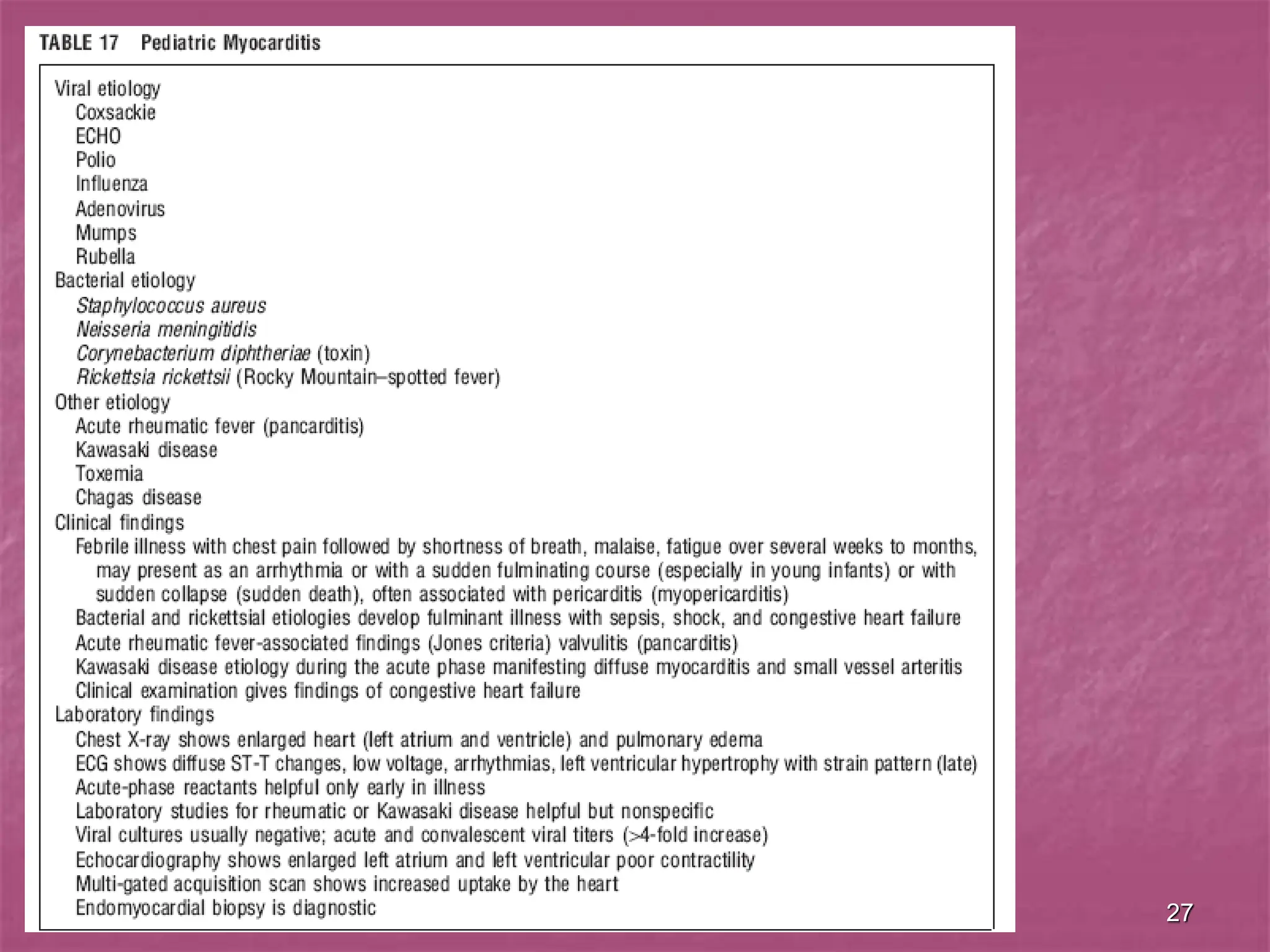
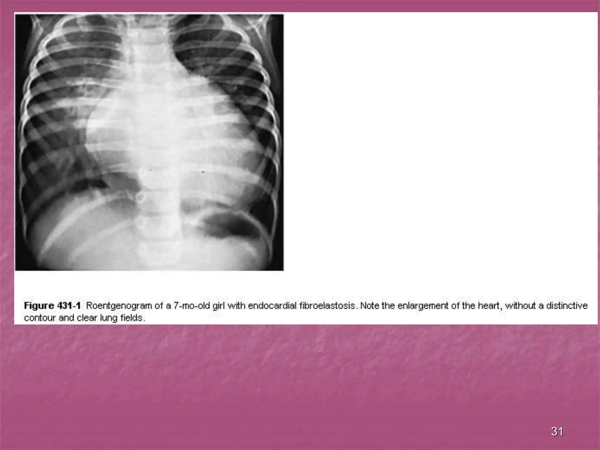
VIRAL MYOCARDITIS,
INFECTIVE ENDOCARDITIS,
AND THE PRIMARY
CARDIOMYOPATHIES
IN CHILDREN
2
Viral Myocarditis
 Myocarditis refers to inflammation,
necrosis, or myocytolysis that may be
caused by many infectious, connective
tissue, granulomatous, toxic, or idiopathic
processes affecting the myocardium with
or without associated systemic
manifestation of the disease process or
involvement of the endocardium or
pericardium.
3
Viral Myocarditis
 Practically, endocardium and pericardium are
involved in the process as well, so the term
"carditis" is preferable.
 Coronary pathology is uniformly absent.
 The most common manifestation is congestive
heart failure, although arrhythmias and sudden
death may be the first detectable signs.
 Viral infections are the most common etiology.
4
Etiology And Epidemiology
 The incidence of viral myocarditis in
children is unknown, as many mild cases
may go undetected.
 The incidence of viral myocarditis in
children is unknown, as many mild cases
may go undetected.
5
Etiology And Epidemiology
 Its manifestations are to some degree age
dependent.
 In early infancy, viral myocarditis often occurs
as an acute, fulminant disease.
 In toddlers and young children, it occurs as an
acute but less fulminant myopericarditis.
 In older children and adolescents, it is often
asymptomatic and comes to clinical attention
primarily as a precursor to idiopathic dilated
cardiomyopathy.
6
Pathophysiology
 Acute viral myocarditis may produce a
fulminant inflammatory process
characterized by
 ellular infiltrates,
 cell degeneration and necrosis, and
 subsequent fibrosis.
7
Pathophysiology
 Viral myocarditis may also become a chronic process
with persistence of viral RNA or DNA (but not infectious
virus particles) in the myocardium.
 Chronic inflammation is then perpetuated by the host
immune response, which includes T lymphocytes
activated against viral-host antigenic alterations.
 Such cytotoxic lymphocytes and natural killer cells,
together with persistent and possibly defective viral
replication, may impair myocyte function without obvious
cytolysis.
8
Pathophysiology
 Alternatively, the persistent viral infection may
alter major histocompatibility complex antigen
expression, with resultant exposure of
neoantigens to the immune system.
 In addition, some viral proteins may share
antigenic epitopes with host cells, resulting in
autoimmune damage to the antigenicaly related
myocyte.
 The net final result of chronic viral-associated
inflammation is often dilated cardiomyopathy.
9
Clinical Manifestations
 The presentation depends on the age and
acute or chronic nature of the infection.
 Connection between the onset of the
disease and acute respiratory infection
sometimes is clear.
 Interval between acute respiratory
infection and the onset of carditis is short
(2-9 days) or it is absent.
10
11
Clinical Manifestations
In The Neonates
 The neonate may present with
 fever,
 severe heart failure,
 respiratory distress,
 cyanosis,
 distant heart sounds,
 weak pulses,
 tachycardia out of proportion to the fever,
 mitral insufficiency caused by dilatation of the valve annulus,
 a gallop rhythm,
 acidosis, and
 shock.
12
Clinical Manifestations
In The Neonates
 In the most fulminant form, death may occur
within 1 -7 days of the onset of symptoms.
 The chest roentgenogram demonstrates an
enormously enlarged heart and pulmonary
edema.
 The electrocardiogram reveals sinus tachycardia,
reduced QRS complex voltage, and ST segment
and T wave abnormalities. Arrhythmias may be
the first clinical manifestation and in the
presence of fever and a large heart strongly
suggest acute myocarditis.
13
Clinical Manifestations
In The Older Patients
 The older patient with acute myocarditis may
also present with acute congestive heart failure;
however, more commonly patients will present
with the gradual onset of congestive heart
failure or the sudden onset of ventricular
arrhythmias.
 In these patients, the acute infectious phase has
usually passed and an idiopathic dilated
cardiomyopathy is present.
14
15
16
Atrial extrasystoles
17
18
Ventricular extrasystoles
19
Ventricular extrasystoles
20
Multifocal ventricular extrasystoles
21
Diagnosis
 The sedimentation rate and heart enzymes
(creatine phosphokinase (CPK), lactate
dehydrogenase (LDH)) may be elevated in acute
or chronic myocarditis.
 Echocardiography will demonstrate poor
ventricular function and often a pericardial
effusion, mitral valve regurgitation.
 The use of the polymerase chain reaction (PCR)
to identify viral RNA or DNA has allowed the
viral etiology of many of these formerly
"idiopathic" cases to be determined.
22
Treatment
 The approach to treating acute myocarditis
involves supportive measures for severe
congestive heart failure.
 Dopamine or epinephrine may be helpful if there
is poor cardiac output with systemic
hypotension.
 However, all inotropic agents, including digoxin,
should be used with caution as patients with
myocarditis may be more susceptible to the
arrhythmogenic properties of these agents.
 Digoxin is often started at half the normal
dosage.
23
Treatment
 Arrhythmias should be treated in the normal fashion.
 The role of corticosteroids for treatment of acute viral
myocarditis is still controversial. In a small series of
pediatric patients, treatment with prednisone (2 mg/kg
daily; tapered to 0.3 mg/kg daily over 3 mo.) was
effective in reducing myocardial inflammation and in
improving cardiac function.
 Corticosteroids are administered in diffuse process with
cardiac failure, subacute onset, with affection of
conductive system.
 However, relapse may occur when immunosuppression
is discontinued.
24
Treatment
 Antibiotics are given during 2-3 weeks and more
for prevention of complications.
 Trials are currently under way evaluating the
efficacy of intravenous gamma globulin.
 Improvement of myocardial metabolism includes
prescription of riboxin, panangine, vitamins,
polarizing mixture i.v.
25
Prognosis
 The outcome of the symptomatic neonate
with acute viral myocarditis remains poor,
with a mortality between 50 % and 70 %.
 Patients with lesser symptoms may have a
somewhat better prognosis, and complete
resolution has been described.
26
Prognosis
 The outcome of older patients with chronic
dilated cardiomyopathy associated with prior
viral infection is also poor without therapy.
 These patients continue to have inflammation,
fibrosis, and deteriorating cardiac function.
 Although spontaneous resolution may occur in
10-20 %, as many as 50 % of untreated older
patients will die within 2 yr. of presentation and
80 % within 8 yr. without cardiac
transplantation.
27
28
29
ENDOCARDIAL FIBROELASTOSIS
 This condition has been called
 fetal endocarditis,
 endocardial fibrosis,
 prenatal fibroelastosis,
 elastic tissue hyperplasia, and
 endocardial sclerosis.
30
Fibroelastosis of the left
ventricle.
M/3 weeks. Death from
cardiac failure. The
endocardial surface of the
left ventricle is lined by a
thick layer of white tissue.
31
32
ENDOCARDIAL FIBROELASTOSIS
 In primary endocardial fibroelastosis (EFE)
there is no apparent predisposing valvular lesion
or other congenital heart abnormality.
 In secondary EFE severe congenital heart
disease of the left-sided obstructive type (e.g.,
aortic stenosis or atresia, forms of hypoplastic
left heart syndrome, or severe coarctation of the
aorta) is present.
33
ENDOCARDIAL FIBROELASTOSIS
 In secondary EFE the ventricular cavity is
often contracted, whereas in the primary
disease a dilated left ventricular chamber
is seen, usually during infancy.
 However, in young adults a contracted
form of primary EFE has been observed.
 No etiology for primary EFE has been
established.
34
Pathology
 Pathologically, there is a white fibroblastic
thickening of the endocardium, virtually
always in the left ventricle, which
frequently obscures the trabeculation of
the inner surfaces of the cardiac chamber.
 The lesion may spread to involve the
valves.
35
Pathology
 Microscopically, the lesion consists of a
fibroelastic thickening of the endocardium and
may result in subendocardial degeneration or
necrosis of muscle with vacuolation of muscle
fibers.
 The involved valve leaflets are characterized by
a myxomatous proliferation with an increase in
collagenous elements.
36
Pathology
 The endocardium may be 5-6 times
thicker than normal and there is
subsequent narrowing of the ventricular
chamber.
 The poor diastolic filling leads to a low
cardiac output resulting ultimately in
forward failure and features of coronary
ischemia.
37
Clinical manifestations
 The clinical manifestations are variable.
 Infants, usually younger than 6 mo. of
age, who apparently had been in good
health, develop severe congestive heart
failure, often precipitated by a respiratory
infection.
38
Clinical manifestations
 Affected infants may manifest
 dyspnea,
 cough,
 anorexia,
 hepatomegaly,
 edema,
 failure to thrive, and
 recurrent pulmonary infections.
39
Clinical manifestations
 Chronic congestive heart failure can be
controlled for some time by digitalis and
diuretics; however, most patients
eventually succumb.
 Infants in whom valvular lesions or
associated congenital cardiovascular
defects are predominant usually expire in
the 1st mo. of life.
40
Clinical manifestations
 Roentgenograms confirm significant
cardiac enlargement.
 The electrocardiogram is abnormal, with
changes indicative of left atrial and left
ventricular hypertrophy with strain.
 The echocardiogram shows a bright-
appearing endocardial surface and a
dilated, poorly functioning left ventricle.
41
Treatment
 It is directed toward alleviation of
congestive heart failure and prevention of
intercurrent infections.
 End-stage EFE, with signs of heart failure
despite a maximal medical regimen, is an
indication for cardiac transplantation.
42
Myocardiodystrophy
 There is a cardiac affection of non-
inflammatory character.
 Myocardiodystrophies are the disorders of
physico-chemical structure of the cardiac
muscle and its metabolism.
 Anamnestic diagnostic criterion is
connection with previous disease.
43
Myocardiodystrophy
 Clinical diagnostic criteria are as follows:
 cardialgias,
 felling of unsatisfactory inspiration,
 dispnea on physical exertion,
 vegetative symptoms.
44
Myocardiodystrophy
 Considerable cardiomegaly,
 steady diastolic murmur,
 severe degree of processes,
 atrio-ventricular tachycardia,
 arrhythmia,
 chronic disorders of blood circulation
 testify against myocardiodystrophy.
45
46
47
Infective endocarditis
Clinical signs
 Fever
 Anaemia and pallor
 Splinter haemorrhages in nailbed
 Clubbing (late)
 Necrotic skin lesions
 Changing cardiac signs
 Splenomegaly
 Neurological signs from cerebral infarction
 Retinal infarcts
 Arthritis/arthralgia
 Haematuria (microscopic).
48
Parasternal long axis view demonstrating thickened leaflets and mobile vegetation
(arrow) adherent to the mitral valve in a patient with infectious endocarditis. This
patient developed endocarditis after a routine dental procedure without antibiotic
prophylaxis in the setting of antecedent mitral valve prolapse. The left ventricular
cavity (LV) and left atrium (LA) are also identified
49
A transesophageal echocardiogram demonstrates the mitral valve (mv) and several
mobile vegetations (v) attached to both the anterior and posterior leaflets. The left
atrium (LA) and left ventricle (LV) are shown for orientation.
Transesophageal echocardiography provides excellent views of the mitral valve and
left atrium in clinical situations in which endocarditis or potential cardiac sources of
embolism (e.g. atrial thrombus) must be evaluated
50
51
Widespread
infected emboli
and infarcts in a
child with
bacterial
endocarditis.
The tip of the
third toe is
gangrenous.
52
Vegetations on the mitral valve in subacute bacterial endocarditis.
These are also a source of peripheral emboli.
53
Bacterial endocarditis.
Rupture of an aortic valve cusp
54
Petechial haemorrhages in the conjunctiva from septic emboli in
bacterial endocarditis.
Note also the linear haemorrhage at the junction between the conjunctiva and
the sclera. This is another feature of peripheral embolizati
55
Fundal
photograph
showing
petechial
haemorrhages
from septic
emboli in
bacterial
endocarditis.
56
57
supraventricular tachycardia
Supraventricular tachycardia
58
Supraventricular tachycardia
59
Supraventricular tachycardia
60
Ventricular tachycardia
61
Ventricular tachycardia
62
Ventricular tachycardia
63
Ventricular tachycardia
Questions
1.Myocarditis refers to inflammation, necrosis, or myocytolysis that
may be caused by many infectious, connective tissue,
granulomatous, toxic, or idiopathic processes affecting the
myocardium with or without associated systemic manifestation of
the disease process or involvement of the endocardium or
pericardium. (true/false).
2. Please, list the Clinical Manifestations in the Neonates.
64

viral myocarditis in pediatrics age group .ppt

  • 1.
    VIRAL MYOCARDITIS, INFECTIVE ENDOCARDITIS, ANDTHE PRIMARY CARDIOMYOPATHIES IN CHILDREN
  • 2.
    2 Viral Myocarditis  Myocarditisrefers to inflammation, necrosis, or myocytolysis that may be caused by many infectious, connective tissue, granulomatous, toxic, or idiopathic processes affecting the myocardium with or without associated systemic manifestation of the disease process or involvement of the endocardium or pericardium.
  • 3.
    3 Viral Myocarditis  Practically,endocardium and pericardium are involved in the process as well, so the term "carditis" is preferable.  Coronary pathology is uniformly absent.  The most common manifestation is congestive heart failure, although arrhythmias and sudden death may be the first detectable signs.  Viral infections are the most common etiology.
  • 4.
    4 Etiology And Epidemiology The incidence of viral myocarditis in children is unknown, as many mild cases may go undetected.  The incidence of viral myocarditis in children is unknown, as many mild cases may go undetected.
  • 5.
    5 Etiology And Epidemiology Its manifestations are to some degree age dependent.  In early infancy, viral myocarditis often occurs as an acute, fulminant disease.  In toddlers and young children, it occurs as an acute but less fulminant myopericarditis.  In older children and adolescents, it is often asymptomatic and comes to clinical attention primarily as a precursor to idiopathic dilated cardiomyopathy.
  • 6.
    6 Pathophysiology  Acute viralmyocarditis may produce a fulminant inflammatory process characterized by  ellular infiltrates,  cell degeneration and necrosis, and  subsequent fibrosis.
  • 7.
    7 Pathophysiology  Viral myocarditismay also become a chronic process with persistence of viral RNA or DNA (but not infectious virus particles) in the myocardium.  Chronic inflammation is then perpetuated by the host immune response, which includes T lymphocytes activated against viral-host antigenic alterations.  Such cytotoxic lymphocytes and natural killer cells, together with persistent and possibly defective viral replication, may impair myocyte function without obvious cytolysis.
  • 8.
    8 Pathophysiology  Alternatively, thepersistent viral infection may alter major histocompatibility complex antigen expression, with resultant exposure of neoantigens to the immune system.  In addition, some viral proteins may share antigenic epitopes with host cells, resulting in autoimmune damage to the antigenicaly related myocyte.  The net final result of chronic viral-associated inflammation is often dilated cardiomyopathy.
  • 9.
    9 Clinical Manifestations  Thepresentation depends on the age and acute or chronic nature of the infection.  Connection between the onset of the disease and acute respiratory infection sometimes is clear.  Interval between acute respiratory infection and the onset of carditis is short (2-9 days) or it is absent.
  • 10.
  • 11.
    11 Clinical Manifestations In TheNeonates  The neonate may present with  fever,  severe heart failure,  respiratory distress,  cyanosis,  distant heart sounds,  weak pulses,  tachycardia out of proportion to the fever,  mitral insufficiency caused by dilatation of the valve annulus,  a gallop rhythm,  acidosis, and  shock.
  • 12.
    12 Clinical Manifestations In TheNeonates  In the most fulminant form, death may occur within 1 -7 days of the onset of symptoms.  The chest roentgenogram demonstrates an enormously enlarged heart and pulmonary edema.  The electrocardiogram reveals sinus tachycardia, reduced QRS complex voltage, and ST segment and T wave abnormalities. Arrhythmias may be the first clinical manifestation and in the presence of fever and a large heart strongly suggest acute myocarditis.
  • 13.
    13 Clinical Manifestations In TheOlder Patients  The older patient with acute myocarditis may also present with acute congestive heart failure; however, more commonly patients will present with the gradual onset of congestive heart failure or the sudden onset of ventricular arrhythmias.  In these patients, the acute infectious phase has usually passed and an idiopathic dilated cardiomyopathy is present.
  • 14.
  • 15.
  • 16.
  • 17.
  • 18.
  • 19.
  • 20.
  • 21.
    21 Diagnosis  The sedimentationrate and heart enzymes (creatine phosphokinase (CPK), lactate dehydrogenase (LDH)) may be elevated in acute or chronic myocarditis.  Echocardiography will demonstrate poor ventricular function and often a pericardial effusion, mitral valve regurgitation.  The use of the polymerase chain reaction (PCR) to identify viral RNA or DNA has allowed the viral etiology of many of these formerly "idiopathic" cases to be determined.
  • 22.
    22 Treatment  The approachto treating acute myocarditis involves supportive measures for severe congestive heart failure.  Dopamine or epinephrine may be helpful if there is poor cardiac output with systemic hypotension.  However, all inotropic agents, including digoxin, should be used with caution as patients with myocarditis may be more susceptible to the arrhythmogenic properties of these agents.  Digoxin is often started at half the normal dosage.
  • 23.
    23 Treatment  Arrhythmias shouldbe treated in the normal fashion.  The role of corticosteroids for treatment of acute viral myocarditis is still controversial. In a small series of pediatric patients, treatment with prednisone (2 mg/kg daily; tapered to 0.3 mg/kg daily over 3 mo.) was effective in reducing myocardial inflammation and in improving cardiac function.  Corticosteroids are administered in diffuse process with cardiac failure, subacute onset, with affection of conductive system.  However, relapse may occur when immunosuppression is discontinued.
  • 24.
    24 Treatment  Antibiotics aregiven during 2-3 weeks and more for prevention of complications.  Trials are currently under way evaluating the efficacy of intravenous gamma globulin.  Improvement of myocardial metabolism includes prescription of riboxin, panangine, vitamins, polarizing mixture i.v.
  • 25.
    25 Prognosis  The outcomeof the symptomatic neonate with acute viral myocarditis remains poor, with a mortality between 50 % and 70 %.  Patients with lesser symptoms may have a somewhat better prognosis, and complete resolution has been described.
  • 26.
    26 Prognosis  The outcomeof older patients with chronic dilated cardiomyopathy associated with prior viral infection is also poor without therapy.  These patients continue to have inflammation, fibrosis, and deteriorating cardiac function.  Although spontaneous resolution may occur in 10-20 %, as many as 50 % of untreated older patients will die within 2 yr. of presentation and 80 % within 8 yr. without cardiac transplantation.
  • 27.
  • 28.
  • 29.
    29 ENDOCARDIAL FIBROELASTOSIS  Thiscondition has been called  fetal endocarditis,  endocardial fibrosis,  prenatal fibroelastosis,  elastic tissue hyperplasia, and  endocardial sclerosis.
  • 30.
    30 Fibroelastosis of theleft ventricle. M/3 weeks. Death from cardiac failure. The endocardial surface of the left ventricle is lined by a thick layer of white tissue.
  • 31.
  • 32.
    32 ENDOCARDIAL FIBROELASTOSIS  Inprimary endocardial fibroelastosis (EFE) there is no apparent predisposing valvular lesion or other congenital heart abnormality.  In secondary EFE severe congenital heart disease of the left-sided obstructive type (e.g., aortic stenosis or atresia, forms of hypoplastic left heart syndrome, or severe coarctation of the aorta) is present.
  • 33.
    33 ENDOCARDIAL FIBROELASTOSIS  Insecondary EFE the ventricular cavity is often contracted, whereas in the primary disease a dilated left ventricular chamber is seen, usually during infancy.  However, in young adults a contracted form of primary EFE has been observed.  No etiology for primary EFE has been established.
  • 34.
    34 Pathology  Pathologically, thereis a white fibroblastic thickening of the endocardium, virtually always in the left ventricle, which frequently obscures the trabeculation of the inner surfaces of the cardiac chamber.  The lesion may spread to involve the valves.
  • 35.
    35 Pathology  Microscopically, thelesion consists of a fibroelastic thickening of the endocardium and may result in subendocardial degeneration or necrosis of muscle with vacuolation of muscle fibers.  The involved valve leaflets are characterized by a myxomatous proliferation with an increase in collagenous elements.
  • 36.
    36 Pathology  The endocardiummay be 5-6 times thicker than normal and there is subsequent narrowing of the ventricular chamber.  The poor diastolic filling leads to a low cardiac output resulting ultimately in forward failure and features of coronary ischemia.
  • 37.
    37 Clinical manifestations  Theclinical manifestations are variable.  Infants, usually younger than 6 mo. of age, who apparently had been in good health, develop severe congestive heart failure, often precipitated by a respiratory infection.
  • 38.
    38 Clinical manifestations  Affectedinfants may manifest  dyspnea,  cough,  anorexia,  hepatomegaly,  edema,  failure to thrive, and  recurrent pulmonary infections.
  • 39.
    39 Clinical manifestations  Chroniccongestive heart failure can be controlled for some time by digitalis and diuretics; however, most patients eventually succumb.  Infants in whom valvular lesions or associated congenital cardiovascular defects are predominant usually expire in the 1st mo. of life.
  • 40.
    40 Clinical manifestations  Roentgenogramsconfirm significant cardiac enlargement.  The electrocardiogram is abnormal, with changes indicative of left atrial and left ventricular hypertrophy with strain.  The echocardiogram shows a bright- appearing endocardial surface and a dilated, poorly functioning left ventricle.
  • 41.
    41 Treatment  It isdirected toward alleviation of congestive heart failure and prevention of intercurrent infections.  End-stage EFE, with signs of heart failure despite a maximal medical regimen, is an indication for cardiac transplantation.
  • 42.
    42 Myocardiodystrophy  There isa cardiac affection of non- inflammatory character.  Myocardiodystrophies are the disorders of physico-chemical structure of the cardiac muscle and its metabolism.  Anamnestic diagnostic criterion is connection with previous disease.
  • 43.
    43 Myocardiodystrophy  Clinical diagnosticcriteria are as follows:  cardialgias,  felling of unsatisfactory inspiration,  dispnea on physical exertion,  vegetative symptoms.
  • 44.
    44 Myocardiodystrophy  Considerable cardiomegaly, steady diastolic murmur,  severe degree of processes,  atrio-ventricular tachycardia,  arrhythmia,  chronic disorders of blood circulation  testify against myocardiodystrophy.
  • 45.
  • 46.
  • 47.
    47 Infective endocarditis Clinical signs Fever  Anaemia and pallor  Splinter haemorrhages in nailbed  Clubbing (late)  Necrotic skin lesions  Changing cardiac signs  Splenomegaly  Neurological signs from cerebral infarction  Retinal infarcts  Arthritis/arthralgia  Haematuria (microscopic).
  • 48.
    48 Parasternal long axisview demonstrating thickened leaflets and mobile vegetation (arrow) adherent to the mitral valve in a patient with infectious endocarditis. This patient developed endocarditis after a routine dental procedure without antibiotic prophylaxis in the setting of antecedent mitral valve prolapse. The left ventricular cavity (LV) and left atrium (LA) are also identified
  • 49.
    49 A transesophageal echocardiogramdemonstrates the mitral valve (mv) and several mobile vegetations (v) attached to both the anterior and posterior leaflets. The left atrium (LA) and left ventricle (LV) are shown for orientation. Transesophageal echocardiography provides excellent views of the mitral valve and left atrium in clinical situations in which endocarditis or potential cardiac sources of embolism (e.g. atrial thrombus) must be evaluated
  • 50.
  • 51.
    51 Widespread infected emboli and infarctsin a child with bacterial endocarditis. The tip of the third toe is gangrenous.
  • 52.
    52 Vegetations on themitral valve in subacute bacterial endocarditis. These are also a source of peripheral emboli.
  • 53.
  • 54.
    54 Petechial haemorrhages inthe conjunctiva from septic emboli in bacterial endocarditis. Note also the linear haemorrhage at the junction between the conjunctiva and the sclera. This is another feature of peripheral embolizati
  • 55.
  • 56.
  • 57.
  • 58.
  • 59.
  • 60.
  • 61.
  • 62.
  • 63.
  • 64.
    Questions 1.Myocarditis refers toinflammation, necrosis, or myocytolysis that may be caused by many infectious, connective tissue, granulomatous, toxic, or idiopathic processes affecting the myocardium with or without associated systemic manifestation of the disease process or involvement of the endocardium or pericardium. (true/false). 2. Please, list the Clinical Manifestations in the Neonates. 64