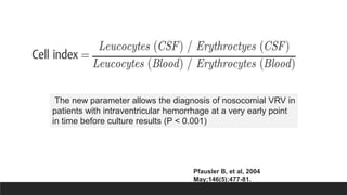
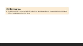
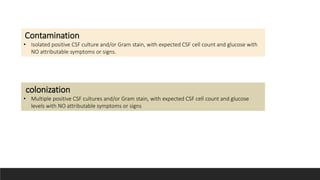
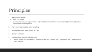
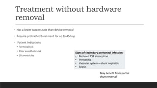
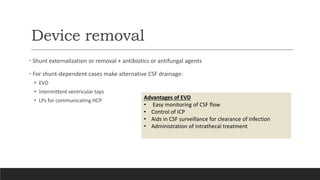
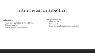
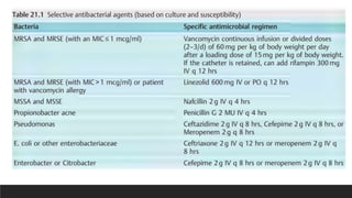
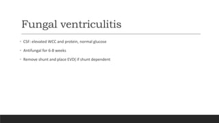
Ventriculitis
Dr. Tim Mutafya
Surgery Resident
Kamuzu Central Hospital, Malawi
Layout
Introduction
Epidemiology
Risk factors
Diagnosis
Principles of management
Introduction
• Ventriculitis is the inflammation of the ependymal lining of the cerebral ventricles, usually
secondary to infection for example meningitis, device-related or a complication of trauma
• AKA Ependymitis, Ventricular empyema, Pyocephalus, and Pyogenic ventriculitis
• It is an indolent but lethal infection and a source of persistent infection following meningitis
treatment.
• Early diagnosis is essential for appropriate treatment.
• It is of particular concern in patients with external ventricular drains (EVDs) or intraventricular shunts.
Epidemiology
• Acceptable shunt infection rate is <5-7%
• Onset of infection after shunt
• Early 3-20% ( on average 7%)
• Over 50% staph infection occur in 2 weeks and 70% < 2mo
• EVD infection incidence is around 9.5%
• Source of infection
• Mainly skin
• 3% CSF infection(therefore need for CSF analysis pre-op)
Risk factors
Shunt
1. Young age: waiting for 2 weeks in MMC
lowers the risk of infection
2. Length of the procedure
3. Open neural tube defect
EVD
1. duration of EVD
2. Site leakage
3. blood in CSF (IVH and SAH)
4. Irrigation and flushing
Etiology
Early infection
• Staphylococus
• epidermidis (coagulase-negative staph): 60–75%
of infections(most common)
• S. Aureus
• Gram-negative bacilli (GNB): 6–20% (may
migrate come from intestinal perforation)
perforation)
• In neonates: E. coli and Strep.
hemolyticus dominate
Late infection (> 6 Mo Post OP)
• 6% Risk per patient.
• Almost all are indolent infections of S.
epidermidis.
• seeding of a vascular shunt during an
episode of septicemia (probably very
rare)
• colonization from an episode of
meningitis
Etiology cont..
Fungal infection
• Candida spp. (majority)
• Usually children < 1 year(1-7%)
Possibly related to the use of prophylactic antibiotics used for ICP monitoring
and CSF drainage
Nguyen MH, Yu VL.
Presentation
• Non-specific syndrome:
• fever, N/V, H/A, lethargy, anorexia, irritability
• Shunt malfunction
• Erythema and tenderness along shunt tubing
• Distal infection of ventriculoperitoneal shunts may
mimic an acute abdomen
•Shunt nephritis
• characterized by proteinuria and hematuria.
Neonates
• apneic episodes
• anemia
• Hepatosplenomegaly
• stiff neck
Gram neg. bacteria cause more severe symptoms with
intermittent fevers.
S. Epidermidis tend to be indolent
Approach
History and exam for:
Meningitis Neck stiffness, seizures, fever
GI symptoms Diarrhea, Vomiting
Otitis media Ear discharge and erythema
Pneumonia SOB, cough, fever
URI Runny nose, fever
Tonsil-pharyngitis Irritability, check the mouth
UTI Irritability, suprapubic tenderness,
Viral syndromes Cough, runny nose, diarrhea, fever
Labs
Bloods
• FBC + differential
• < 10K (25%)
• > 20K (30%)
• Acute phase reactants:
• ESR & CRP
• Blood cultures
Urine
• Proteinuria
• Hematuria
CSF
• Usually <100k
• Gram + ≈ 50%(Lower yield S. Epid)
• Elevated protein
• Glucose normal or low
• Lactate
• PCR
• Serum procalcitonin
• Cultures
• Gram -ve in 40%
• Higher yield for > 20K
Neonatal cranial USS
• Increased periventricular echogenicity
and irregularity of the ventricular surface.
• Choroid plexus irregularity
• Echogenic intraventricular debris and
well-delineated intraventricular septations
CT
• usually unhelpful in diagnosing infection.
• Ependymal enhancement when it occurs is
is diagnostic of ventriculitis.
• CT may demonstrate shunt malfunction
layering debris in the occipital horns.
Role of LP
• usually NOT recommended!
• May be hazardous in obstructive hydrocephalus (HCP) with a nonfunctioning shunt.
• Often does not yield the pathogen even in communicating HCP, especially if the infection is limited
to ventriculitis.
• If positive, may obviate a shunt tap
Classification
MODIFIED LOZIER’S DEFINITIONS
The new parameter allows the diagnosis of nosocomial VRV in
patients with intraventricular hemorrhage at a very early point
in time before culture results (P < 0.001)
Pfausler B, et al, 2004
May;146(5):477-81.
Possible ventriculitis
• Progressive rise in cell index or progressive decrease in CSF: blood glucose ratio or an extreme
value for CSF WBC count (> 1000/micro L) OR
• CSF: blood glucose ratio (< 0.2), with attributable symptoms and signs, but NEGATIVE Gram stain &
& cultures
Definitive ventriculitis
• Progressive rise in cell index or progressive decrease in CSF: blood glucose ratio or an extreme
value for CSF WBC count (> 1000/micro L) OR
• CSF: blood glucose ratio (< 0.2), with attributable symptoms or signs and a POSITIVE Gram stain &
cultures
Probable ventriculitis
• CSF WBC count or CSF: blood glucose ratio MORE abnormal than expected, but NOT an extreme value (CSF
WBC count 1000/micro L OR
• CSF: blood glucose ratio < 0.2) and stable (not progressively worsening) attributable symptoms and signs and
POSITIVE Gram stain & cultures
colonization
• Multiple positive CSF cultures and/or Gram stain, with expected CSF cell count and glucose
levels with NO attributable symptoms or signs
Contamination
• Isolated positive CSF culture and/or Gram stain, with expected CSF cell count and glucose with
NO attributable symptoms or signs.
colonization
• Multiple positive CSF cultures and/or Gram stain, with expected CSF cell count and glucose
levels with NO attributable symptoms or signs
Contamination
• Isolated positive CSF culture and/or Gram stain, with expected CSF cell count and glucose with
NO attributable symptoms or signs.
Management
Principles
1. High dose of agents
• Blood: CSF barrier
• Some hospital-acquired organisms have higher MICs (minimal inhibitory concentration) for antimicrobials than
community-acquired organisms
2. Start empiric antibiotics after sampling
3. Modify treatment agent based on C&S
4. Remove catheter
5. Individualized Duration of treatment
• Rule of thumb: treat for 2 weeks if the infection was with S. aureus and S. epidermidis, and 3 weeks if it was
gram-negative
Empiric antibiotics
• Vancomycin for MRSA coverage + meropenem 2 g q 8h to cover gram-negative pathogens.
• Intraventricular injection of preservative-free antibiotics may be used in addition to IV therapy.
• Clamp EVD for one hour after injection
• Streamline therapy based on culture and sensitivity results
Treatment steps
Treatment without hardware
removal
• Has a lower success rate than device removal
• Require protracted treatment for up to 45days
• Patient Indications
• Terminally ill
• Poor anesthetic risk
• Slit ventricles
Signs of secondary peritoneal infection
• Reduced CSF absorption
• Peritonitis
• Vascular system—shunt nephritis
• Sepsis
May benefit from partial
shunt reversal
Device removal
• Shunt externalization or removal + antibiotics or antifungal agents
• For shunt-dependent cases make alternative CSF drainage:
• EVD
• Intermittent ventricular taps
• LPs for communicating HCP
Advantages of EVD
• Easy monitoring of CSF flow
• Control of ICP
• Aids in CSF surveillance for clearance of infection
• Administration of intrathecal treatment
Intrathecal antibiotics
Indications
- Failure to respond to systemic antibiotics
- Resistant organism
- Choose based on susceptibility
Dosage depends on
- Ventricular size
- Drain output
- Concentration of causative microorganism
Fungal ventriculitis
• CSF: elevated WCC and protein, normal glucose
• Antifungal for 6-8 weeks
• Remove shunt and place EVD( if shunt dependent
When to reimplant
1. Negative CSF cultures and functional parameter
2. Virulence factors
• Low: normal CSF as early as day 3
abnormal CSF after day 7
• High: wait until 7-10days after culture is sterile(??regardless)
Period of antibiotics does not indicate the clearing of infection
Response monitoring
• clinical improvement
• Improved CSF parameters and cultures become negative + clinical improvement
Daily CSF cultures & analysis are not recommended unless EVD is in
place (not practical here)
Primary prevention of ventriculitis
1. Pre-Op antibiotics
2. Tunneling >5cm away from burrhole
3. Use antbx-coated catheters( eg Rifampicin+minocycline)
Both routine catheter changes on day 5 and prolonged antibiotics don’t
actually reduce the rate of infection
References
• Greenberg’s handbook of neurosurgery 10th Ed
• Nguyen MH, Yu VL. Meningitis caused by Candida species: an emerging problem in neurosurgical patients. Clin Infect
Dis. 1995 Aug;21(2):323-7. doi: 10.1093/clinids/21.2.323. PMID: 8562739.
• Luque-Paz D, Revest M, Eugène F, Boukthir S, Dejoies L, Tattevin P, Le Reste PJ. Ventriculitis: A Severe Complication of
Central Nervous System Infections. Open Forum Infect Dis. 2021 Apr 29;8(6):ofab216. doi: 10.1093/ofid/ofab216. PMID:
34095339; PMCID: PMC8176394.
•https://www.thelancet.com/journals/ebiom/article/PIIS2352-3964%2822%2900115-3/fulltext#

Ventriculitis.pptx

  • 1.
    Ventriculitis Dr. Tim Mutafya SurgeryResident Kamuzu Central Hospital, Malawi
  • 2.
  • 4.
    Introduction • Ventriculitis isthe inflammation of the ependymal lining of the cerebral ventricles, usually secondary to infection for example meningitis, device-related or a complication of trauma • AKA Ependymitis, Ventricular empyema, Pyocephalus, and Pyogenic ventriculitis • It is an indolent but lethal infection and a source of persistent infection following meningitis treatment. • Early diagnosis is essential for appropriate treatment. • It is of particular concern in patients with external ventricular drains (EVDs) or intraventricular shunts.
  • 5.
    Epidemiology • Acceptable shuntinfection rate is <5-7% • Onset of infection after shunt • Early 3-20% ( on average 7%) • Over 50% staph infection occur in 2 weeks and 70% < 2mo • EVD infection incidence is around 9.5% • Source of infection • Mainly skin • 3% CSF infection(therefore need for CSF analysis pre-op)
  • 6.
    Risk factors Shunt 1. Youngage: waiting for 2 weeks in MMC lowers the risk of infection 2. Length of the procedure 3. Open neural tube defect EVD 1. duration of EVD 2. Site leakage 3. blood in CSF (IVH and SAH) 4. Irrigation and flushing
  • 7.
    Etiology Early infection • Staphylococus •epidermidis (coagulase-negative staph): 60–75% of infections(most common) • S. Aureus • Gram-negative bacilli (GNB): 6–20% (may migrate come from intestinal perforation) perforation) • In neonates: E. coli and Strep. hemolyticus dominate Late infection (> 6 Mo Post OP) • 6% Risk per patient. • Almost all are indolent infections of S. epidermidis. • seeding of a vascular shunt during an episode of septicemia (probably very rare) • colonization from an episode of meningitis
  • 8.
    Etiology cont.. Fungal infection •Candida spp. (majority) • Usually children < 1 year(1-7%) Possibly related to the use of prophylactic antibiotics used for ICP monitoring and CSF drainage Nguyen MH, Yu VL.
  • 9.
    Presentation • Non-specific syndrome: •fever, N/V, H/A, lethargy, anorexia, irritability • Shunt malfunction • Erythema and tenderness along shunt tubing • Distal infection of ventriculoperitoneal shunts may mimic an acute abdomen •Shunt nephritis • characterized by proteinuria and hematuria. Neonates • apneic episodes • anemia • Hepatosplenomegaly • stiff neck Gram neg. bacteria cause more severe symptoms with intermittent fevers. S. Epidermidis tend to be indolent
  • 10.
  • 11.
    History and examfor: Meningitis Neck stiffness, seizures, fever GI symptoms Diarrhea, Vomiting Otitis media Ear discharge and erythema Pneumonia SOB, cough, fever URI Runny nose, fever Tonsil-pharyngitis Irritability, check the mouth UTI Irritability, suprapubic tenderness, Viral syndromes Cough, runny nose, diarrhea, fever
  • 12.
    Labs Bloods • FBC +differential • < 10K (25%) • > 20K (30%) • Acute phase reactants: • ESR & CRP • Blood cultures Urine • Proteinuria • Hematuria CSF • Usually <100k • Gram + ≈ 50%(Lower yield S. Epid) • Elevated protein • Glucose normal or low • Lactate • PCR • Serum procalcitonin • Cultures • Gram -ve in 40% • Higher yield for > 20K
  • 13.
    Neonatal cranial USS •Increased periventricular echogenicity and irregularity of the ventricular surface. • Choroid plexus irregularity • Echogenic intraventricular debris and well-delineated intraventricular septations
  • 14.
    CT • usually unhelpfulin diagnosing infection. • Ependymal enhancement when it occurs is is diagnostic of ventriculitis. • CT may demonstrate shunt malfunction
  • 15.
    layering debris inthe occipital horns.
  • 16.
    Role of LP •usually NOT recommended! • May be hazardous in obstructive hydrocephalus (HCP) with a nonfunctioning shunt. • Often does not yield the pathogen even in communicating HCP, especially if the infection is limited to ventriculitis. • If positive, may obviate a shunt tap
  • 17.
  • 18.
    The new parameterallows the diagnosis of nosocomial VRV in patients with intraventricular hemorrhage at a very early point in time before culture results (P < 0.001) Pfausler B, et al, 2004 May;146(5):477-81.
  • 19.
    Possible ventriculitis • Progressiverise in cell index or progressive decrease in CSF: blood glucose ratio or an extreme value for CSF WBC count (> 1000/micro L) OR • CSF: blood glucose ratio (< 0.2), with attributable symptoms and signs, but NEGATIVE Gram stain & & cultures Definitive ventriculitis • Progressive rise in cell index or progressive decrease in CSF: blood glucose ratio or an extreme value for CSF WBC count (> 1000/micro L) OR • CSF: blood glucose ratio (< 0.2), with attributable symptoms or signs and a POSITIVE Gram stain & cultures Probable ventriculitis • CSF WBC count or CSF: blood glucose ratio MORE abnormal than expected, but NOT an extreme value (CSF WBC count 1000/micro L OR • CSF: blood glucose ratio < 0.2) and stable (not progressively worsening) attributable symptoms and signs and POSITIVE Gram stain & cultures
  • 20.
    Possible ventriculitis • Progressiverise in cell index or progressive decrease in CSF: blood glucose ratio or an extreme value for CSF WBC count (> 1000/micro L) OR • CSF: blood glucose ratio (< 0.2), with attributable symptoms and signs, but NEGATIVE Gram stain & & cultures Definitive ventriculitis • Progressive rise in cell index or progressive decrease in CSF: blood glucose ratio or an extreme value for CSF WBC count (> 1000/micro L) OR • CSF: blood glucose ratio (< 0.2), with attributable symptoms or signs and a POSITIVE Gram stain & cultures Probable ventriculitis • CSF WBC count or CSF: blood glucose ratio MORE abnormal than expected, but NOT an extreme value (CSF WBC count 1000/micro L OR • CSF: blood glucose ratio < 0.2) and stable (not progressively worsening) attributable symptoms and signs and POSITIVE Gram stain & cultures
  • 21.
    Possible ventriculitis • Progressiverise in cell index or progressive decrease in CSF: blood glucose ratio or an extreme value for CSF WBC count (> 1000/micro L) OR • CSF: blood glucose ratio (< 0.2), with attributable symptoms and signs, but NEGATIVE Gram stain & & cultures Definitive ventriculitis • Progressive rise in cell index or progressive decrease in CSF: blood glucose ratio or an extreme value for CSF WBC count (> 1000/micro L) OR • CSF: blood glucose ratio (< 0.2), with attributable symptoms or signs and a POSITIVE Gram stain & cultures Probable ventriculitis • CSF WBC count or CSF: blood glucose ratio MORE abnormal than expected, but NOT an extreme value (CSF WBC count 1000/micro L OR • CSF: blood glucose ratio < 0.2) and stable (not progressively worsening) attributable symptoms and signs and POSITIVE Gram stain & cultures
  • 22.
    colonization • Multiple positiveCSF cultures and/or Gram stain, with expected CSF cell count and glucose levels with NO attributable symptoms or signs Contamination • Isolated positive CSF culture and/or Gram stain, with expected CSF cell count and glucose with NO attributable symptoms or signs.
  • 23.
    colonization • Multiple positiveCSF cultures and/or Gram stain, with expected CSF cell count and glucose levels with NO attributable symptoms or signs Contamination • Isolated positive CSF culture and/or Gram stain, with expected CSF cell count and glucose with NO attributable symptoms or signs.
  • 24.
  • 25.
    Principles 1. High doseof agents • Blood: CSF barrier • Some hospital-acquired organisms have higher MICs (minimal inhibitory concentration) for antimicrobials than community-acquired organisms 2. Start empiric antibiotics after sampling 3. Modify treatment agent based on C&S 4. Remove catheter 5. Individualized Duration of treatment • Rule of thumb: treat for 2 weeks if the infection was with S. aureus and S. epidermidis, and 3 weeks if it was gram-negative
  • 26.
    Empiric antibiotics • Vancomycinfor MRSA coverage + meropenem 2 g q 8h to cover gram-negative pathogens. • Intraventricular injection of preservative-free antibiotics may be used in addition to IV therapy. • Clamp EVD for one hour after injection • Streamline therapy based on culture and sensitivity results
  • 27.
  • 28.
    Treatment without hardware removal •Has a lower success rate than device removal • Require protracted treatment for up to 45days • Patient Indications • Terminally ill • Poor anesthetic risk • Slit ventricles Signs of secondary peritoneal infection • Reduced CSF absorption • Peritonitis • Vascular system—shunt nephritis • Sepsis May benefit from partial shunt reversal
  • 29.
    Device removal • Shuntexternalization or removal + antibiotics or antifungal agents • For shunt-dependent cases make alternative CSF drainage: • EVD • Intermittent ventricular taps • LPs for communicating HCP Advantages of EVD • Easy monitoring of CSF flow • Control of ICP • Aids in CSF surveillance for clearance of infection • Administration of intrathecal treatment
  • 30.
    Intrathecal antibiotics Indications - Failureto respond to systemic antibiotics - Resistant organism - Choose based on susceptibility Dosage depends on - Ventricular size - Drain output - Concentration of causative microorganism
  • 32.
    Fungal ventriculitis • CSF:elevated WCC and protein, normal glucose • Antifungal for 6-8 weeks • Remove shunt and place EVD( if shunt dependent
  • 33.
    When to reimplant 1.Negative CSF cultures and functional parameter 2. Virulence factors • Low: normal CSF as early as day 3 abnormal CSF after day 7 • High: wait until 7-10days after culture is sterile(??regardless) Period of antibiotics does not indicate the clearing of infection
  • 34.
    Response monitoring • clinicalimprovement • Improved CSF parameters and cultures become negative + clinical improvement Daily CSF cultures & analysis are not recommended unless EVD is in place (not practical here)
  • 35.
    Primary prevention ofventriculitis 1. Pre-Op antibiotics 2. Tunneling >5cm away from burrhole 3. Use antbx-coated catheters( eg Rifampicin+minocycline) Both routine catheter changes on day 5 and prolonged antibiotics don’t actually reduce the rate of infection
  • 36.
    References • Greenberg’s handbookof neurosurgery 10th Ed • Nguyen MH, Yu VL. Meningitis caused by Candida species: an emerging problem in neurosurgical patients. Clin Infect Dis. 1995 Aug;21(2):323-7. doi: 10.1093/clinids/21.2.323. PMID: 8562739. • Luque-Paz D, Revest M, Eugène F, Boukthir S, Dejoies L, Tattevin P, Le Reste PJ. Ventriculitis: A Severe Complication of Central Nervous System Infections. Open Forum Infect Dis. 2021 Apr 29;8(6):ofab216. doi: 10.1093/ofid/ofab216. PMID: 34095339; PMCID: PMC8176394. •https://www.thelancet.com/journals/ebiom/article/PIIS2352-3964%2822%2900115-3/fulltext#