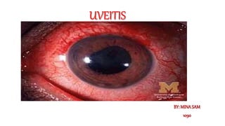
uveitis
•Download as PPTX, PDF•
4 likes•177 views

medical presentation regarding ophthalmology assignment at the faculty of medicine in alexandria

Recommended
More Related Content
Similar to uveitis
Similar to uveitis (20)
More from mina sam eskander
More from mina sam eskander (20)
Recently uploaded
Recently uploaded (20)
uveitis
- 2. 1-Applied anatomy. 2-Definition of uveitis. 3-Classification of uveitis.
- 3. I. IRIS • It is a coloured, free, circular diaphragm with a central aperture—the pupil. • When pupil is constricted, more of the posterior surface of the iris is in contact with the lens capsule. • When pupil is fully dilated, the iris may not touch the lens. • It divides the space between the cornea and lens into the anterior and posterior chambers of eye. • At the periphery, the iris is attached to the middle of anterior surface of the ciliary body 1-Applied anatomy.
- 4. • II. CILIARY BODY • The shape of the ciliary body is like a isosceles triangle with base forwards. • Parts Ciliary body has two parts namely:- i. Pars plicata—The anterior one-third of ciliary body (about 2 mm) is known as pars plicata. ii. Pars plana—The posterior two-third of ciliary body (about 4 mm) is known as pars plana. • Functions i. Pars plicata part of the ciliary body secretes aqueous humour. ii. The ciliary muscle helps in accommodation of the lens for seeing near objects.
- 5. III. CHOROID . It is a dark brown, highly vascular layer situated in between the sclera and retina. . It extends from the ora serrata upto the optic nerve aperture. . The outer layers of retina are dependent for their nutrition upon the choroid. . The inflammation of choroid always involves the retina.
- 6. 2- INFLAMMATIONOF THE UVEALTRACT (UVEITIS) The term uveitis strictly means inflammation of the uveal tissue only. However, there is always associated inflammation of the adjacent structures such as retina, vitreous, scleral and cornea. 3-Classification 1. Anatomical classification. 2. Clinical classification. 3. Pathological classification. 4. Etiological classification (Duke Elder’s).
- 7. 1-Anatomical classification i. Anterior uveitis—It can be divided as follows: • Iritis—The inflammation mainly affects the iris. • Iridocyclitis—Iris and pars plicata part of the ciliary body are involved. • Cyclitis—Pars plicata part of the ciliary body is affected predominantly. ii. Intermediate uveitis—There is inflammation of pars plana part of the ciliary body and peripheral retina and underlying choroid. It is also called “pars planitis”. iii. Posterior uveitis—There is inflammation of the choroid (choroiditis). There is associated inflammation of adjacent retina and hence the term “chorioretinitis” is used. iv. Panuveitis—There is inflammation of the whole uveal tract.
- 8. 2- Clinical classification: • i. Acute uveitis—The onset is sudden and it usually lasts for less than 3 weeks. ii. Chronic uveitis—The onset is insidious and the duration is more than 3 weeks. iii. Recurrent uveitis—The uveitis keeps recurring periodically. 3-Pathological classification • Uveitis can be further divided according to the pathological lesions which can be of two types: i. Granulomatous uveitis—It is infective in nature. Inflammation is insidious in onset, chronic in nature with minimum clinical features. ii. Non-granulomatous uveitis—It is usually due to allergic or immune related reaction. • It is of acute onset and of short duration.
- 9. 3- Etiological classification (Duke Elder’s): i. Infective uveitis ii. Allergic or immune related uveitis iii. Toxic uveitis iv. Traumatic uveitis v. Uveitis associated with non-infective systemic diseases vi. Idiopathic uveitis.
- 10. Etiology In most cases, uveitis is not due to direct infection. It is usually due to allergy or hypersensitivity reaction to an infectious agent. 1. Exogenous infection—It occurs due to a perforating wound or corneal ulcer. It causes acute purulent iridocyclitis and panophthalmitis. 2. Endogenous infection—Organisms lodged in some other organ of the body reach the eye through the bloodstream. I. Bacterial • Septicaemia due to Streptococcus, Staphylococcus, Meningococcus, Pneumococcus, etc. • Tuberculosis, syphilis, gonorrhoea, etc. ii. Viral Mumps, measles, influenza, herpes, etc. iii. Protoza Toxoplasma, toxocara, cysticercosis.
- 11. . Allergic inflammation It occurs in a sensitized ocular tissue which comes in contact again with the same organism or its protein (antigen-antibody reaction), e.g. tubercular lesion in lymph nodes, streptococcal and other infections in teeth, tonsils, paranasal sinuses, urinary and genital tract. . Hypersensitivity reaction It occurs due to hypersensitivity reaction to autologous tissue components (autoimmune reaction). Therefore uveitis occurs commonly in association with: rheumatoid arthritis, systemic lupus erythematosus, sarcoidosis, ankylosing spondylitis,Reiter’s disease, Behcet’s syndrome.
- 12. 1- secondary: -to corneal ulcer. -to keratitis. -to scleritis. -to sublaxtion of lens. -phacoanahylaxis. -hypermature cataract. -retinal detachement. -Inta-ocular FB. -Inta-ocular tumor. We can classify it to:- 2- primary: A-infectious: exogenous and endogenous. B-non-infectious: -allergic(autoimmune). -traumatic. -metabolic. -toxic. -idiopathic. -complicating syndromes. -Complicating systemic disease.