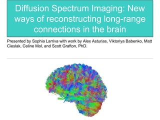
1. Diffusion Spectrum Imaging: New
ways of reconstructing long-range
connections in the brain
Presented by Sophia Larriva with work by Alex Asturias, Viktoriya Babenko, Matt
Cieslak, Celine Mol, and Scott Grafton, PhD.
2. What is all this?
• Brain has many overlapping connections
• Neurons
• White matter
• Different connections are
different!
Split Brain: Corpus Callosum
3. Left Lateral Occipital to
Left Inferior Temporal
• Diffusion MRI looks at water
movement
• Constrained by axon bundles
• Maps out connections (speech, visual
pathways, etc.)
• Clinical uses for strokes and certain
types of injuries
Right Superior
Parietal to Brain
Stem
Some Background
5. A Comparison of Scans
• Center for Magnetic Resonance Research at University of Minnesota
(CMRR)
• Siemens Corporation
• Examined differences in signal quality
6. Results
CMRR Wins! (almost outright)
Neuronal fiber tracts (axons) where Siemens
appeared better were statistically
insignificant/not real
Right Paracentral Gyrus
to Left Paracentral Gyrus
Right Entorhinal Cortex to Brain Stem
-whats a brain made out of
-neurons, white matter,
-patient with split brain surgery
-surgery with person who has no ability to speak
-diffusion mri looks at all water diffusion, when you put a brain within the Diffusion scanner is constrained by axon bundles
-white matter, brain connections
-neuron cartoon, connections of computers
Examined areas with significant differences in the strength of directional diffusion
-ratio of the peak of the ODF to the isotropic component
-connections looked at
in regions with difference in signal quality
-showed an advantage for CMRR
even sometimes with antatomically incorrect signals