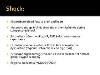
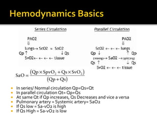
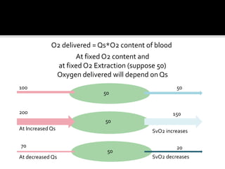
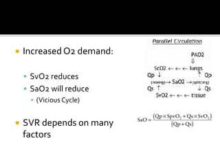
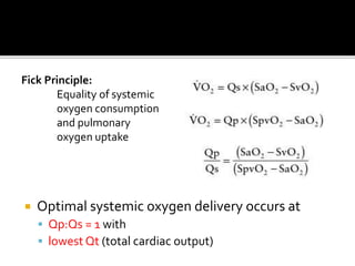
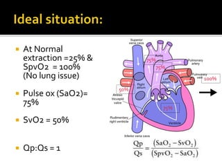
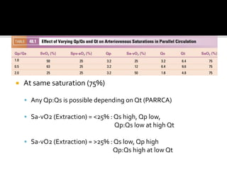
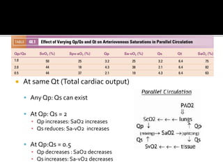
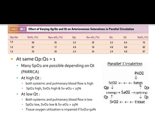
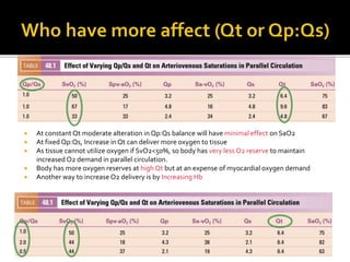
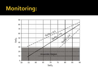
Dr.AbhinavAgarwal
 Cardiac output affected by
ď‚§ Preload
ď‚§ Afterload
ď‚§ Rate
ď‚§ Rhythm
ď‚§ Contractility
ď‚§ AP shunts
 Regional Resistance
ď‚§ Neurohumoral Factors (Inflammation, sympathetic
nervous system)
ď‚§ Local Factors (Autoregulation)
 Redistribute Blood flow to brain and heart
 Mesentric and splanchnic circulation- silent ischemia during
compensated shock
 Baroreflex - Contractility, HR, SVR & decreases venous
capacitance
 Often body impairs systemic flow in face of myocardial
dysfunction (regional ischaemia due to high SVR)
 Ischaemic organ damage can occur even in presence of normal
global oxygen economy
 Regional Ischaemia →MODS→Death
 In series/ Normal circulation Qp=Qs=Qt
 In parallel circulation Qt= Qp+Qs
 At same Qt if Qp increases, Qs Decreases and vice a versa
 Pulmonary artery = Systemic artery= SaO2
 If Qs low = Sa-vO2 is high
 If Qs High = Sa-vO2 is low
200
At Increased Qs
At decreased Qs
150
2070
50
50
100
50
50
SvO2 decreases
SvO2 increases
O2 delivered = Qs*O2 content of blood
At fixed O2 content and
at fixed O2 Extraction (suppose 50)
Oxygen delivered will depend on Qs
 Increased O2 demand:
ď‚§ SvO2 reduces
ď‚§ SaO2 will reduce
â–Ş (Vicious Cycle)
 SVR depends on many
factors
 Optimal systemic oxygen delivery occurs at
ď‚§ Qp:Qs = 1 with
ď‚§ lowest Qt (total cardiac output)
Fick Principle:
Equality of systemic
oxygen consumption
and pulmonary
oxygen uptake
 At Normal
extraction =25% &
SpvO2 = 100%
(No lung issue)
 Pulse ox (SaO2)=
75%
 SvO2 = 50%
 Qp:Qs = 1
75%
75%
75%
50%
100%
 At same saturation (75%)
ď‚§ Any Qp:Qs is possible depending on Qt (PARRCA)
ď‚§ Sa-vO2 (Extraction) = <25% : Qs high, Qp low,
Qp:Qs low at high Qt
ď‚§ Sa-vO2 (Extraction) = >25% : Qs low, Qp high
Qp:Qs high at low Qt
 At same Sa-vO2: (Systemic flow is fixed)
ď‚§ Any SpO2 is possible depending on Qt (PARRCA)
ď‚§ If SaO2 High: Qp is high and Qp:Qs = 2 at high Qt
ď‚§ If SaO2 Low: Qp is low and Qp:Qs = 0.5 at low Qt
 At same Qt (Total cardiac output)
ď‚§ Any Qp: Qs can exist
ď‚§ At Qp: Qs = 2
â–Ş Qp increases: SaO2 increases
â–Ş Qs reduces: Sa-vO2 increases
ď‚§ At Qp:Qs = 0.5
â–Ş Qp decreases : SaO2 decreases
â–Ş Qs increases: Sa-vO2 decreases
 At same Qp:Qs = 1
ď‚§ Many SpO2 are possible depending on Qt
(PARRCA)
ď‚§ At high Qt :
â–Ş both systemic and pulmonary blood flow is high
â–Ş SpO2 high, SvO2 high & Sv-aO2 < 25%
ď‚§ At low Qt :
â–Ş Both systemic and pulmonary blood flow is low
â–Ş SpO2 low, SvO2 low & Sv-aO2 > 25%
â–Ş Tissue oxygen utilization is impaired if SvO2<50%
 At constant Qt moderate alteration in Qp:Qs balance will have minimal effect on SaO2
 At fixed Qp:Qs, Increase in Qt can deliver more oxygen to tissue
 As tissue cannot utilize oxygen if SvO2<50%, so body has very less O2 reserve to maintain
increased O2 demand in parallel circulation.
 Body has more oxygen reserves at high Qt but at an expense of myocardial oxygen demand
 Another way to increase O2 delivery is by Increasing Hb
 Optimization of SaO2 alone will result in acute hemodynamic
collapse unexpectedly in an apparently stable child.
 Gas manipulation of PVR
ď‚§ Inspired CO2: Increased PVR, decreased SVR, Increased O2 delivery
(esp. brain)
ď‚§ Subatmospheric FiO2: Raises PVR
 Controlled PPV while avoiding hypervenilation (PEEP)
 O2 can be used if:
ď‚§ Respiratory pathology is present
ď‚§ Restrictive communications
 Control of elevated SVR was more effective than increasing PVR
ď‚§ Inotropes increase SVR at high doses
ď‚§ Inodilators preferred (while preventing significant hypotension)
ď‚§ Morphine reduces Sympathetic outflow
 Regional saturations of brain, liver, kidney, gut and muscle can be
measured to rule out regional ischaemia
 Increase Hct > 50% increases O2 carrying capacity
 Pulmonary venous SpvO2 = 100%
 Variablity in Arteriovenous saturation
difference
 Not possible to obtain true sytemic venous
mixed venous saturation
 Thank you

Univentricular circulation

  • 1.
  • 2.
     Cardiac outputaffected by  Preload  Afterload  Rate  Rhythm  Contractility  AP shunts  Regional Resistance  Neurohumoral Factors (Inflammation, sympathetic nervous system)  Local Factors (Autoregulation)
  • 3.
     Redistribute Bloodflow to brain and heart  Mesentric and splanchnic circulation- silent ischemia during compensated shock  Baroreflex - Contractility, HR, SVR & decreases venous capacitance  Often body impairs systemic flow in face of myocardial dysfunction (regional ischaemia due to high SVR)  Ischaemic organ damage can occur even in presence of normal global oxygen economy  Regional Ischaemia →MODS→Death
  • 4.
     In series/Normal circulation Qp=Qs=Qt  In parallel circulation Qt= Qp+Qs  At same Qt if Qp increases, Qs Decreases and vice a versa  Pulmonary artery = Systemic artery= SaO2  If Qs low = Sa-vO2 is high  If Qs High = Sa-vO2 is low
  • 5.
    200 At Increased Qs Atdecreased Qs 150 2070 50 50 100 50 50 SvO2 decreases SvO2 increases O2 delivered = Qs*O2 content of blood At fixed O2 content and at fixed O2 Extraction (suppose 50) Oxygen delivered will depend on Qs
  • 6.
     Increased O2demand:  SvO2 reduces  SaO2 will reduce ▪ (Vicious Cycle)  SVR depends on many factors
  • 7.
     Optimal systemicoxygen delivery occurs at  Qp:Qs = 1 with  lowest Qt (total cardiac output) Fick Principle: Equality of systemic oxygen consumption and pulmonary oxygen uptake
  • 8.
     At Normal extraction=25% & SpvO2 = 100% (No lung issue)  Pulse ox (SaO2)= 75%  SvO2 = 50%  Qp:Qs = 1 75% 75% 75% 50% 100%
  • 9.
     At samesaturation (75%)  Any Qp:Qs is possible depending on Qt (PARRCA)  Sa-vO2 (Extraction) = <25% : Qs high, Qp low, Qp:Qs low at high Qt  Sa-vO2 (Extraction) = >25% : Qs low, Qp high Qp:Qs high at low Qt
  • 10.
     At sameSa-vO2: (Systemic flow is fixed)  Any SpO2 is possible depending on Qt (PARRCA)  If SaO2 High: Qp is high and Qp:Qs = 2 at high Qt  If SaO2 Low: Qp is low and Qp:Qs = 0.5 at low Qt
  • 11.
     At sameQt (Total cardiac output)  Any Qp: Qs can exist  At Qp: Qs = 2 ▪ Qp increases: SaO2 increases ▪ Qs reduces: Sa-vO2 increases  At Qp:Qs = 0.5 ▪ Qp decreases : SaO2 decreases ▪ Qs increases: Sa-vO2 decreases
  • 12.
     At sameQp:Qs = 1  Many SpO2 are possible depending on Qt (PARRCA)  At high Qt : ▪ both systemic and pulmonary blood flow is high ▪ SpO2 high, SvO2 high & Sv-aO2 < 25%  At low Qt : ▪ Both systemic and pulmonary blood flow is low ▪ SpO2 low, SvO2 low & Sv-aO2 > 25% ▪ Tissue oxygen utilization is impaired if SvO2<50%
  • 13.
     At constantQt moderate alteration in Qp:Qs balance will have minimal effect on SaO2  At fixed Qp:Qs, Increase in Qt can deliver more oxygen to tissue  As tissue cannot utilize oxygen if SvO2<50%, so body has very less O2 reserve to maintain increased O2 demand in parallel circulation.  Body has more oxygen reserves at high Qt but at an expense of myocardial oxygen demand  Another way to increase O2 delivery is by Increasing Hb
  • 15.
     Optimization ofSaO2 alone will result in acute hemodynamic collapse unexpectedly in an apparently stable child.
  • 16.
     Gas manipulationof PVR  Inspired CO2: Increased PVR, decreased SVR, Increased O2 delivery (esp. brain)  Subatmospheric FiO2: Raises PVR  Controlled PPV while avoiding hypervenilation (PEEP)  O2 can be used if:  Respiratory pathology is present  Restrictive communications  Control of elevated SVR was more effective than increasing PVR  Inotropes increase SVR at high doses  Inodilators preferred (while preventing significant hypotension)  Morphine reduces Sympathetic outflow  Regional saturations of brain, liver, kidney, gut and muscle can be measured to rule out regional ischaemia  Increase Hct > 50% increases O2 carrying capacity
  • 17.
     Pulmonary venousSpvO2 = 100%  Variablity in Arteriovenous saturation difference  Not possible to obtain true sytemic venous mixed venous saturation
  • 18.