
Recommended
More Related Content
What's hot
What's hot (20)
Similar to Diabetic Ketoacidosis
Similar to Diabetic Ketoacidosis (20)
More from Dr. Abhinav Agarwal
Recently uploaded
Recently uploaded (20)
Diabetic Ketoacidosis
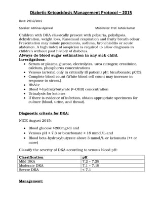
- 1. Diabetic Ketoacidosis Management Protocol – 2015 Date: 29/10/2015 Speaker: Abhinav Agarwal Moderator: Prof. Ashok Kumar Children with DKA classically present with polyuria, polydipsia, dehydration, weight loss, Kussmaul respiration and fruity breath odour. Presentation may mimic pneumonia, asthma, bronchiolitis or acute abdomen. A high index of suspicion is required to allow diagnosis in children without past history of diabetes. Always do blood sugar estimation in any sick child. Investigations: • Serum or plasma glucose, electrolytes, urea nitrogen; creatinine, calcium, phosphorus concentrations • Venous (arterial only in critically ill patient) pH; bicarbonate; pCO2 • Complete blood count (White blood cell count may increase in response to stress.) • HbA1c • Blood •-hydroxybutyrate (•-OHB) concentration • Urinalysis for ketones • If there is evidence of infection, obtain appropriate specimens for culture (blood, urine, and throat). Diagnostic criteria for DKA: NICE August 2015: • Blood glucose >200mg/dl and • Venous pH < 7.3 or bicarbonate < 18 mmol/L and • Blood beta-hydroxybutyrate above 3 mmol/L or ketonuria (++ or more) Classify the severity of DKA according to venous blood pH: Classification pH Mild DKA 7.2 – 7.29 Moderate DKA 7.1 – 7.19 Severe DKA < 7.1 Management:
- 2. Page | 2 1. SUPPORTIVE: • Secure the airway and empty the stomach by continuous nasogastric suction to prevent pulmonary aspiration, in case there is deterioration in conscious level. • Perform continuous electrocardiographic monitoring to assess T- waves for evidence of hyper- or hypokalemia • Give oxygen to patients with severe circulatory impairment or shock • Give antibiotics to febrile patients after obtaining appropriate cultures of body fluids • Catheterize the bladder if the child is unconscious or unable to void on demand. (e.g., infants and very ill young children) • Secure two peripheral i.v. lines. 2. INITIAL FLUID BOLUS: Given only if shock is present as indicated by poor peripheral pulses, poor capillary filling time and hypotension. • No fluid bolus in mild to moderate DKA (pH • 7.1) • Do not routinely give fluid bolus in severe DKA (pH < 7.1). o However, one fluid bolus may be considered in severe DKA. o Do not give more than one fluid bolus in severe DKA without discussion with the consultant • If patient is in shock bolus can be given until restoration of circulation volume. o Fluid: Normal Saline o Amount: 10ml/kg o Duration: as quickly as possible o No of boluses: until restoration of circulatory volume. Generally two boluses are sufficient in most cases. Inotropes can be used if patient is in hypotensive shock. 3. FLUID MANAGEMENT: Once circulating volume is restored calculate fluid requirement as follows: Fluid requirement: Deficit + Maintenance Distribute total fluid requirement over 48 hours. Type of fluid: Normal saline NICE August 2015:
- 3. Page | 3 Clinical assessment of the degree of dehydration is difficult in DKA. Therefore use venous blood pH to decide fluid deficit. Deficit Calculation: pH Deficit Amount (Normal saline) Mild to Mod. DKA 7.1 – 7.29 5% 50 ml/kg Severe DKA < 7.1 10% 100 ml/kg Maintenance Fluid: Weight < 10 kg 2 ml/kg/hr Weight between 10 and 40 kg 1 ml/kg/hr Weight >40 kg Fixed volume of 40 ml/hr • These maintenance rates are lower than standard fluid maintenance rates because large fluid volumes are associated with an increased rate of cerebral edema. • Don’t subtract any fluid boluses given upto 20ml/kg from fluid calculation i.e. if 30 ml/kg has been given subtract 10 ml/kg from the calculations. Hourly rate = (deficit – bolus over 20ml/kg) + maintenance per hour 48 • Use 0.9% sodium chloride with 40 meq/L potassium chloride calculated for 48 hr. • If oral fluids are given before the 48hr rehydration period is completed, the IV infusion needs to be reduced to take account of the oral intake. • If a massive diuresis continues for several hours fluid input may need to be increased. If large volumes of gastric aspirate continue, these will need to be replaced with 0.45% saline with KCl. 4. POTASSIUM SUPPLEMENTATION: • All fluids (except any initial bolus) contain 40 meq/l potassium chloride, unless there is evidence of renal failure. • At admission, if hypokalemia, start potassium replacement at the time of initial volume expansion and before starting insulin therapy. (20 meq/L).
- 4. Page | 4 • If potassium levels normal, start potassium replacement after initial volume expansion. • If hyperkalemia, defer potassium replacement until urine output is documented. • If immediate serum potassium levels are not available, obtain ECG to document hypo or hyperkalemia. Serum K (meq/L) K Supplementation < 3 meq/l 60 – 80 meq/L, don’t start or temporarily suspend insulin infusion. 3 – 5.5 meq/L 40 meq/L >5.5 meq/L Withhold until urine output adequate. 5. INSULIN INFUSION: • Begin insulin infusion 1 – 2 hr after starting intravenous fluid therapy. • A recent RCT has shown that o Low dose insulin (0.05U/kg/hr) is as effective as standard dose insulin (0.1 U/kg/hr) and more safe. (JAMA Pediatr.doi:10.1001/jamapediatrics.2014.1211) • Do not give bolus insulin. • Regular insulin (40U/ml) dissolve in 39 ml NS to make concentration of 1U/ml • Before starting infusion flush syringe and tubing with insulin solution. • Don’t start insulin if hypokalemia is present (K < 3 meq/L) • Start an intravenous insulin infusion 1-2 hours after beginning intravenous fluid therapy • Insulin infusion started @ 0.05 U/kg/hr • If the blood beta-hydroxybutyrate level is not falling within 6–8 hours, increase the insulin dosage to 0.1 U/kg/hr or greater. 6. BICARBONATE THERAPY: • Not indicated in DKA regardless of blood pH. • May be beneficial in life threatening hyperkalemia. (cardiac arrhythmia or respiratory insufficiency). • Dose: 1 – 2 meq/kg over 1 hour. 7. PHOSPHATE: • Plasma phosphate levels fall after starting treatment.
- 5. Page | 5 • Patients usually do not have symptoms until plasma phosphate is <1mg/dL. • Manifestations include: o Metabolic encephalopathy (irritability, paresthesias, confusion, seizures, coma) o Impaired myocardial contractility o Respiratory failure o Muscle dysfunction o Hematologic effects include hemolysis, decreased phagocytosis and granulocyte chemotaxis, defective clot retraction and thrombocytopenia. • For treatment, IV solution that contains a 50:50 mixture of potassium phosphate and another suitable potassium salt (potassium chloride or potassium acetate) should be used. 8. MONITORING SHEET: • Strict fluid balance including oral fluids and urine output • Continuous ECG and Pulse oximetry • HOURLY: o Capillary blood glucose measurements o Capillary blood ketone levels o Vitals o Modified Glasgow coma score • 4 HOURLY: o Glucose (laboratory measurement) o ABGA o RFT o Blood ketones (beta-hydroxybutyrate). • Twice daily weight; can be helpful in assessing fluid balance 9. CONTINUING MANAGEMENT: Serum Glucose NICE 2015 >250 mg/dl NS with KCl Insulin @ 0.05 – 0.1 U/kg/hr <250 mg/dl with S. Ketones<3mmol/L S. Ketones>3mmol/L S. Ketones not 5% DNS with KCl Insulin @ 0.05 U/kg/hr 10% DNS with KCl Insulin@ >0.05 U/kg/hr 5% DNS with KCl
- 6. Page | 6 available Insulin @ 0.05 – 0.1 U/kg/hr <100 mg/dl Increase the glucose concentration of i.v.f. DO not reduce insulin below 0.05U/kg/hr if ketones still present. <70 mg/dl Give a bolus of 2 ml/kg of 10% glucose. Increase the glucose concentration of the infusion Reduce insulin for 1 hour If acidosis is not correcting, consider the following: Wrong diagnosis Insufficient insulin to switch off ketones Inadequate resuscitation Sepsis Hyperchloraemic acidosis Once all these causes of acidosis have been excluded, and if ketones are falling gradually, then residual acidosis is likely to be due to hyperchloraemia, and this can be left to resolve on its own, and does not require any treatment. 10. CORRECTED SODIUM: Corrected sodium = measured Na + 1.6 ([plasma glucose -100]/100) (mg/dL) • The measured serum sodium concentration is an unreliable index of the degree of ECF contraction. • Corrected sodium levels should rise as blood glucose levels fall during treatment. (increase by 1.6 meq/L for each 100 mg/dL decline in blood glucose) 11. CEREBRAL EDEMA: Suspicion Sign - Headache - agitation or irritability - unexpected fall in heart rate - increased blood pressure - deterioration in level of consciousness - abnormalities of breathing pattern - oculomotor palsies - pupillary inequality or dilatation • Treat fever • Elevate head end to 30•
- 7. Page | 7 • Restrict i.v. fluids by 1/2. • 20% Mannitol 2.5 – 5 ml/kg (0.5 – 1 g/kg) over 10 – 15 min or 3% NaCl 3.5 – 5 ml/kg over 10 - 15 min. • A repeated dose of Mannitol or 3% NaCl may be required after 2 hours if no response 12. SHIFT TO SUBCUTANEOUS INSULIN: • Ketosis is resolving (blood beta-OHB < 1.0 mmol/litre) or pH normal. • Patient is alert. • Tolerating fluids without nausea or vomiting. Urinary ketones may still be present after resolution of DKA. Start subcutaneous insulin at least 30 min before stopping intravenous insulin. Prepubertal Total Daily Dose 0.75–1.0 unit/kg Pubertal Total Daily Dose 1.0–1.2 unit/kg Split-mix regimen: Before breakfast Two-thirds of TDD One-third short-acting insulin Two-thirds intermediate-acting insulin Before dinner One-third of TDD One-third to one-half as short- acting insulin One-half to two-thirds as intermediate-acting insulin Basal Bolus Regimen: Long acting insulin One-half of the TDD Short-acting insulin One-half of the TDD; the dose before each meal comprises 15–20% of the TDD 13. TECHNIQUE OF SUBCUTANEOUS INSULIN: • Rapid-acting insulin analogs should be injected within 15 min before a meal or immediately after a meal • The hands and the injection site should be clean.
- 8. Page | 8 • The top of the insulin vial should be wiped with alcohol swab. • The vial should be gently rolled in the palms of the hands (not shaken) 20 times to resuspend the insulin. • An amount of air equal to the dose of insulin required should first be drawn up and injected into the vial to avoid creating a vacuum. • For a mixed dose, putting sufficient air into both bottles before drawing up the dose is important. • When mixing rapid- or short-acting insulin with intermediate- or long-acting insulin, the clear rapid- or short-acting insulin should be drawn into the syringe first. • Lightly grasp a fold of skin and inject at a 90° angle. • Release the piched skin, count to 5 and pull the needle straight out. • If an injection seems painful or if blood or clear fluid is seen after withdrawing the needle, the patient should apply pressure for 5–8s without rubbing. • Insulin may be injected into the subcutaneous tissue of the upper arm and the anterior and lateral aspects of the thigh, buttocks, and abdomen. • An injection at a certain hour should always be given in the same anatomical site. However, it is important to rotate within a site each day, moving one finger width from the site of the previous injection or alternating from left to right, to avoid the build-up of lipohypertrophies. • Rotating within one area is recommended rather than rotating to a different area with each injection. 14. EXAMPLE: a. 30 kg 8 year old girl with a pH of 6.9 with shock. and who was given 30 ml/kg 0.9% sodium chloride for circulatory collapse. Timeline NICE 2015 Resuscitation / Bolus 300 ml NS 3 times to restore circulatory volume. Deficit 3000 ml (10%) Maintenance 30 ml/hr Duration of correction 48 hr Total fluid to be given 3000 – 300 = 2700 + 30ml/hr 48
- 9. Page | 9 Hourly Rate 2700/48 + 30 = 86.25 ml/hr Fluid NS with KCl Insulin @ 0.05 U/kg/hr b. 10 kg 2 year old girl with a pH of 7.2 . Timeline NICE 2015 Resuscitation / Bolus Not required Deficit 500ml (5%) Maintenance 10 ml/hr Duration of correction 48 hr Total fluid to be given 500 – 0 = 500 + 10ml/hr 48 Hourly Rate 500/48 + 10 = 20.4 ml/hr Fluid NS with KCl Insulin @ 0.05 U/kg/hr References:References:References:References: • Diabetes (type 1 and type 2) in children and young people: diagnosis and management; National Institute for Health and Care Excellence Guideline Published: 26 August 2015 • Diabetic ketoacidosis and hyperglycemic hyperosmolar state; ISPAD Clinical Practice Consensus Guidelines 2014 Compendium. Pediatric Diabetes 2014: 15(Suppl. 20): 154–179 • British Society For Pediatric Endocrinology and Diabetes Recommended Guideline for the Management of Children and Young People under the age of 18 years with Diabetic Ketoacidosis 2015 • Diabetic Ketoacidosis in Infants, Children, and Adolescents: A consensus statement from the American Diabetes Association. Diabetes care 2006: 29(5) • Sivanandan S, Sinha A, Jain V, Lodha R. Symposium on PICU protocols of AIIMS: Management of Diabetic Ketoacidosis. The Indian Journal of Pediatrics 2011; 78(5): 576-84 • Alemzadeh R, Ali O. Diabetes Mellitus. In: Nelson textbook of pediatrics. Eds. Kliegman RM, Stanton BF, Schor NF, Geme JWS, Behrman RE. 20th Edn. Elsevier, Philadelphia, USA. 2015: pp. 2760-90. • Ravikumar KG. Acute and chronic complications of Diabetes Mellitus. In: PG Textbook of Pediatrics. Eds. Gupta P, Menon PSN, Ramji S, Lodha R. 1st Edn. Jaypee, New Delhi, India 2015: pp. 2384-8 • Nallasamy K, Jayashree M, Singhi S. Low-Dose vs Standard-Dose Insulin Pediatric Diabetic Ketoacidosis. JAMA Pediatr.doi:10.1001/jamapediatrics.2014.1211
- 10. Page | 10 Algorithm for Management of Acute Diabetic Ketoacidosis: