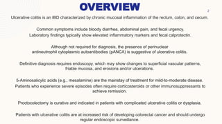
ULCERATIVE
COLITIS
OVERVIEW 2
Ulcerative colitis is an IBD characterized by chronic mucosal inflammation of the rectum, colon, and cecum.
Common symptoms include bloody diarrhea, abdominal pain, and fecal urgency.
Laboratory findings typically show elevated inflammatory markers and fecal calprotectin.
Although not required for diagnosis, the presence of perinuclear
antineutrophil cytoplasmic autoantibodies (pANCA) is suggestive of ulcerative colitis.
Definitive diagnosis requires endoscopy, which may show changes to superficial vascular patterns,
friable mucosa, and erosions and/or ulcerations.
5-Aminosalicylic acids (e.g., mesalamine) are the mainstay of treatment for mild-to-moderate disease.
Patients who experience severe episodes often require corticosteroids or other immunosuppressants to
achieve remission.
Proctocolectomy is curative and indicated in patients with complicated ulcerative colitis or dysplasia.
Patients with ulcerative colitis are at increased risk of developing colorectal cancer and should undergo
regular endoscopic surveillance.
EPIDEMIOLOGY
Prevalence
•Higher in caucasian populations than in Black, Hispanic, or Asian
populations.
•Highest among individuals of Ashkenazi Jewish descent.
Peak incidence
• 15–35 years of age
• Another smaller peak may be observed in individuals > 55 years of age.
• Similar for men and women
3
ETIOLOGY
Risk factors
 Genetic predisposition (e.g., HLA-B27 association)
 Ethnicity (White populations, individuals of Ashkenazi Jewish descent)
 Family history of inflammatory bowel disease
 Episodes of previous intestinal infection
 Increased fat intake
 Oral contraceptive use
 NSAIDs may exacerbate ulcerative colitis.
Protective factors
 Appendectomy
 Smoking
CLASSIFICATION
PATHOPHYSIOLOGY
6
The exact mechanism is unknown but studies suggest that ulcerative colitis is caused by abnormal interactions
between host immune cells and commensal bacteria.
1. Dysregulation of intestinal epithelium.
2. Dysregulation of the immune system.
CLINICAL FEATURES
Intestinal symptoms
• Bloody diarrhea with mucus
• Fecal urgency
• Abdominal pain and cramps
• Tenesmus
7
8
Extra- Intestinal symptoms
• General: fatigue, fever
• Skeletal: osteoarthritis, ankylosing spondylitis, sacroiliitis
• Ocular: uveitis, episcleritis, iritis
• Biliary: primary sclerosing cholangitis
• Cutaneous:Erythema nodosum, Pyoderma gangrenosum, Aphthous stomatitis, Pyostomatitis vegetans
• In children and adolescents: growth retardation and delayed puberty
CLINICAL FEATURES
APPROACH
 Evaluate patients with hematochezia and fecal urgency for
ulcerative colitis.
 Rule out infectious gastroenteritis.
 Consult gastroenterology for ileocolonoscopy with histological
examination to definitively diagnose ulcerative colitis
 Determine the extent of disease and severity
 Identify signs of acute severe ulcerative colitis
 Consider CT or MRI abdomen if direct endoscopy is
contraindicated.
 Assess for the presence of extraintestinal symptoms of ulcerative
colitis.
9
INVESTIGATIONS
Stool testing for causes of gastroenteritis is indicated in all patients. Blood tests are not routinely
required for diagnosis but help assess disease activity and severity.
Blood tests
 CBC: anemia, leukocytosis, thrombocytosis
 ESR, CRP: Elevated levels may indicate active ulcerative colitis and often correlates with disease
severity.
 Hypoalbuminemia: associated with a poor prognosis and more severe disease
 ALP, GGT: elevated in patients with concurrent PSC
 Perinuclear ANCA (pANCA)
INVESTIGATIONS
11
Stool diagnostic studies
 Stool test for Clostridioides difficile infection
 PCR panel for other enteric infections: depending on the patient's symptoms and risk factors for diarrhea
 Stool culture and microscopy: to assess for bacteria and ova and parasites if a stool PCR panel is not available
 Fecal calprotectin: can help assess for mucosal inflammation
ENDOSCOPY
ileocolonoscopy
• Recommended method for diagnosis and disease monitoring
Sigmoidoscopy
• Initially used as an alternative to colonoscopy, e.g., in ASUC
• Monitoring treatment response
• Findings are similar to colonoscopy findings.
EGD
• recommended for patients with upper gastrointestinal symptoms to rule out
Crohn disease
12
ENDOSCOPY CHANGES
13
IMAGING
14
Used as an adjunct to endoscopy, particularly for the detection of complications, or if endoscopy is not
possible.
On abdominal xray:
• Typically normal in mild-to-moderate disease
• Loss of colonic haustra (lead pipe appearance) may be seen in severe cases
• May show signs of complications, e.g toxic megacolon: massive distention
• Ulceration: segmental dilation with irregular edges outlined by gas
• Perforation: pneumoperitoneum
OTHER IMAGING
MODALITIES
CT/MRI abdomen
• Granular appearance of the mucosa
• Deep ulcerations
• Loss of haustra
• Pseudopolyps that appear as filling defects
Abdominal ultrasound
 increased bowel wall thickness
16
Late Stages
• Lymphocyte infiltration
• Mucosal atrophy
• Altered crypt architecture
• Branching of crypts: Irregularities in size and shape
• Epithelial dysplasia
HISTOLOGICAL FINDINGS
Early stages
• Granulocyte (neutrophil) infiltration: limited to mucosa and submucosa
• Crypt abscesses: an infiltration of neutrophils into the lumen of intestinal crypts due to a breakdown of the crypt epithelium
DDX
• Crohn disease
• Exudative-inflammatory diarrhea
• Diverticular disease
• Appendicitis
• Ischemic colitis
• Infectious colitis
• Radiation colitis
• Celiac disease
• Inflammatory diarrhoea
17
TREATMENT
18
MEDICAL
• Pharmacological therapy is used to
induce and maintain disease remission
SURGICAL
• Ulcerative colitis can be cured
surgically. Surgical treatment also
reduces the risk of colorectal cancer.
PHARMACOLOGICAL
TREATMENT
19
LONG TERM
MANAGEMENT
Disease monitoring
•Assess treatment response using endoscopy or fecal calprotectin if endoscopy is not possible.
•Flexible sigmoidoscopy is recommended 3–6 months after starting a new treatment.
•Follow-up every 3 months until remission has been achieved, then every 6–12 months.
Colorectal cancer screening
•Start screening 8–10 years after the initial diagnosis or at the time of diagnosis of PSC.
•Modality: ileocolonoscopy with biopsies
•Interval: every 1–5 years
21
REFERENCES
1. Vanga R, Long MD. Contemporary Management of Ulcerative Colitis. Curr Gastroenterol Rep. 2018; 20(3). doi:
10.1007/s11894-018-0622-0.| Open in Read by QxMD
2.Rubin et al. ACG Clinical Guideline: Ulcerative Colitis in Adults. Am J Gastroenterol. 2019; 114(3): p.384-413. doi:
10.14309/ajg.0000000000000152.| Open in Read by QxMD
3.Peppercorn MA, Cheifetz AS. Definition, epidemiology, and risk factors in inflammatory bowel disease. In: Post TW, ed.
UpToDate .Waltham, MA: UpToDate.https://www.uptodate.com/contents/definition-epidemiology-and-risk-factors-in-inflammatory-
bowel-disease.Last updated November 8, 2016. Accessed February 8, 2017.
4.Ungaro R, Mehandru S, Allen PB, Peyrin-Biroulet L, Colombel J-F. Ulcerative colitis. The Lancet. 2017; 389(10080): p.1756-
1770. doi: 10.1016/s0140-6736(16)32126-2.| Open in Read by QxMD
5.Ordas et al.. Ulcerative colitis. The Lancet. 2012; 380(9853): p.1606-1619. doi: 10.1016/S0140-6736(12)60150-0.| Open in
Read by QxMD
6.Ulcerative colitis - knowledge @ amboss (no date) ambossIcon. Available at:
https://www.amboss.com/us/knowledge/Ulcerative_colitis (Accessed: 11 May 2023).
22
THANK YOU

ulcerative colitis.pptx

  • 1.
  • 2.
    OVERVIEW 2 Ulcerative colitisis an IBD characterized by chronic mucosal inflammation of the rectum, colon, and cecum. Common symptoms include bloody diarrhea, abdominal pain, and fecal urgency. Laboratory findings typically show elevated inflammatory markers and fecal calprotectin. Although not required for diagnosis, the presence of perinuclear antineutrophil cytoplasmic autoantibodies (pANCA) is suggestive of ulcerative colitis. Definitive diagnosis requires endoscopy, which may show changes to superficial vascular patterns, friable mucosa, and erosions and/or ulcerations. 5-Aminosalicylic acids (e.g., mesalamine) are the mainstay of treatment for mild-to-moderate disease. Patients who experience severe episodes often require corticosteroids or other immunosuppressants to achieve remission. Proctocolectomy is curative and indicated in patients with complicated ulcerative colitis or dysplasia. Patients with ulcerative colitis are at increased risk of developing colorectal cancer and should undergo regular endoscopic surveillance.
  • 3.
    EPIDEMIOLOGY Prevalence •Higher in caucasianpopulations than in Black, Hispanic, or Asian populations. •Highest among individuals of Ashkenazi Jewish descent. Peak incidence • 15–35 years of age • Another smaller peak may be observed in individuals > 55 years of age. • Similar for men and women 3
  • 4.
    ETIOLOGY Risk factors  Geneticpredisposition (e.g., HLA-B27 association)  Ethnicity (White populations, individuals of Ashkenazi Jewish descent)  Family history of inflammatory bowel disease  Episodes of previous intestinal infection  Increased fat intake  Oral contraceptive use  NSAIDs may exacerbate ulcerative colitis. Protective factors  Appendectomy  Smoking
  • 5.
  • 6.
    PATHOPHYSIOLOGY 6 The exact mechanismis unknown but studies suggest that ulcerative colitis is caused by abnormal interactions between host immune cells and commensal bacteria. 1. Dysregulation of intestinal epithelium. 2. Dysregulation of the immune system.
  • 7.
    CLINICAL FEATURES Intestinal symptoms •Bloody diarrhea with mucus • Fecal urgency • Abdominal pain and cramps • Tenesmus 7
  • 8.
    8 Extra- Intestinal symptoms •General: fatigue, fever • Skeletal: osteoarthritis, ankylosing spondylitis, sacroiliitis • Ocular: uveitis, episcleritis, iritis • Biliary: primary sclerosing cholangitis • Cutaneous:Erythema nodosum, Pyoderma gangrenosum, Aphthous stomatitis, Pyostomatitis vegetans • In children and adolescents: growth retardation and delayed puberty CLINICAL FEATURES
  • 9.
    APPROACH  Evaluate patientswith hematochezia and fecal urgency for ulcerative colitis.  Rule out infectious gastroenteritis.  Consult gastroenterology for ileocolonoscopy with histological examination to definitively diagnose ulcerative colitis  Determine the extent of disease and severity  Identify signs of acute severe ulcerative colitis  Consider CT or MRI abdomen if direct endoscopy is contraindicated.  Assess for the presence of extraintestinal symptoms of ulcerative colitis. 9
  • 10.
    INVESTIGATIONS Stool testing forcauses of gastroenteritis is indicated in all patients. Blood tests are not routinely required for diagnosis but help assess disease activity and severity. Blood tests  CBC: anemia, leukocytosis, thrombocytosis  ESR, CRP: Elevated levels may indicate active ulcerative colitis and often correlates with disease severity.  Hypoalbuminemia: associated with a poor prognosis and more severe disease  ALP, GGT: elevated in patients with concurrent PSC  Perinuclear ANCA (pANCA)
  • 11.
    INVESTIGATIONS 11 Stool diagnostic studies Stool test for Clostridioides difficile infection  PCR panel for other enteric infections: depending on the patient's symptoms and risk factors for diarrhea  Stool culture and microscopy: to assess for bacteria and ova and parasites if a stool PCR panel is not available  Fecal calprotectin: can help assess for mucosal inflammation
  • 12.
    ENDOSCOPY ileocolonoscopy • Recommended methodfor diagnosis and disease monitoring Sigmoidoscopy • Initially used as an alternative to colonoscopy, e.g., in ASUC • Monitoring treatment response • Findings are similar to colonoscopy findings. EGD • recommended for patients with upper gastrointestinal symptoms to rule out Crohn disease 12
  • 13.
  • 14.
    IMAGING 14 Used as anadjunct to endoscopy, particularly for the detection of complications, or if endoscopy is not possible. On abdominal xray: • Typically normal in mild-to-moderate disease • Loss of colonic haustra (lead pipe appearance) may be seen in severe cases • May show signs of complications, e.g toxic megacolon: massive distention • Ulceration: segmental dilation with irregular edges outlined by gas • Perforation: pneumoperitoneum
  • 15.
    OTHER IMAGING MODALITIES CT/MRI abdomen •Granular appearance of the mucosa • Deep ulcerations • Loss of haustra • Pseudopolyps that appear as filling defects Abdominal ultrasound  increased bowel wall thickness
  • 16.
    16 Late Stages • Lymphocyteinfiltration • Mucosal atrophy • Altered crypt architecture • Branching of crypts: Irregularities in size and shape • Epithelial dysplasia HISTOLOGICAL FINDINGS Early stages • Granulocyte (neutrophil) infiltration: limited to mucosa and submucosa • Crypt abscesses: an infiltration of neutrophils into the lumen of intestinal crypts due to a breakdown of the crypt epithelium
  • 17.
    DDX • Crohn disease •Exudative-inflammatory diarrhea • Diverticular disease • Appendicitis • Ischemic colitis • Infectious colitis • Radiation colitis • Celiac disease • Inflammatory diarrhoea 17
  • 18.
    TREATMENT 18 MEDICAL • Pharmacological therapyis used to induce and maintain disease remission SURGICAL • Ulcerative colitis can be cured surgically. Surgical treatment also reduces the risk of colorectal cancer.
  • 19.
  • 20.
    LONG TERM MANAGEMENT Disease monitoring •Assesstreatment response using endoscopy or fecal calprotectin if endoscopy is not possible. •Flexible sigmoidoscopy is recommended 3–6 months after starting a new treatment. •Follow-up every 3 months until remission has been achieved, then every 6–12 months. Colorectal cancer screening •Start screening 8–10 years after the initial diagnosis or at the time of diagnosis of PSC. •Modality: ileocolonoscopy with biopsies •Interval: every 1–5 years
  • 21.
  • 22.
    REFERENCES 1. Vanga R,Long MD. Contemporary Management of Ulcerative Colitis. Curr Gastroenterol Rep. 2018; 20(3). doi: 10.1007/s11894-018-0622-0.| Open in Read by QxMD 2.Rubin et al. ACG Clinical Guideline: Ulcerative Colitis in Adults. Am J Gastroenterol. 2019; 114(3): p.384-413. doi: 10.14309/ajg.0000000000000152.| Open in Read by QxMD 3.Peppercorn MA, Cheifetz AS. Definition, epidemiology, and risk factors in inflammatory bowel disease. In: Post TW, ed. UpToDate .Waltham, MA: UpToDate.https://www.uptodate.com/contents/definition-epidemiology-and-risk-factors-in-inflammatory- bowel-disease.Last updated November 8, 2016. Accessed February 8, 2017. 4.Ungaro R, Mehandru S, Allen PB, Peyrin-Biroulet L, Colombel J-F. Ulcerative colitis. The Lancet. 2017; 389(10080): p.1756- 1770. doi: 10.1016/s0140-6736(16)32126-2.| Open in Read by QxMD 5.Ordas et al.. Ulcerative colitis. The Lancet. 2012; 380(9853): p.1606-1619. doi: 10.1016/S0140-6736(12)60150-0.| Open in Read by QxMD 6.Ulcerative colitis - knowledge @ amboss (no date) ambossIcon. Available at: https://www.amboss.com/us/knowledge/Ulcerative_colitis (Accessed: 11 May 2023). 22
  • 23.