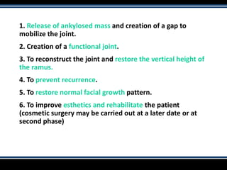
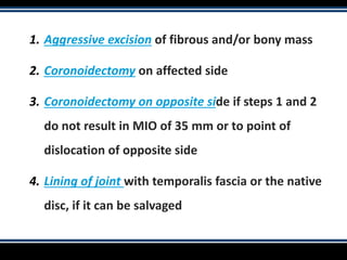
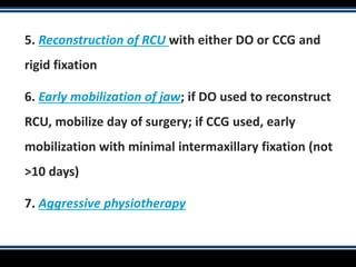
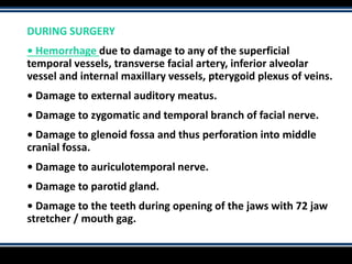
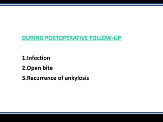
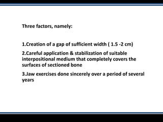
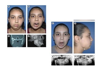
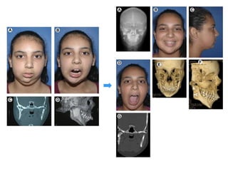
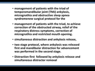
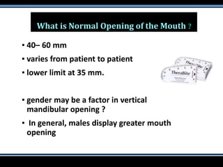
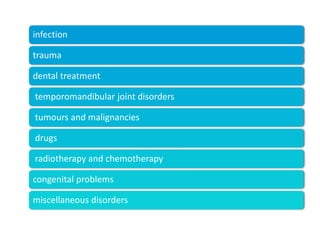
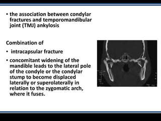
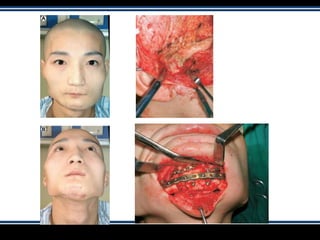
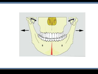
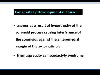
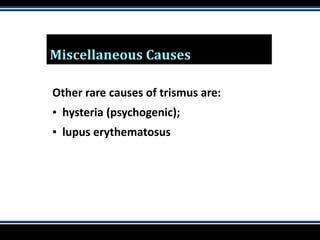
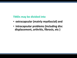
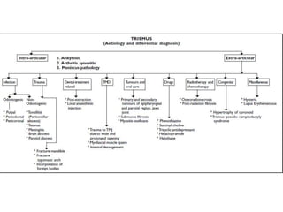
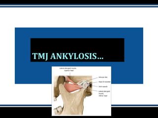
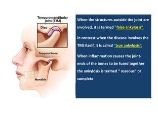
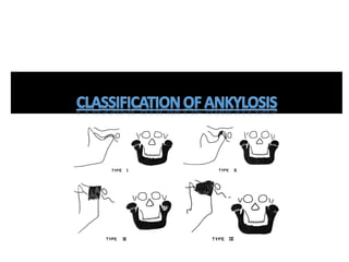
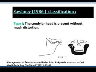
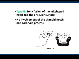
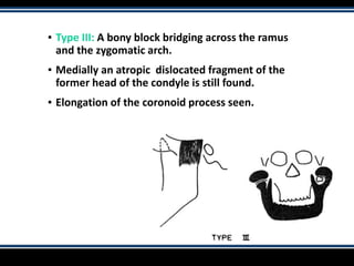
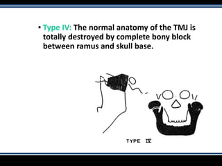
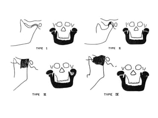
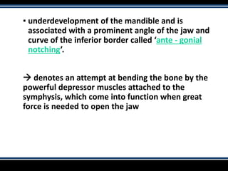
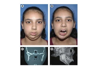
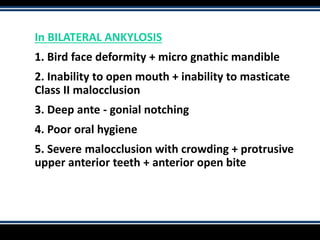
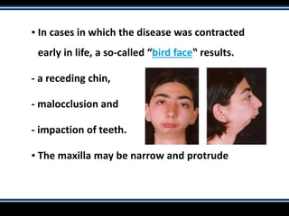
SEMINAR BY TEJASWINI PSS
TRISMUS & TMJ
ANKYLOSIS
Contents :
▪ Trismus
▪ Etiology of trismus
▪ TMJ ankylosis
▪ Etiology of TMJ ankylosis
▪ Classification
▪ Clinical manifestations
▪ Sequlae
▪ Management of TMJ ankylosis
▪ Conservative
▪ Surgical
Trismus
“inability to open the mouth “
What is Normal Opening of the Mouth ?
▪ 40– 60 mm
▪ varies from patient to patient
▪ lower limit at 35 mm.
▪ gender may be a factor in vertical
mandibular opening ?
▪ In general, males display greater mouth
opening
infection
trauma
dental treatment
temporomandibular joint disorders
tumours and malignancies
drugs
radiotherapy and chemotherapy
congenital problems
miscellaneous disorders
TRISMUS RELATED TO DENTAL
PROCEDURES
▪ Following extraction of teeth
▪ inflammation involving the muscles of mastication
or direct trauma to the TMJ
▪ 2–5 days after a mandibular block
▪ medial pterygoid muscle is accidentally penetrated
or
▪ A vessel is punctured - small bleed haematoma
 organize  fibrosis
INFECTIONS
INFECTIONS – TRISMUS
▪ The hallmark of a masticatory space infection
▪ odontogenic or non-odontogenic nature
▪ Odontogenic infections - 3 major origins: pulpal,
periodontal and pericoronal
▪ if unchecked  can spread to various facial
spaces of the head and neck and  serious
complications such as cervical cellulitis or
mediastinitis
Non-odontogenic infections :
▪ tonsillitis,
▪ tetanus,
▪ meningitis,
▪ Parotid abscess and brain abscess
TRAUMA - TRISMUS
▪ Mandible fractures – common
▪ accidental incorporation of foreign bodies
because of external traumatic injury.
▪ trauma of the zygomatic arch and
zygomaticomaxillary complex (ZMC), which
interferes with the movement of the coronoid
process
Etiology of Temporomandibular Joint
Ankylosis Secondary to Condylar Fractures:
The Role of Concomitant Mandibular
Fractures
Dongmei He, DDS, MD, PhD,* Edward Ellis III, DDS, Yi Zhang,
DDS, MD, PhD‡ J Oral Maxillofac Surg 66:77-84, 2008
▪ the association between condylar
fractures and temporomandibular
joint (TMJ) ankylosis
Combination of
▪ intracapsular fracture
▪ concomitant widening of the
mandible leads to the lateral pole
of the condyle or the condylar
stump to become displaced
laterally or superolaterally in
relation to the zygomatic arch,
where it fuses.
TUMOURS AND MALIGNANCIES…
Tumours and Oral Malignancies
▪ risk of misdiagnosing
▪ Thorough clinical and radiographic examination
to rule out neoplastic disease primary or
metastatic, in the epipharyngeal region, parotid
gland, jaws or TMJ
▪ Oral submucous fibrosis - precancerous
condition
DRUG THERAPY
Drug Therapy
Some drugs cause trismus as a secondary effect
▪ succinyl choline
▪ phenothiazines and
▪ Tricyclic antidepressants
▪ Trismus can be seen as an extrapyramidal side-effect of :
▪ metaclopramide, phenothiazines and other medications.
RADIOTHERAPY
&
CHEMOTHERAPY
Radiotherapy / Chemotherapy
▪ Radiotherapy is commonly used to treat squamous cell
carcinoma of the head and neck and regional lymphomas
▪ muscles of mastication within the field of radiation 
fibrosis  trismus
▪ Ischaemia caused by endarteritis obliterans
▪ Trismus complicates post-radiation dental care
HOW TO MINIMIZE THESE ILL-EFFECTS ?
▪ protective stents,
▪ jaw exercises and
▪ hyperbaric oxygen to increase neovascularization.
Congenital / Developmental Causes
▪ trismus as a result of hypertrophy of the
coronoid process causing interference of
the coronoids against the anteromedial
margin of the zygomatic arch.
▪ Trismuspseudo- camptodactyly syndrome
Miscellaneous Causes
Other rare causes of trismus are:
▪ hysteria (psychogenic);
▪ lupus erythematosus
TMDs may be divided into
▪ extracapsular (mainly myofascial) and
▪ intracapsular problems (including disc
displacement, arthritis, fibrosis, etc )
TMJ ANKYLOSIS…
▪ Ankylosis, or Anchylosis ( Greek –bent /
crooked )
▪ Ankylosis is a stiffness of a joint, as a result
of injury or disease and results in
hypomobiliy or no mobility.
When the structures outside the joint are
involved, it is termed "false ankylosis”.
in contrast when the disease involves the
TMJ itself, it is called "true ankylosis”.
When inflammation causes the joint-
ends of the bones to be fused together
the ankylosis is termed “ osseous” or
complete
Sawhney (1986 ) classification :
Type I: The condylar head is present without
much distortion.
Management of Temporomandibular Joint Ankylosis Reza Movahed, DMD Oral
Maxillofacial Surg Clin N Am 27 (2015) 27–35
▪ Type II: Bony fusion of the misshaped
head and the articular surface.
▪ No involvement of the sigmoid notch
and coronoid process.
▪ Type III: A bony block bridging across the ramus
and the zygomatic arch.
▪ Medially an atropic dislocated fragment of the
former head of the condyle is still found.
▪ Elongation of the coronoid process seen.
▪ Type IV: The normal anatomy of the TMJ is
totally destroyed by complete bony block
between ramus and skull base.
ETIOPATHOLOGY OF TMJ FALSE
ANKYLOSIS
▪ False ankylosis results from pathological condition
outside the joint and leads to limited mandibular
mobility.
CAUSES OF FALSE ANKYLOSIS
1. MUSCULAR TRISMUS
pericoronitis,
submasseteric, pterygomandibular, infra - temporal
or submandibular spaces infection
MUSCULAR FIBROSIS
Muscular fibrosis from any long standing
dysfunction like arthritis and myositis.
MYOSITIS OSSIFICANS
When there is progressive ossification after injury
and hematoma formation especially of the
masseter muscle, inability to open the mouth
develops. This can be confirmed radiologically as
well
TETANY
▪ When there is hypocalcaemia, the spasms in the
muscles are produced hampering the opening of
the mouth.
TETANUS
▪ Acute infectious disease caused by Clostridium
tetani is represented by an early symptom of
lock-jaw because of persistent tonic spasm of the
muscles
NEUROGENIC CAUSES
Neurogenic causes like epilepsy, brain tumour and embolic
hemorrhage in medulla oblongata are also represented by
hypomobility of the jaw.
TRISMUS HYSTERICUS
• It is disease of psychogenic origin.
DRUG INDUCED SPASMS
• Drug induced spasms like in drug poisoning
MECHANICAL BLOCKADE
Mechanical blockade on account of osteoma or
elongation of the coronoid process of the mandible
there by reducing it's movement under the zygomatic
arch.
FRACTURE OF THE ZYGOMATIC ARCH
• Fracture of the zygomatic arch with inward buckling.
FRACTURE OF THE MANDIBLE
• Trauma causing fracture of the mandible leads to
reflex spasm of the muscles and hence trismus
SCARS AND BURNS OF THE FACE
▪ Scars and burns of the face also, restrict the
movements of the jaw.
▪ Post irradiation fibrosis lead to hypomobility of
the mandible.
▪ CLEFT PALATE OPERATIONS can produce
fibrosis of the pterygomandibular raphe and,
consequently, limitation of mouth opening
ETIOPATHOLOGY of true
ankylosis
Birth Trauma
• Birth trauma producing so-called congenital ankylosis and
occurs in cases of difficult delivery, particularly forceps delivery
• At times, other bones are fractured as well.
• The injury of the jaw caused by the use of forceps may not be
discovered until later when it is noticed that the child could
open his jaw only slightly.
▪ Haemarthrosis
Haemarthrosis is another cause of ankylosis.
It is generally, due to fracture of the base of skull
extending through the mandibular fossa - may also
be caused by an intracapsular injury.
▪ Cortical bone in a child is very thin - as a result of
intracapsular fracture of TMJ, bleeding takes place
within the joint – known as hemarthrosis
▪ The hematoma within the joint organises slowly
which is then converted to fibrous tissue and then
bone resulting in bony ankylosis
Suppurative arthritis
• Suppurative arthritis, may be due to infection
of the ear or mastoiditis or it may be of
hematogenous origin leading to ankylosi
Rheumatoid arthritis
• Rheumatoid arthritis, may cause great limitation
of motion or complete ankylosis
• There is associated atrophy of the muscles
generally accompaning ankylosis, if contracted
early in life.
Osteomyelitis
▪ Osteomyelitis affecting the mandibular condyle
without involving the joint itself frequently
results in limitation of motion +
▪ muscular trismus + peri-articular swelling, and
suppuration often results in fistula formation
▪ Fracture of the condyle especially comminuted
fractures of the head of the condyle, may cause
ankylosis.
▪ Trauma to the condyle in children is more likely to
cause ankylosis than adults
Clinical manifestations vary according
to:
(a) Severity of ankylosis,
(b) Time of onset of ankylosis,
(c) Duration.
1. Early joint involvement - less than 15 years:
Severe facial deformity and loss of function.
2. Later joint involvement after the age of 15 years:
Facial deformity marginal or nil. But, functional loss
severe.
Those patients in whom the ankylosis develops
after full growth completion have no facial
deformity
CLINICAL MANIFESTATIONS
• Pain
▪ Healed chin laceration
▪ Reduced interincisal mouth opening or No mouth opening at all
▪ neglected oral hygiene
▪ impacted / malposed /carious teeth.
• Inability to open the jaw and difficulty or inability to masticate
food
UNILATERAL ANKYLOSIS :
1. Facial asymmetry with affected side appearing normal & the
opposite side appearing flat.
2. Chin deviated to the ankylosed side.
3. Ante-gonial notch on the affected side
4. Minimal condylar movements on palpation.
5. Class II malocclusion on affected side and cross bite may be
seen
▪ underdevelopment of the mandible and is
associated with a prominent angle of the jaw and
curve of the inferior border called ‘ante - gonial
notching’.
 denotes an attempt at bending the bone by the
powerful depressor muscles attached to the
symphysis, which come into function when great
force is needed to open the jaw
In BILATERAL ANKYLOSIS
1. Bird face deformity + micro gnathic mandible
2. Inability to open mouth + inability to masticate
Class II malocclusion
3. Deep ante - gonial notching
4. Poor oral hygiene
5. Severe malocclusion with crowding + protrusive
upper anterior teeth + anterior open bite
▪ In cases in which the disease was contracted
early in life, a so-called “bird face‟ results.
- a receding chin,
- malocclusion and
- impaction of teeth.
▪ The maxilla may be narrow and protrude
PROBLEMS ASSOCIATED
WITH ANKYLOSIS
1. Interferes with the mastication of food and with
nutrition
2. Prevents oral hygiene and prophylactic care, and
treatment of dental caries
There is destruction of the growth center [situated in the
condyle] and with absence of functional stimulation
prevents normal development of the jaw prevents
normal eruption of the teeth and causes micrognathia
Diagnosis
1. History of infection or trauma (birth trauma + falls
+ previous infection of the ear)
2. Findings at clinical examination (reduced
interincisal opening + diminished/no TMJ
movements + scar on the chin due to trauma)
3. Radiological findings
Radiographic Examination
▪ Orthopantomograph: OPG will show both the joints for
comparision – important in unilateral cases –will also
reveal ante-gonial notching.
• PA view will show the mediolateral extent of the bony
mass – also reveal any mandibular asymmetry.
• Lateral oblique – will demonstrate the anteroposterior
extent of the bony mass and the elongation of the
coronoid process
▪ CT Scan/3D CT Scan
– gives relationship to the middle cranial fossa and
internal carotid artery (carotid canal) medially to the
ankylotic mass – usually not seen in conventional
radiographs.
3D CT SCAN showing Bony Ankylosis
Coronal CT Scan showing Bilateral TM Joint Ankylosis
▪ Radiographic changes are of extreme value in diagnosis In
fibrous ankylosis
1.evidence of destructive + proliferative changes seen in
bony compartments of TMJ +
2.haziness or narrowing of joint space
In bony ankylosis
- overall obliteration of joint space.
- antegonial notching anterior to the angle of mandible
- elongation of coronoid process
Imaging of temporomandibular joint ankylosis. A new
radiographic classification- IE El-Hakim
Dentomaxillofacial Radiology (2002) 31, 19 ± 23
Coronal CT showing bony exostoses in the glenoid fossa
superiorly as well as medial on the condylar head, resulting in
bony ankylosis
3D CT of an ankylosed joint with an elongated coronoid
process (c) that projects clearly under the zygomatic arch (z)
Post-contrast coronal CT scan of an ankylosed joint
showing its relationship to the maxillary artery (M)
Axial (a) and coronal (b) CT scans of an ankylosed joint
showing an elongated lateral pterygoid plate (P) which appears fused
to the bony exostosis
What Happens If Ankylosis Is Left
Untreated..
▪ Normal growth & development of face is affected
▪ Nutritional impairment
▪ Speech impairment
▪ Sleep apnoea ( tongue falls back in sleep) in Bilateral
Ankylosis.
▪ Malocclusion
▪ Poor and neglected oral hygiene
▪ Multiple carious and impacted teeth
MANAGEMENT OF
ANKYLOSIS
CONSERVATIVE THERAPY
ANKYLOSIS OF THE TMJ
V.H.KAZANJIAN DMD, MD SURGERY, GYNECOLOGY OBSTETRICS, SEPTEMBER
1938, VOL 36
▪ Treatment of Ankylosis is Surgical & should
be corrected at the earliest  TO ENSURE
EFFECTIVE FUNCTION AND GROWTH
Surgical treatment options will depend on:
1. Age of onset of ankylosis
2. Whether unilateral or bilateral ankylosis
3. Extent of ankylosis and
4. Any associated facial deformity
Aims and Objectives of Surgery
1. Release of ankylosed mass and creation of a gap to
mobilize the joint.
2. Creation of a functional joint.
3. To reconstruct the joint and restore the vertical height of
the ramus.
4. To prevent recurrence.
5. To restore normal facial growth pattern.
6. To improve esthetics and rehabilitate the patient
(cosmetic surgery may be carried out at a later date or at
second phase)
Surgical Approaches to the TMJ
1. Preauricular incision with
modifications
2. Submandibular incision
3. Post auricular
4. Post ramal
5. Endaural incision
6. Coronal incision
APPROACHES TO TMJ
TIMING OF SURGERY
Surgery for Ankylosis can be done in 2 stages:
• In the first stage surgery - release of ankylosis to
bring about jaw mobility and growth.
• In the second stage surgery an orthognathic surgery
can be planned to restore facial esthetics.
• single stage procedure where release of ankylosis and
esthetic correction are done in a single stage in adults or
after cessation of growth spurts in children
TYPES OF SURGICAL
PROCEDURES
1. Condylectomy
2. Gap arthroplasty
3. Interpositional Arthroplasty.
4. Ramus Osteotomy
5. Angle’s Osteotomy
▪ Use of Brisement Force •
The forced opening of the jaw with the patient under general
anesthesia is the oldest method that has been employed.
• The jaw is forced open by means of a mouth gag and
mobilized as much as possible by forceful manipulation.
• In FIBROUS ANKYLOSIS or immobility caused by muscle
spasms or fibrosis, this method may yield good results.
• After the jaw has been mobilized, the patient is further
benefited by exercises
Condylectomy and Arthroplasty
• In both condylectomy and osteoarthrotomy the
condyle is removed.
• The two methods do not differ a great deal, except
for the amount of bone excised.
• In a condylectomy the condyle is detached and
removed, whereas an osteoarthrotomy includes the
excision of the entire joint and / or an adequate
amount of bone below the joint
Condylectomy
Condylectomy consists of excision of the condyle.
Indications :
1.severe arthrosis,
2.various types of arthritis,
3.in malunited condylar fractures with limitation of
motion and pain during mastication.
4. cases of fibrous ankylosis where the articular
space has not been completely eliminated.
Complications / Disadvantages of Condylectomy
- Loss of vertical height of the ramus.
- In case of bilateral condylectomy, it may create
an anterior open bite.
- In unilateral cases, there may be deviation of
the jaw on opening
Osteoarthrotomy (Gap Arthoplasty)
• In bony ankylosis - the condyle is completely fused to the
base of the skull - the coronoid is small size, often elongated
• Because the joint is completely eliminated by the disease, or
bony mass - an osteoarthrotomy or a double osteotomy is
performed to remove a slice of bone, which is known as gap
arthroplasty ‟.
• In gap arthroplasty, a piece of bone about 1.5 to 2 cm in
width is removed.
• tendency for ankylosis to recur, some autogenous /
alloplastic material interposed between cut bony ends
▪ The mouth is forced open with the help of a mouth gag
to check the mouth opening -a gap of 1.5 - 2 cms is
created & not interposed with any material.
• Post-op, this gap is maintained by active physiotherapy
to prevent re-ankylosis.
• When adequate movement and mouth opening cannot
be achieved, it may be necessary to osteotomise the
coronoid process also.
Interpositional Arthroplasty
• similar to the gap arthroplasty.
Placing a interpositional material between the
two bony cut ends avoids contact between them
and thus minimises chances of re-ankylosis
▪ Various interpositional materials have been
used which can be either alloplastic materials or
autogenous.
• ALLOPLASTIC MATERIALS such as silastic, acrylic
Another excellent material is Teflon such as is
used for vascular grafts.
• AUTOGENOUS MATERIALS such as temporalis
muscle, temporalis fascia, skin grafts, auricular
cartilage, fascia lata etc; have been attempted
CORONOIDECTOMY
• In most of these cases there is always an elongated
coronoid process.
• Ipsilateral coronoidectomy and temporalis
myotomy
After carrying out gap arthroplasty, coronoidectomy on
the same side should be carried out either separately or
in combination with the gap arthroplasty from the same
extra-oral incision.
• The coronoid process is cut at the level of sigmoid
notch till the anterior border of the ramus + the
temporalis muscle attachments are severed by carrying
out temporalis myotomy
▪ The interincisal opening > 35 mm  then there is no need to
carry out contralateral coronoidectomy.
• maximum inter-incisal opening < 35 mm  coronoidectomy
+ temporalis myotomy on the uninvolved side
Lining of the glenoid fossa is done with temporalis muscle /
fascia.
RAMUS OSTEOTOMY:
▪ An operation in which osteotomy of ramus is
done and a pully like structure is inserted
between the fragments.
ANGLES OSTEOTOMY: Angle osteotomy in which
masseter and medial pterygoid muscle are
interposed between the fragment to prevent
relapse
Kaban, Bouchard, and Troulis. Management of Pediatric
TMJ Ankylosis. J Oral Maxillofac Surg 2009.
1. Aggressive excision of fibrous and/or bony mass
2. Coronoidectomy on affected side
3. Coronoidectomy on opposite side if steps 1 and 2
do not result in MIO of 35 mm or to point of
dislocation of opposite side
4. Lining of joint with temporalis fascia or the native
disc, if it can be salvaged
5. Reconstruction of RCU with either DO or CCG and
rigid fixation
6. Early mobilization of jaw; if DO used to reconstruct
RCU, mobilize day of surgery; if CCG used, early
mobilization with minimal intermaxillary fixation (not
>10 days)
7. Aggressive physiotherapy
USE OF COSTOCHONDRAL GRAFT
• In children, after the release of the ankylosis. it is necessary
to place a material that will allow growth to occur.
• A costochondral graft is harvested from the 5th 6th or 7th
rib.
•A costo-chondral junction of about 1.5 cm is harvested and
attached to the lateral surface of the ramus of the mandible
to reconstruct the ramus.
 functional and anatomical joint is created.
• Cosmetic surgery is carried out at the later date when the
growth of the patient is completed
Temporalis flap outlined (malachite green),
B, dissected, and C, then elevated and rotated
over the zygomatic arch.
D, Flap lining glenoid fossa and sutured to medial
soft tissue.
TM, deep portion of temporal
muscle; TF, temporalis flap; GF, medial aspect of
glenoid fossa.
Kaban, Bouchard, and Troulis. Management of
Pediatric TMJ Ankylosis. J Oral Maxillofac Surg
2009.
Complications of costochondral grafting procedure
1. Second surgical site
2. Donor site complications such as pleuritic pain,
pneumothorax
3. Excessive growth of the graft beyond what is
required.
This can be minimised by taking not more than 1.5
cm of costochondral graft.
Aftercare
1. After arthroplasty, especially gap arthroplasty, patient
requires a splint inserted at the time of operation to
prevent scar contracture
2. Scar formation - tendency to pull the ramus up, which
causes malocclusion and cross-bite
3. The use of a splint  raise the bite  increases the
gap between the bone edges  so that after the healing
is complete there is a slack which allows the patient to
bring the teeth into occlusion when the splint is
removed and permits free opening of the jaw.
4. In unilateral cases it is also important to guide the jaw
into normal position by means of a guide plane
PHYSIOTHERAPY
Postoperative Physiotherapy
Physiotherapy is as important as the surgery itself….
• active exercises of the jaws as soon as it can be tolerated.
• Pressure with finger or simple finger exercises to gently force
the mouth open initially; ice cream sticks / tongue blades /
acrylic screw / jaw exerciser.
• A mouth gag can be used for forceful mouth opening at a later
stage.
COMPLICATIONS DURING
TM JOINT ANKYLOSIS
SURGERY
DURING ANAESTHESIA
1.As the patient cannot open the mouth, awake blind
intubation has to be done, especially where patients
cooperation is required, which is very difficult to obtain
from younger patients.
2.Because of small mandible and altered position of the
larynx, intubation poses a problem
3.Aspiration of blood clot, tooth or foreign body during
extubation as throat cannot be packed prior to surgery.
4.Danger of falling back of tongue and obstructing airway
is always after extubation
DURING SURGERY
DURING SURGERY
• Hemorrhage due to damage to any of the superficial
temporal vessels, transverse facial artery, inferior alveolar
vessel and internal maxillary vessels, pterygoid plexus of veins.
• Damage to external auditory meatus.
• Damage to zygomatic and temporal branch of facial nerve.
• Damage to glenoid fossa and thus perforation into middle
cranial fossa.
• Damage to auriculotemporal nerve.
• Damage to parotid gland.
• Damage to the teeth during opening of the jaws with 72 jaw
stretcher / mouth gag.
DURING POSTOPERATIVE FOLLOW-UP
1.Infection
2.Open bite
3.Recurrence of ankylosis
Three factors, namely:
1.Creation of a gap of sufficient width ( 1.5 -2 cm)
2.Careful application & stabilization of suitable
interpositional medium that completely covers the
surfaces of sectioned bone
3.Jaw exercises done sincerely over a period of several
years
Review of literature
A Protocol for Management of Temporomandibular Joint
Ankylosis in Children Leonard B. Kaban, DMD, MD,* Carl Bouchard,
DMD, MSc, FRCD(C),† and Maria J. Troulis, DDS, MSc‡
J Oral Maxillofac Surg 67:1966-1978, 2009
Ten-year-old girl with left TMJ ankylosis secondary to local infection (otitis
media). A, Frontal photograph demonstrating maximal mouth opening of 0 mm.
B, Three-dimensional computed tomographic scan reconstruction demonstrating
bony ankylosis between condyle and cranial base and coronoid hyperplasia.
New protocol to prevent TMJ
reankylosis and potentially life
threatening complications in
triad patients
N. N. Andrade, R. Kalra, S. P. Shetye: New protocol to prevent TMJ
reankylosis and potentially life threatening complications in triad
patients. Int. J. Oral Maxillofac. Surg. 2012; 41: 1495–1500.
▪ management of patients with the triad of
temporomandibular joint (TMJ) ankylosis,
micrognathia and obstructive sleep apnea
syndromenew surgical protocol for the
▪ management of patients with the triad, to achieve
correction of the obstructed airway, relief of the
respiratory distress symptoms, correction of
micrognathia and restricted mouth opening.
▪ simultaneous distraction and ankylosis release,
▪ two stage protocol, where ankylosis was released
first and mandibular distraction for advancement
was performed in the second stage.
▪ Distraction first followed by ankylosis release and
simultaneous distractor removal
Surgical Management of
Temporomandibular
Joint Ankylosis
Maki et al J Craniofac Surg. 2008 Nov;19(6):1583-8
Key points
▪ Sufficient exposure
▪ Choosing the correct plane of dissection
▪ The size and extensions of the bony mass will
govern the type of bony resections
▪ aggressive resections
▪ proper interposition materials
▪ strict postoperative physiotherapy
Thank you..

Trismus

  • 1.
    SEMINAR BY TEJASWINIPSS TRISMUS & TMJ ANKYLOSIS
  • 2.
    Contents : ▪ Trismus ▪Etiology of trismus ▪ TMJ ankylosis ▪ Etiology of TMJ ankylosis ▪ Classification ▪ Clinical manifestations ▪ Sequlae ▪ Management of TMJ ankylosis ▪ Conservative ▪ Surgical
  • 3.
  • 4.
    What is NormalOpening of the Mouth ? ▪ 40– 60 mm ▪ varies from patient to patient ▪ lower limit at 35 mm. ▪ gender may be a factor in vertical mandibular opening ? ▪ In general, males display greater mouth opening
  • 5.
    infection trauma dental treatment temporomandibular jointdisorders tumours and malignancies drugs radiotherapy and chemotherapy congenital problems miscellaneous disorders
  • 6.
    TRISMUS RELATED TODENTAL PROCEDURES
  • 7.
    ▪ Following extractionof teeth ▪ inflammation involving the muscles of mastication or direct trauma to the TMJ ▪ 2–5 days after a mandibular block ▪ medial pterygoid muscle is accidentally penetrated or ▪ A vessel is punctured - small bleed haematoma  organize  fibrosis
  • 8.
  • 9.
    INFECTIONS – TRISMUS ▪The hallmark of a masticatory space infection ▪ odontogenic or non-odontogenic nature ▪ Odontogenic infections - 3 major origins: pulpal, periodontal and pericoronal ▪ if unchecked  can spread to various facial spaces of the head and neck and  serious complications such as cervical cellulitis or mediastinitis
  • 10.
    Non-odontogenic infections : ▪tonsillitis, ▪ tetanus, ▪ meningitis, ▪ Parotid abscess and brain abscess
  • 11.
  • 12.
    ▪ Mandible fractures– common ▪ accidental incorporation of foreign bodies because of external traumatic injury. ▪ trauma of the zygomatic arch and zygomaticomaxillary complex (ZMC), which interferes with the movement of the coronoid process
  • 13.
    Etiology of TemporomandibularJoint Ankylosis Secondary to Condylar Fractures: The Role of Concomitant Mandibular Fractures Dongmei He, DDS, MD, PhD,* Edward Ellis III, DDS, Yi Zhang, DDS, MD, PhD‡ J Oral Maxillofac Surg 66:77-84, 2008
  • 14.
    ▪ the associationbetween condylar fractures and temporomandibular joint (TMJ) ankylosis Combination of ▪ intracapsular fracture ▪ concomitant widening of the mandible leads to the lateral pole of the condyle or the condylar stump to become displaced laterally or superolaterally in relation to the zygomatic arch, where it fuses.
  • 17.
  • 18.
    Tumours and OralMalignancies ▪ risk of misdiagnosing ▪ Thorough clinical and radiographic examination to rule out neoplastic disease primary or metastatic, in the epipharyngeal region, parotid gland, jaws or TMJ ▪ Oral submucous fibrosis - precancerous condition
  • 19.
  • 20.
    Drug Therapy Some drugscause trismus as a secondary effect ▪ succinyl choline ▪ phenothiazines and ▪ Tricyclic antidepressants ▪ Trismus can be seen as an extrapyramidal side-effect of : ▪ metaclopramide, phenothiazines and other medications.
  • 21.
  • 22.
    Radiotherapy / Chemotherapy ▪Radiotherapy is commonly used to treat squamous cell carcinoma of the head and neck and regional lymphomas ▪ muscles of mastication within the field of radiation  fibrosis  trismus ▪ Ischaemia caused by endarteritis obliterans ▪ Trismus complicates post-radiation dental care HOW TO MINIMIZE THESE ILL-EFFECTS ? ▪ protective stents, ▪ jaw exercises and ▪ hyperbaric oxygen to increase neovascularization.
  • 23.
    Congenital / DevelopmentalCauses ▪ trismus as a result of hypertrophy of the coronoid process causing interference of the coronoids against the anteromedial margin of the zygomatic arch. ▪ Trismuspseudo- camptodactyly syndrome
  • 24.
    Miscellaneous Causes Other rarecauses of trismus are: ▪ hysteria (psychogenic); ▪ lupus erythematosus
  • 25.
    TMDs may bedivided into ▪ extracapsular (mainly myofascial) and ▪ intracapsular problems (including disc displacement, arthritis, fibrosis, etc )
  • 27.
  • 28.
    ▪ Ankylosis, orAnchylosis ( Greek –bent / crooked ) ▪ Ankylosis is a stiffness of a joint, as a result of injury or disease and results in hypomobiliy or no mobility.
  • 29.
    When the structuresoutside the joint are involved, it is termed "false ankylosis”. in contrast when the disease involves the TMJ itself, it is called "true ankylosis”. When inflammation causes the joint- ends of the bones to be fused together the ankylosis is termed “ osseous” or complete
  • 31.
    Sawhney (1986 )classification : Type I: The condylar head is present without much distortion. Management of Temporomandibular Joint Ankylosis Reza Movahed, DMD Oral Maxillofacial Surg Clin N Am 27 (2015) 27–35
  • 32.
    ▪ Type II:Bony fusion of the misshaped head and the articular surface. ▪ No involvement of the sigmoid notch and coronoid process.
  • 33.
    ▪ Type III:A bony block bridging across the ramus and the zygomatic arch. ▪ Medially an atropic dislocated fragment of the former head of the condyle is still found. ▪ Elongation of the coronoid process seen.
  • 34.
    ▪ Type IV:The normal anatomy of the TMJ is totally destroyed by complete bony block between ramus and skull base.
  • 36.
    ETIOPATHOLOGY OF TMJFALSE ANKYLOSIS
  • 37.
    ▪ False ankylosisresults from pathological condition outside the joint and leads to limited mandibular mobility. CAUSES OF FALSE ANKYLOSIS 1. MUSCULAR TRISMUS pericoronitis, submasseteric, pterygomandibular, infra - temporal or submandibular spaces infection
  • 38.
    MUSCULAR FIBROSIS Muscular fibrosisfrom any long standing dysfunction like arthritis and myositis. MYOSITIS OSSIFICANS When there is progressive ossification after injury and hematoma formation especially of the masseter muscle, inability to open the mouth develops. This can be confirmed radiologically as well
  • 39.
    TETANY ▪ When thereis hypocalcaemia, the spasms in the muscles are produced hampering the opening of the mouth. TETANUS ▪ Acute infectious disease caused by Clostridium tetani is represented by an early symptom of lock-jaw because of persistent tonic spasm of the muscles
  • 40.
    NEUROGENIC CAUSES Neurogenic causeslike epilepsy, brain tumour and embolic hemorrhage in medulla oblongata are also represented by hypomobility of the jaw. TRISMUS HYSTERICUS • It is disease of psychogenic origin. DRUG INDUCED SPASMS • Drug induced spasms like in drug poisoning
  • 41.
    MECHANICAL BLOCKADE Mechanical blockadeon account of osteoma or elongation of the coronoid process of the mandible there by reducing it's movement under the zygomatic arch. FRACTURE OF THE ZYGOMATIC ARCH • Fracture of the zygomatic arch with inward buckling. FRACTURE OF THE MANDIBLE • Trauma causing fracture of the mandible leads to reflex spasm of the muscles and hence trismus
  • 42.
    SCARS AND BURNSOF THE FACE ▪ Scars and burns of the face also, restrict the movements of the jaw. ▪ Post irradiation fibrosis lead to hypomobility of the mandible. ▪ CLEFT PALATE OPERATIONS can produce fibrosis of the pterygomandibular raphe and, consequently, limitation of mouth opening
  • 43.
  • 44.
    Birth Trauma • Birthtrauma producing so-called congenital ankylosis and occurs in cases of difficult delivery, particularly forceps delivery • At times, other bones are fractured as well. • The injury of the jaw caused by the use of forceps may not be discovered until later when it is noticed that the child could open his jaw only slightly.
  • 45.
    ▪ Haemarthrosis Haemarthrosis isanother cause of ankylosis. It is generally, due to fracture of the base of skull extending through the mandibular fossa - may also be caused by an intracapsular injury.
  • 46.
    ▪ Cortical bonein a child is very thin - as a result of intracapsular fracture of TMJ, bleeding takes place within the joint – known as hemarthrosis ▪ The hematoma within the joint organises slowly which is then converted to fibrous tissue and then bone resulting in bony ankylosis
  • 47.
    Suppurative arthritis • Suppurativearthritis, may be due to infection of the ear or mastoiditis or it may be of hematogenous origin leading to ankylosi
  • 48.
    Rheumatoid arthritis • Rheumatoidarthritis, may cause great limitation of motion or complete ankylosis • There is associated atrophy of the muscles generally accompaning ankylosis, if contracted early in life.
  • 49.
    Osteomyelitis ▪ Osteomyelitis affectingthe mandibular condyle without involving the joint itself frequently results in limitation of motion + ▪ muscular trismus + peri-articular swelling, and suppuration often results in fistula formation
  • 50.
    ▪ Fracture ofthe condyle especially comminuted fractures of the head of the condyle, may cause ankylosis. ▪ Trauma to the condyle in children is more likely to cause ankylosis than adults
  • 51.
    Clinical manifestations varyaccording to: (a) Severity of ankylosis, (b) Time of onset of ankylosis, (c) Duration.
  • 52.
    1. Early jointinvolvement - less than 15 years: Severe facial deformity and loss of function. 2. Later joint involvement after the age of 15 years: Facial deformity marginal or nil. But, functional loss severe. Those patients in whom the ankylosis develops after full growth completion have no facial deformity
  • 53.
  • 54.
    • Pain ▪ Healedchin laceration ▪ Reduced interincisal mouth opening or No mouth opening at all ▪ neglected oral hygiene ▪ impacted / malposed /carious teeth. • Inability to open the jaw and difficulty or inability to masticate food
  • 55.
    UNILATERAL ANKYLOSIS : 1.Facial asymmetry with affected side appearing normal & the opposite side appearing flat. 2. Chin deviated to the ankylosed side. 3. Ante-gonial notch on the affected side 4. Minimal condylar movements on palpation. 5. Class II malocclusion on affected side and cross bite may be seen
  • 56.
    ▪ underdevelopment ofthe mandible and is associated with a prominent angle of the jaw and curve of the inferior border called ‘ante - gonial notching’.  denotes an attempt at bending the bone by the powerful depressor muscles attached to the symphysis, which come into function when great force is needed to open the jaw
  • 58.
    In BILATERAL ANKYLOSIS 1.Bird face deformity + micro gnathic mandible 2. Inability to open mouth + inability to masticate Class II malocclusion 3. Deep ante - gonial notching 4. Poor oral hygiene 5. Severe malocclusion with crowding + protrusive upper anterior teeth + anterior open bite
  • 59.
    ▪ In casesin which the disease was contracted early in life, a so-called “bird face‟ results. - a receding chin, - malocclusion and - impaction of teeth. ▪ The maxilla may be narrow and protrude
  • 60.
  • 61.
    1. Interferes withthe mastication of food and with nutrition 2. Prevents oral hygiene and prophylactic care, and treatment of dental caries There is destruction of the growth center [situated in the condyle] and with absence of functional stimulation prevents normal development of the jaw prevents normal eruption of the teeth and causes micrognathia
  • 62.
  • 63.
    1. History ofinfection or trauma (birth trauma + falls + previous infection of the ear) 2. Findings at clinical examination (reduced interincisal opening + diminished/no TMJ movements + scar on the chin due to trauma) 3. Radiological findings
  • 64.
  • 65.
    ▪ Orthopantomograph: OPGwill show both the joints for comparision – important in unilateral cases –will also reveal ante-gonial notching. • PA view will show the mediolateral extent of the bony mass – also reveal any mandibular asymmetry. • Lateral oblique – will demonstrate the anteroposterior extent of the bony mass and the elongation of the coronoid process
  • 66.
    ▪ CT Scan/3DCT Scan – gives relationship to the middle cranial fossa and internal carotid artery (carotid canal) medially to the ankylotic mass – usually not seen in conventional radiographs. 3D CT SCAN showing Bony Ankylosis Coronal CT Scan showing Bilateral TM Joint Ankylosis
  • 67.
    ▪ Radiographic changesare of extreme value in diagnosis In fibrous ankylosis 1.evidence of destructive + proliferative changes seen in bony compartments of TMJ + 2.haziness or narrowing of joint space In bony ankylosis - overall obliteration of joint space. - antegonial notching anterior to the angle of mandible - elongation of coronoid process
  • 68.
    Imaging of temporomandibularjoint ankylosis. A new radiographic classification- IE El-Hakim Dentomaxillofacial Radiology (2002) 31, 19 ± 23 Coronal CT showing bony exostoses in the glenoid fossa superiorly as well as medial on the condylar head, resulting in bony ankylosis
  • 69.
    3D CT ofan ankylosed joint with an elongated coronoid process (c) that projects clearly under the zygomatic arch (z)
  • 70.
    Post-contrast coronal CTscan of an ankylosed joint showing its relationship to the maxillary artery (M)
  • 71.
    Axial (a) andcoronal (b) CT scans of an ankylosed joint showing an elongated lateral pterygoid plate (P) which appears fused to the bony exostosis
  • 72.
    What Happens IfAnkylosis Is Left Untreated..
  • 73.
    ▪ Normal growth& development of face is affected ▪ Nutritional impairment ▪ Speech impairment ▪ Sleep apnoea ( tongue falls back in sleep) in Bilateral Ankylosis. ▪ Malocclusion ▪ Poor and neglected oral hygiene ▪ Multiple carious and impacted teeth
  • 74.
  • 75.
    CONSERVATIVE THERAPY ANKYLOSIS OFTHE TMJ V.H.KAZANJIAN DMD, MD SURGERY, GYNECOLOGY OBSTETRICS, SEPTEMBER 1938, VOL 36
  • 76.
    ▪ Treatment ofAnkylosis is Surgical & should be corrected at the earliest  TO ENSURE EFFECTIVE FUNCTION AND GROWTH Surgical treatment options will depend on: 1. Age of onset of ankylosis 2. Whether unilateral or bilateral ankylosis 3. Extent of ankylosis and 4. Any associated facial deformity
  • 77.
  • 78.
    1. Release ofankylosed mass and creation of a gap to mobilize the joint. 2. Creation of a functional joint. 3. To reconstruct the joint and restore the vertical height of the ramus. 4. To prevent recurrence. 5. To restore normal facial growth pattern. 6. To improve esthetics and rehabilitate the patient (cosmetic surgery may be carried out at a later date or at second phase)
  • 79.
    Surgical Approaches tothe TMJ 1. Preauricular incision with modifications 2. Submandibular incision 3. Post auricular 4. Post ramal 5. Endaural incision 6. Coronal incision
  • 80.
  • 81.
  • 82.
    Surgery for Ankylosiscan be done in 2 stages: • In the first stage surgery - release of ankylosis to bring about jaw mobility and growth. • In the second stage surgery an orthognathic surgery can be planned to restore facial esthetics. • single stage procedure where release of ankylosis and esthetic correction are done in a single stage in adults or after cessation of growth spurts in children
  • 83.
  • 84.
    1. Condylectomy 2. Gaparthroplasty 3. Interpositional Arthroplasty. 4. Ramus Osteotomy 5. Angle’s Osteotomy
  • 85.
    ▪ Use ofBrisement Force • The forced opening of the jaw with the patient under general anesthesia is the oldest method that has been employed. • The jaw is forced open by means of a mouth gag and mobilized as much as possible by forceful manipulation. • In FIBROUS ANKYLOSIS or immobility caused by muscle spasms or fibrosis, this method may yield good results. • After the jaw has been mobilized, the patient is further benefited by exercises
  • 86.
    Condylectomy and Arthroplasty •In both condylectomy and osteoarthrotomy the condyle is removed. • The two methods do not differ a great deal, except for the amount of bone excised. • In a condylectomy the condyle is detached and removed, whereas an osteoarthrotomy includes the excision of the entire joint and / or an adequate amount of bone below the joint
  • 87.
    Condylectomy Condylectomy consists ofexcision of the condyle. Indications : 1.severe arthrosis, 2.various types of arthritis, 3.in malunited condylar fractures with limitation of motion and pain during mastication. 4. cases of fibrous ankylosis where the articular space has not been completely eliminated.
  • 88.
    Complications / Disadvantagesof Condylectomy - Loss of vertical height of the ramus. - In case of bilateral condylectomy, it may create an anterior open bite. - In unilateral cases, there may be deviation of the jaw on opening
  • 89.
    Osteoarthrotomy (Gap Arthoplasty) •In bony ankylosis - the condyle is completely fused to the base of the skull - the coronoid is small size, often elongated • Because the joint is completely eliminated by the disease, or bony mass - an osteoarthrotomy or a double osteotomy is performed to remove a slice of bone, which is known as gap arthroplasty ‟. • In gap arthroplasty, a piece of bone about 1.5 to 2 cm in width is removed. • tendency for ankylosis to recur, some autogenous / alloplastic material interposed between cut bony ends
  • 90.
    ▪ The mouthis forced open with the help of a mouth gag to check the mouth opening -a gap of 1.5 - 2 cms is created & not interposed with any material. • Post-op, this gap is maintained by active physiotherapy to prevent re-ankylosis. • When adequate movement and mouth opening cannot be achieved, it may be necessary to osteotomise the coronoid process also.
  • 91.
    Interpositional Arthroplasty • similarto the gap arthroplasty. Placing a interpositional material between the two bony cut ends avoids contact between them and thus minimises chances of re-ankylosis
  • 92.
    ▪ Various interpositionalmaterials have been used which can be either alloplastic materials or autogenous. • ALLOPLASTIC MATERIALS such as silastic, acrylic Another excellent material is Teflon such as is used for vascular grafts. • AUTOGENOUS MATERIALS such as temporalis muscle, temporalis fascia, skin grafts, auricular cartilage, fascia lata etc; have been attempted
  • 93.
    CORONOIDECTOMY • In mostof these cases there is always an elongated coronoid process. • Ipsilateral coronoidectomy and temporalis myotomy
  • 94.
    After carrying outgap arthroplasty, coronoidectomy on the same side should be carried out either separately or in combination with the gap arthroplasty from the same extra-oral incision. • The coronoid process is cut at the level of sigmoid notch till the anterior border of the ramus + the temporalis muscle attachments are severed by carrying out temporalis myotomy
  • 95.
    ▪ The interincisalopening > 35 mm  then there is no need to carry out contralateral coronoidectomy. • maximum inter-incisal opening < 35 mm  coronoidectomy + temporalis myotomy on the uninvolved side Lining of the glenoid fossa is done with temporalis muscle / fascia.
  • 96.
    RAMUS OSTEOTOMY: ▪ Anoperation in which osteotomy of ramus is done and a pully like structure is inserted between the fragments. ANGLES OSTEOTOMY: Angle osteotomy in which masseter and medial pterygoid muscle are interposed between the fragment to prevent relapse
  • 97.
    Kaban, Bouchard, andTroulis. Management of Pediatric TMJ Ankylosis. J Oral Maxillofac Surg 2009.
  • 98.
    1. Aggressive excisionof fibrous and/or bony mass 2. Coronoidectomy on affected side 3. Coronoidectomy on opposite side if steps 1 and 2 do not result in MIO of 35 mm or to point of dislocation of opposite side 4. Lining of joint with temporalis fascia or the native disc, if it can be salvaged
  • 99.
    5. Reconstruction ofRCU with either DO or CCG and rigid fixation 6. Early mobilization of jaw; if DO used to reconstruct RCU, mobilize day of surgery; if CCG used, early mobilization with minimal intermaxillary fixation (not >10 days) 7. Aggressive physiotherapy
  • 100.
    USE OF COSTOCHONDRALGRAFT • In children, after the release of the ankylosis. it is necessary to place a material that will allow growth to occur. • A costochondral graft is harvested from the 5th 6th or 7th rib. •A costo-chondral junction of about 1.5 cm is harvested and attached to the lateral surface of the ramus of the mandible to reconstruct the ramus.  functional and anatomical joint is created. • Cosmetic surgery is carried out at the later date when the growth of the patient is completed
  • 101.
    Temporalis flap outlined(malachite green), B, dissected, and C, then elevated and rotated over the zygomatic arch. D, Flap lining glenoid fossa and sutured to medial soft tissue. TM, deep portion of temporal muscle; TF, temporalis flap; GF, medial aspect of glenoid fossa. Kaban, Bouchard, and Troulis. Management of Pediatric TMJ Ankylosis. J Oral Maxillofac Surg 2009.
  • 102.
    Complications of costochondralgrafting procedure 1. Second surgical site 2. Donor site complications such as pleuritic pain, pneumothorax 3. Excessive growth of the graft beyond what is required. This can be minimised by taking not more than 1.5 cm of costochondral graft.
  • 103.
    Aftercare 1. After arthroplasty,especially gap arthroplasty, patient requires a splint inserted at the time of operation to prevent scar contracture 2. Scar formation - tendency to pull the ramus up, which causes malocclusion and cross-bite 3. The use of a splint  raise the bite  increases the gap between the bone edges  so that after the healing is complete there is a slack which allows the patient to bring the teeth into occlusion when the splint is removed and permits free opening of the jaw. 4. In unilateral cases it is also important to guide the jaw into normal position by means of a guide plane
  • 104.
  • 105.
    Postoperative Physiotherapy Physiotherapy isas important as the surgery itself…. • active exercises of the jaws as soon as it can be tolerated. • Pressure with finger or simple finger exercises to gently force the mouth open initially; ice cream sticks / tongue blades / acrylic screw / jaw exerciser. • A mouth gag can be used for forceful mouth opening at a later stage.
  • 107.
  • 108.
  • 109.
    1.As the patientcannot open the mouth, awake blind intubation has to be done, especially where patients cooperation is required, which is very difficult to obtain from younger patients. 2.Because of small mandible and altered position of the larynx, intubation poses a problem 3.Aspiration of blood clot, tooth or foreign body during extubation as throat cannot be packed prior to surgery. 4.Danger of falling back of tongue and obstructing airway is always after extubation
  • 110.
  • 111.
    DURING SURGERY • Hemorrhagedue to damage to any of the superficial temporal vessels, transverse facial artery, inferior alveolar vessel and internal maxillary vessels, pterygoid plexus of veins. • Damage to external auditory meatus. • Damage to zygomatic and temporal branch of facial nerve. • Damage to glenoid fossa and thus perforation into middle cranial fossa. • Damage to auriculotemporal nerve. • Damage to parotid gland. • Damage to the teeth during opening of the jaws with 72 jaw stretcher / mouth gag.
  • 112.
  • 113.
    Three factors, namely: 1.Creationof a gap of sufficient width ( 1.5 -2 cm) 2.Careful application & stabilization of suitable interpositional medium that completely covers the surfaces of sectioned bone 3.Jaw exercises done sincerely over a period of several years
  • 114.
  • 115.
    A Protocol forManagement of Temporomandibular Joint Ankylosis in Children Leonard B. Kaban, DMD, MD,* Carl Bouchard, DMD, MSc, FRCD(C),† and Maria J. Troulis, DDS, MSc‡ J Oral Maxillofac Surg 67:1966-1978, 2009 Ten-year-old girl with left TMJ ankylosis secondary to local infection (otitis media). A, Frontal photograph demonstrating maximal mouth opening of 0 mm. B, Three-dimensional computed tomographic scan reconstruction demonstrating bony ankylosis between condyle and cranial base and coronoid hyperplasia.
  • 118.
    New protocol toprevent TMJ reankylosis and potentially life threatening complications in triad patients N. N. Andrade, R. Kalra, S. P. Shetye: New protocol to prevent TMJ reankylosis and potentially life threatening complications in triad patients. Int. J. Oral Maxillofac. Surg. 2012; 41: 1495–1500.
  • 119.
    ▪ management ofpatients with the triad of temporomandibular joint (TMJ) ankylosis, micrognathia and obstructive sleep apnea syndromenew surgical protocol for the ▪ management of patients with the triad, to achieve correction of the obstructed airway, relief of the respiratory distress symptoms, correction of micrognathia and restricted mouth opening. ▪ simultaneous distraction and ankylosis release, ▪ two stage protocol, where ankylosis was released first and mandibular distraction for advancement was performed in the second stage. ▪ Distraction first followed by ankylosis release and simultaneous distractor removal
  • 120.
    Surgical Management of Temporomandibular JointAnkylosis Maki et al J Craniofac Surg. 2008 Nov;19(6):1583-8
  • 121.
    Key points ▪ Sufficientexposure ▪ Choosing the correct plane of dissection ▪ The size and extensions of the bony mass will govern the type of bony resections ▪ aggressive resections ▪ proper interposition materials ▪ strict postoperative physiotherapy
  • 122.

Editor's Notes

  • #26 Intracapsular problems are often caused by trauma. Pain upon palpation, lateral to the joint capsule Clicking may indicate anterior disc displacement. Painless clicking alone  no treatment. fibrosis or unilateral condylar hyperplasia  surgical consultation and treatment
  • #28 This presentation demonstrates the new capabilities of PowerPoint and it is best viewed in Slide Show. These slides are designed to give you great ideas for the presentations you’ll create in PowerPoint 2010! For more sample templates, click the File tab, and then on the New tab, click Sample Templates.