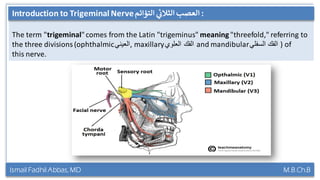
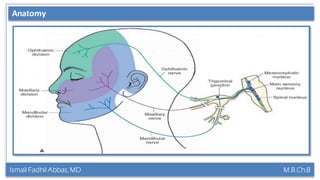
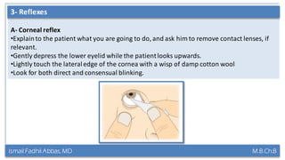
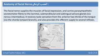
Lecture Title:
Trigeminal & Facial Nerve Clinically
Ismail Fadhil Abbas
M.B.Ch.B / Kirkuk Medicine College
Certified International Medical Education Trainer (TMET)
Certified International Trainer (TNT)
Certified Local Human Rights Trainer (TNHRT)
Ismail Fadhil Abbas, MD M.B.Ch.B
Contents of the lecture:
1. Introductionto Trigeminalnerve.
2. Anatomyof Trigeminalnerve.
3. Examinationof Trigeminalnerve.
4. Trigeminalnerve Clinically.
5. Anatomyof Facial nerve.
6. Examinationof Facial nerve.
7. Facial nerve Clinically.
Contents
Ismail Fadhil Abbas, MD M.B.Ch.B
Introduction to Trigeminal nerve :‫ي‬‫ثالث‬ ‫عصب‬‫التؤائم‬
The term "trigeminal"comes from the Latin "trigeminus" meaning "threefold," referring to
the three divisions (ophthalmic‫,العيني‬ maxillary‫العلوي‬ ‫الفك‬ and mandibular‫السفلي‬ ‫الفك‬ ) of
this nerve.
Introduction to Trigeminal Nerve :‫ي‬‫الثالث‬ ‫العصب‬‫التؤائم‬
Ismail Fadhil Abbas, MD M.B.Ch.B
Anatomy of Trigeminal nerve
It provides sensationto the face, mouth and part of the dura, and motor supply to the
muscles of mastication.There are three major branches of the nerve:
• Ophthalmic(V1): conveyssensory informationfrom the scalp, forehead, upper parts of
the sinuses, upper eyelid, cornea of the eye & bridge of the nose.
• Maxillary(V2): transmits sensory informationfrom the lower eyelid,middle part of the
sinuses, nasal cavity and middle part of the nose, cheeks, upper lip & roof of the mouth.
• Mandibular(V3): communicates sensory information from the outer part of the ear, lower
part of the mouth, front and middle parts of the tongue, lower lip & chin. It also stimulates
movement of the muscles in the jaw (masticationmuscles) and some of the muscles within
the inner ear.
Anatomy
Ismail Fadhil Abbas, MD M.B.Ch.B
Anatomy of Trigeminal nerveAnatomy
Ismail Fadhil Abbas, MD M.B.Ch.B
Anatomy of Trigeminal nerve
Remember:
1. Always do the examination Bilaterally.
2. Always ask patientto compare.
3. C5 examinationcontainsSensory, motor and two reflexes.
Examination
Ismail Fadhil Abbas, MD M.B.Ch.B
Anatomy of Trigeminal nerve
A- Ask the patient to close his eyes and say ‘yes’ each time he feels you lightly touch them
using a cotton wool tip.
1- Sensory Examination
Ismail Fadhil Abbas, MD M.B.Ch.B
Anatomy of Trigeminal nerve
B- Repeat using a fresh neurologicalpin,e.g. Neurotip,to test superficial pain.
1- Sensory Examination
Ismail Fadhil Abbas, MD M.B.Ch.B
Anatomy of Trigeminal nerve
C- Nasaltickle test: use a wisp of cotton wool to ‘tickle’ the inside of each nostril.
1- Sensory Examination
Ismail Fadhil Abbas, MD M.B.Ch.B
Anatomy of Trigeminal nerve
A- Inspect for wasting of the muscles of mastication(most apparentin temporalis).
2- Motor Examination
Ismail Fadhil Abbas, MD M.B.Ch.B
Anatomy of Trigeminal nerve
B- Ask the patient to clench his teeth; feel the masseters & temporalis.
2- Motor Examination
Ismail Fadhil Abbas, MD M.B.Ch.B
Anatomy of Trigeminal nerve
C- Place your hand under the jaw to provide resistance; ask the patientto open his jaw.
2- Motor Examination
Ismail Fadhil Abbas, MD M.B.Ch.B
Anatomy of Trigeminal nerve
A- Corneal reflex
•Explainto the patient what you are going to do, and ask him to remove contact lenses, if
relevant.
•Gently depress the lower eyelid while the patientlooks upwards.
•Lightly touch the lateraledge of the cornea with a wisp of damp cotton wool
•Look for both direct and consensual blinking.
3- Reflexes
Ismail Fadhil Abbas, MD M.B.Ch.B
Anatomy of Trigeminal nerve
B- Jaw jerk
•Ask the patientto let his mouth hang loosely open.
•Place your forefinger in the midline between lower lip and chin.
•Percuss your finger gently with the tendon hammer in a downwards direction noting any
reflex closing of the jaw. An absent, or just present, reflex is normal.
3- Reflexes
Ismail Fadhil Abbas, MD M.B.Ch.B
Anatomy of Trigeminal nerveMacleod’s Clinical Examination
Ismail Fadhil Abbas, MD M.B.Ch.B
Anatomy of Trigeminal nerve
1- Unilateralloss of sensation:may result from direct injury in associationwith facial
fractures (particularly V2) or localinvasionby cancer.
2- Loss of the corneal reflex: Lesions in the cavernous sinus
3- Facial numbness and pain: could be due to
A- Trigeminalneuralgiacauses severe, lancinating(‫)طعنة‬ pain typicallyin distributionof V2
or V3, and is often due to neurovascularcompression.
B- Reactivation ofherpes varicellazoster virus (chickenpox) and it affects typicallyV1.
4- Weakness of the muscles of mastication:myastheniagravis (skeletal muscle weakness,
neuromuscular disease).
5- Brisk jaw jerk (hyper-reflexia) occurs in pseudobulbarpalsy(upper motor neuron lesion)
in cases of stroke, etc.
Clinical Points
Ismail Fadhil Abbas, MD M.B.Ch.B
Anatomy of Trigeminal nerve
1- Unilateralloss of sensation:may result from direct injury in associationwith facial
fractures (particularly V2) or localinvasionby cancer.
Clinical Points
Ismail Fadhil Abbas, MD M.B.Ch.B
Anatomy of Trigeminal nerve
2- Loss of the corneal reflex: Lesions in the cavernous sinus
Clinical Points
Ismail Fadhil Abbas, MD M.B.Ch.B
Anatomy of Trigeminal nerve
3- Facial numbness and pain: could be due to
A- Trigeminalneuralgiacauses severe, lancinating(‫)طعنة‬ pain typicallyin distributionof V2
or V3, and is often due to neurovascularcompression.
Clinical Points
Ismail Fadhil Abbas, MD M.B.Ch.B
Anatomy of Trigeminal nerve
3- Facial numbness and pain: could be due to
B- Reactivation ofherpes varicellazoster virus (chickenpox) and it affects typicallyV1.
Clinical Points
Ismail Fadhil Abbas, MD M.B.Ch.B
Anatomy of Trigeminal nerve
4- Weakness of the muscles of mastication:myastheniagravis (skeletal muscle weakness,
neuromuscular disease).
Clinical Points
Ismail Fadhil Abbas, MD M.B.Ch.B
Anatomy of Trigeminal nerve
5- Brisk jaw jerk (hyper-reflexia) occurs in pseudobulbarpalsy(upper motor neuron lesion)
in cases of stroke, etc.
Clinical Points
Ismail Fadhil Abbas, MD M.B.Ch.B
Introduction to Trigeminal nerve :‫ي‬‫ثالث‬ ‫عصب‬‫التؤائم‬
Ismail Fadhil Abbas, MD M.B.Ch.B
Introduction to Trigeminal nerve :‫ي‬‫ثالث‬ ‫عصب‬‫التؤائم‬
The facialnerve supplies the muscles of facial expression, and carries parasympathetic
secretomotor fibres to the lacrimal, submandibularand sublingualsalivaryglands (via
nervus intermedius). It receives taste sensation from the anterior two-thirds of the tongue
(via the chorda tympani branch), and also provides the efferent supply to several reflexes.
Anatomy of Facial Nerve :‫ي‬‫الوجه‬ ‫العصب‬
Ismail Fadhil Abbas, MD M.B.Ch.B
Anatomy of Trigeminal nerve
Remember:
1. Always do the examination Bilaterally.
2. Always compare.
3. C7 Examinationis usuallyconfined to motor function; taste is rarely tested.
Motor Examination
Ismail Fadhil Abbas, MD M.B.Ch.B
Anatomy of Trigeminal nerve
A- Inspect the face for asymmetry or differences in blinkingor eye closure on one side.
Note that minor facialasymmetry is common and rarely pathological.
B- Watch for spontaneousor involuntarymovement.
Motor Examination
Ismail Fadhil Abbas, MD M.B.Ch.B
Anatomy of Trigeminal nerve
C- Ask the patient to raise the eyebrows and observe for symmetrical wrinkling of the
forehead
Motor Examination
Ismail Fadhil Abbas, MD M.B.Ch.B
Anatomy of Trigeminal nerve
D- Demonstratebaring your teeth and ask the patientto mimic you. Look for asymmetry
Motor Examination
Ismail Fadhil Abbas, MD M.B.Ch.B
Anatomy of Trigeminal nerve
E- Test power by saying:‘Screw your eyes tightly shut and stop me from opening them
Motor Examination
Ismail Fadhil Abbas, MD M.B.Ch.B
Anatomy of Trigeminal nerve
F- Test power by saying: ‘Blow out your cheeks with your mouth closed’
Motor Examination
Ismail Fadhil Abbas, MD M.B.Ch.B
Anatomy of Trigeminal nerveMacleod’s Clinical Examination
Ismail Fadhil Abbas, MD M.B.Ch.B
Anatomy of Trigeminal nerve
1. Weakness of both upper and lower facialmuscles: unilaterallower motor neurone VII
nerve lesion (bell’s palsy, Ramsay Hunt syndrome, cerebellopontineangle tumours, e.g.
acoustic neuroma, trauma and parotidtumours.)
2. Weakness in the lower facial muscles only with relativesparing of the upper face:
unilateral VIInerve upper motor neurone lesions (because there is bilateralcortical
innervationof the upper facial muscles and unilateralcorticalinnervationof the lower
facial muscles).
3. Weakness Bilaterally:less common and occur in cases of Guillain–Barrésyndrome,
sarcoidosis, Lyme disease and HIV infection.
Clinical Points
Ismail Fadhil Abbas, MD M.B.Ch.B
Anatomy of Trigeminal nerve
1- Weakness of both upper and lower facialmuscles: unilaterallower motor neurone VII
nerve lesion (bell’s palsy) in which there is the patientis unable
to close his eye. As he tries, the eyeballrolls upwards, exposing the conjunctivabelow the
cornea.
Clinical Points
Ismail Fadhil Abbas, MD M.B.Ch.B
Anatomy of Trigeminal nerve
2- Weakness in the lower facial muscles only with relativesparing of the upper face:
unilateral VIInerve upper motor neurone lesions (because there is bilateralcortical
innervationof the upper facial muscles and unilateralcorticalinnervationof the lower
facial muscles).
Clinical Points
Ismail Fadhil Abbas, MD M.B.Ch.B
Anatomy of Trigeminal nerve
3- Weakness Bilaterally:less common and occur in cases of Guillain–Barrésyndrome,
sarcoidosis, Lyme disease and HIV infection.
Clinical Points
Ismail Fadhil Abbas, MD M.B.Ch.B
Introduction to Trigeminal nerve :‫ي‬‫ثالث‬ ‫عصب‬‫التؤائم‬
Ismail Fadhil Abbas, MD M.B.Ch.B
Introduction to Trigeminal nerve :‫ي‬‫ثالث‬ ‫عصب‬‫التؤائم‬
References:
1. Macleod's Clinical Examination, 13rd Edition.
2. KenHub.
3. Pictures from Macleod & Google.
For more questions contact me:
Mail: ismailfadhil1996@yahoo.com
Facebook: Ismail Fadhil Abbas
Instagram: Ismail_Fadhil96

Trigeminal & Facial Nerve Clinically

  • 1.
    Lecture Title: Trigeminal &Facial Nerve Clinically Ismail Fadhil Abbas M.B.Ch.B / Kirkuk Medicine College Certified International Medical Education Trainer (TMET) Certified International Trainer (TNT) Certified Local Human Rights Trainer (TNHRT)
  • 2.
    Ismail Fadhil Abbas,MD M.B.Ch.B Contents of the lecture: 1. Introductionto Trigeminalnerve. 2. Anatomyof Trigeminalnerve. 3. Examinationof Trigeminalnerve. 4. Trigeminalnerve Clinically. 5. Anatomyof Facial nerve. 6. Examinationof Facial nerve. 7. Facial nerve Clinically. Contents
  • 3.
    Ismail Fadhil Abbas,MD M.B.Ch.B Introduction to Trigeminal nerve :‫ي‬‫ثالث‬ ‫عصب‬‫التؤائم‬ The term "trigeminal"comes from the Latin "trigeminus" meaning "threefold," referring to the three divisions (ophthalmic‫,العيني‬ maxillary‫العلوي‬ ‫الفك‬ and mandibular‫السفلي‬ ‫الفك‬ ) of this nerve. Introduction to Trigeminal Nerve :‫ي‬‫الثالث‬ ‫العصب‬‫التؤائم‬
  • 4.
    Ismail Fadhil Abbas,MD M.B.Ch.B Anatomy of Trigeminal nerve It provides sensationto the face, mouth and part of the dura, and motor supply to the muscles of mastication.There are three major branches of the nerve: • Ophthalmic(V1): conveyssensory informationfrom the scalp, forehead, upper parts of the sinuses, upper eyelid, cornea of the eye & bridge of the nose. • Maxillary(V2): transmits sensory informationfrom the lower eyelid,middle part of the sinuses, nasal cavity and middle part of the nose, cheeks, upper lip & roof of the mouth. • Mandibular(V3): communicates sensory information from the outer part of the ear, lower part of the mouth, front and middle parts of the tongue, lower lip & chin. It also stimulates movement of the muscles in the jaw (masticationmuscles) and some of the muscles within the inner ear. Anatomy
  • 5.
    Ismail Fadhil Abbas,MD M.B.Ch.B Anatomy of Trigeminal nerveAnatomy
  • 6.
    Ismail Fadhil Abbas,MD M.B.Ch.B Anatomy of Trigeminal nerve Remember: 1. Always do the examination Bilaterally. 2. Always ask patientto compare. 3. C5 examinationcontainsSensory, motor and two reflexes. Examination
  • 7.
    Ismail Fadhil Abbas,MD M.B.Ch.B Anatomy of Trigeminal nerve A- Ask the patient to close his eyes and say ‘yes’ each time he feels you lightly touch them using a cotton wool tip. 1- Sensory Examination
  • 8.
    Ismail Fadhil Abbas,MD M.B.Ch.B Anatomy of Trigeminal nerve B- Repeat using a fresh neurologicalpin,e.g. Neurotip,to test superficial pain. 1- Sensory Examination
  • 9.
    Ismail Fadhil Abbas,MD M.B.Ch.B Anatomy of Trigeminal nerve C- Nasaltickle test: use a wisp of cotton wool to ‘tickle’ the inside of each nostril. 1- Sensory Examination
  • 10.
    Ismail Fadhil Abbas,MD M.B.Ch.B Anatomy of Trigeminal nerve A- Inspect for wasting of the muscles of mastication(most apparentin temporalis). 2- Motor Examination
  • 11.
    Ismail Fadhil Abbas,MD M.B.Ch.B Anatomy of Trigeminal nerve B- Ask the patient to clench his teeth; feel the masseters & temporalis. 2- Motor Examination
  • 12.
    Ismail Fadhil Abbas,MD M.B.Ch.B Anatomy of Trigeminal nerve C- Place your hand under the jaw to provide resistance; ask the patientto open his jaw. 2- Motor Examination
  • 13.
    Ismail Fadhil Abbas,MD M.B.Ch.B Anatomy of Trigeminal nerve A- Corneal reflex •Explainto the patient what you are going to do, and ask him to remove contact lenses, if relevant. •Gently depress the lower eyelid while the patientlooks upwards. •Lightly touch the lateraledge of the cornea with a wisp of damp cotton wool •Look for both direct and consensual blinking. 3- Reflexes
  • 14.
    Ismail Fadhil Abbas,MD M.B.Ch.B Anatomy of Trigeminal nerve B- Jaw jerk •Ask the patientto let his mouth hang loosely open. •Place your forefinger in the midline between lower lip and chin. •Percuss your finger gently with the tendon hammer in a downwards direction noting any reflex closing of the jaw. An absent, or just present, reflex is normal. 3- Reflexes
  • 15.
    Ismail Fadhil Abbas,MD M.B.Ch.B Anatomy of Trigeminal nerveMacleod’s Clinical Examination
  • 16.
    Ismail Fadhil Abbas,MD M.B.Ch.B Anatomy of Trigeminal nerve 1- Unilateralloss of sensation:may result from direct injury in associationwith facial fractures (particularly V2) or localinvasionby cancer. 2- Loss of the corneal reflex: Lesions in the cavernous sinus 3- Facial numbness and pain: could be due to A- Trigeminalneuralgiacauses severe, lancinating(‫)طعنة‬ pain typicallyin distributionof V2 or V3, and is often due to neurovascularcompression. B- Reactivation ofherpes varicellazoster virus (chickenpox) and it affects typicallyV1. 4- Weakness of the muscles of mastication:myastheniagravis (skeletal muscle weakness, neuromuscular disease). 5- Brisk jaw jerk (hyper-reflexia) occurs in pseudobulbarpalsy(upper motor neuron lesion) in cases of stroke, etc. Clinical Points
  • 17.
    Ismail Fadhil Abbas,MD M.B.Ch.B Anatomy of Trigeminal nerve 1- Unilateralloss of sensation:may result from direct injury in associationwith facial fractures (particularly V2) or localinvasionby cancer. Clinical Points
  • 18.
    Ismail Fadhil Abbas,MD M.B.Ch.B Anatomy of Trigeminal nerve 2- Loss of the corneal reflex: Lesions in the cavernous sinus Clinical Points
  • 19.
    Ismail Fadhil Abbas,MD M.B.Ch.B Anatomy of Trigeminal nerve 3- Facial numbness and pain: could be due to A- Trigeminalneuralgiacauses severe, lancinating(‫)طعنة‬ pain typicallyin distributionof V2 or V3, and is often due to neurovascularcompression. Clinical Points
  • 20.
    Ismail Fadhil Abbas,MD M.B.Ch.B Anatomy of Trigeminal nerve 3- Facial numbness and pain: could be due to B- Reactivation ofherpes varicellazoster virus (chickenpox) and it affects typicallyV1. Clinical Points
  • 21.
    Ismail Fadhil Abbas,MD M.B.Ch.B Anatomy of Trigeminal nerve 4- Weakness of the muscles of mastication:myastheniagravis (skeletal muscle weakness, neuromuscular disease). Clinical Points
  • 22.
    Ismail Fadhil Abbas,MD M.B.Ch.B Anatomy of Trigeminal nerve 5- Brisk jaw jerk (hyper-reflexia) occurs in pseudobulbarpalsy(upper motor neuron lesion) in cases of stroke, etc. Clinical Points
  • 23.
    Ismail Fadhil Abbas,MD M.B.Ch.B Introduction to Trigeminal nerve :‫ي‬‫ثالث‬ ‫عصب‬‫التؤائم‬
  • 24.
    Ismail Fadhil Abbas,MD M.B.Ch.B Introduction to Trigeminal nerve :‫ي‬‫ثالث‬ ‫عصب‬‫التؤائم‬ The facialnerve supplies the muscles of facial expression, and carries parasympathetic secretomotor fibres to the lacrimal, submandibularand sublingualsalivaryglands (via nervus intermedius). It receives taste sensation from the anterior two-thirds of the tongue (via the chorda tympani branch), and also provides the efferent supply to several reflexes. Anatomy of Facial Nerve :‫ي‬‫الوجه‬ ‫العصب‬
  • 25.
    Ismail Fadhil Abbas,MD M.B.Ch.B Anatomy of Trigeminal nerve Remember: 1. Always do the examination Bilaterally. 2. Always compare. 3. C7 Examinationis usuallyconfined to motor function; taste is rarely tested. Motor Examination
  • 26.
    Ismail Fadhil Abbas,MD M.B.Ch.B Anatomy of Trigeminal nerve A- Inspect the face for asymmetry or differences in blinkingor eye closure on one side. Note that minor facialasymmetry is common and rarely pathological. B- Watch for spontaneousor involuntarymovement. Motor Examination
  • 27.
    Ismail Fadhil Abbas,MD M.B.Ch.B Anatomy of Trigeminal nerve C- Ask the patient to raise the eyebrows and observe for symmetrical wrinkling of the forehead Motor Examination
  • 28.
    Ismail Fadhil Abbas,MD M.B.Ch.B Anatomy of Trigeminal nerve D- Demonstratebaring your teeth and ask the patientto mimic you. Look for asymmetry Motor Examination
  • 29.
    Ismail Fadhil Abbas,MD M.B.Ch.B Anatomy of Trigeminal nerve E- Test power by saying:‘Screw your eyes tightly shut and stop me from opening them Motor Examination
  • 30.
    Ismail Fadhil Abbas,MD M.B.Ch.B Anatomy of Trigeminal nerve F- Test power by saying: ‘Blow out your cheeks with your mouth closed’ Motor Examination
  • 31.
    Ismail Fadhil Abbas,MD M.B.Ch.B Anatomy of Trigeminal nerveMacleod’s Clinical Examination
  • 32.
    Ismail Fadhil Abbas,MD M.B.Ch.B Anatomy of Trigeminal nerve 1. Weakness of both upper and lower facialmuscles: unilaterallower motor neurone VII nerve lesion (bell’s palsy, Ramsay Hunt syndrome, cerebellopontineangle tumours, e.g. acoustic neuroma, trauma and parotidtumours.) 2. Weakness in the lower facial muscles only with relativesparing of the upper face: unilateral VIInerve upper motor neurone lesions (because there is bilateralcortical innervationof the upper facial muscles and unilateralcorticalinnervationof the lower facial muscles). 3. Weakness Bilaterally:less common and occur in cases of Guillain–Barrésyndrome, sarcoidosis, Lyme disease and HIV infection. Clinical Points
  • 33.
    Ismail Fadhil Abbas,MD M.B.Ch.B Anatomy of Trigeminal nerve 1- Weakness of both upper and lower facialmuscles: unilaterallower motor neurone VII nerve lesion (bell’s palsy) in which there is the patientis unable to close his eye. As he tries, the eyeballrolls upwards, exposing the conjunctivabelow the cornea. Clinical Points
  • 34.
    Ismail Fadhil Abbas,MD M.B.Ch.B Anatomy of Trigeminal nerve 2- Weakness in the lower facial muscles only with relativesparing of the upper face: unilateral VIInerve upper motor neurone lesions (because there is bilateralcortical innervationof the upper facial muscles and unilateralcorticalinnervationof the lower facial muscles). Clinical Points
  • 35.
    Ismail Fadhil Abbas,MD M.B.Ch.B Anatomy of Trigeminal nerve 3- Weakness Bilaterally:less common and occur in cases of Guillain–Barrésyndrome, sarcoidosis, Lyme disease and HIV infection. Clinical Points
  • 36.
    Ismail Fadhil Abbas,MD M.B.Ch.B Introduction to Trigeminal nerve :‫ي‬‫ثالث‬ ‫عصب‬‫التؤائم‬
  • 37.
    Ismail Fadhil Abbas,MD M.B.Ch.B Introduction to Trigeminal nerve :‫ي‬‫ثالث‬ ‫عصب‬‫التؤائم‬
  • 38.
    References: 1. Macleod's ClinicalExamination, 13rd Edition. 2. KenHub. 3. Pictures from Macleod & Google.
  • 39.
    For more questionscontact me: Mail: ismailfadhil1996@yahoo.com Facebook: Ismail Fadhil Abbas Instagram: Ismail_Fadhil96