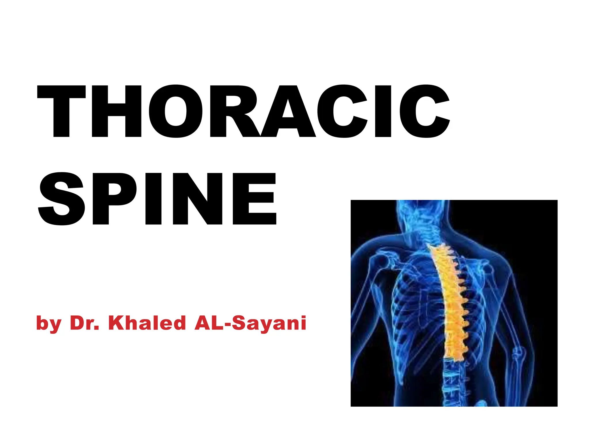
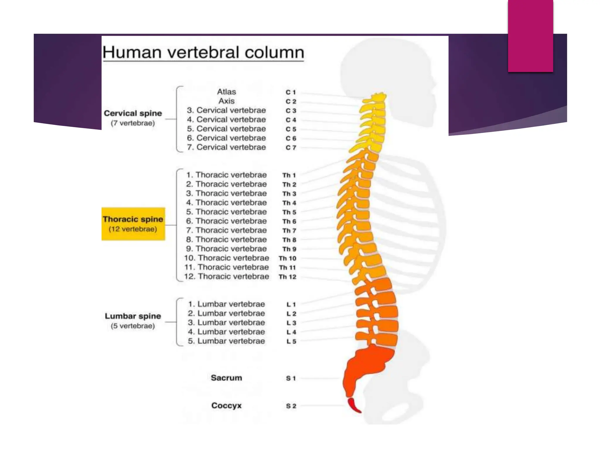
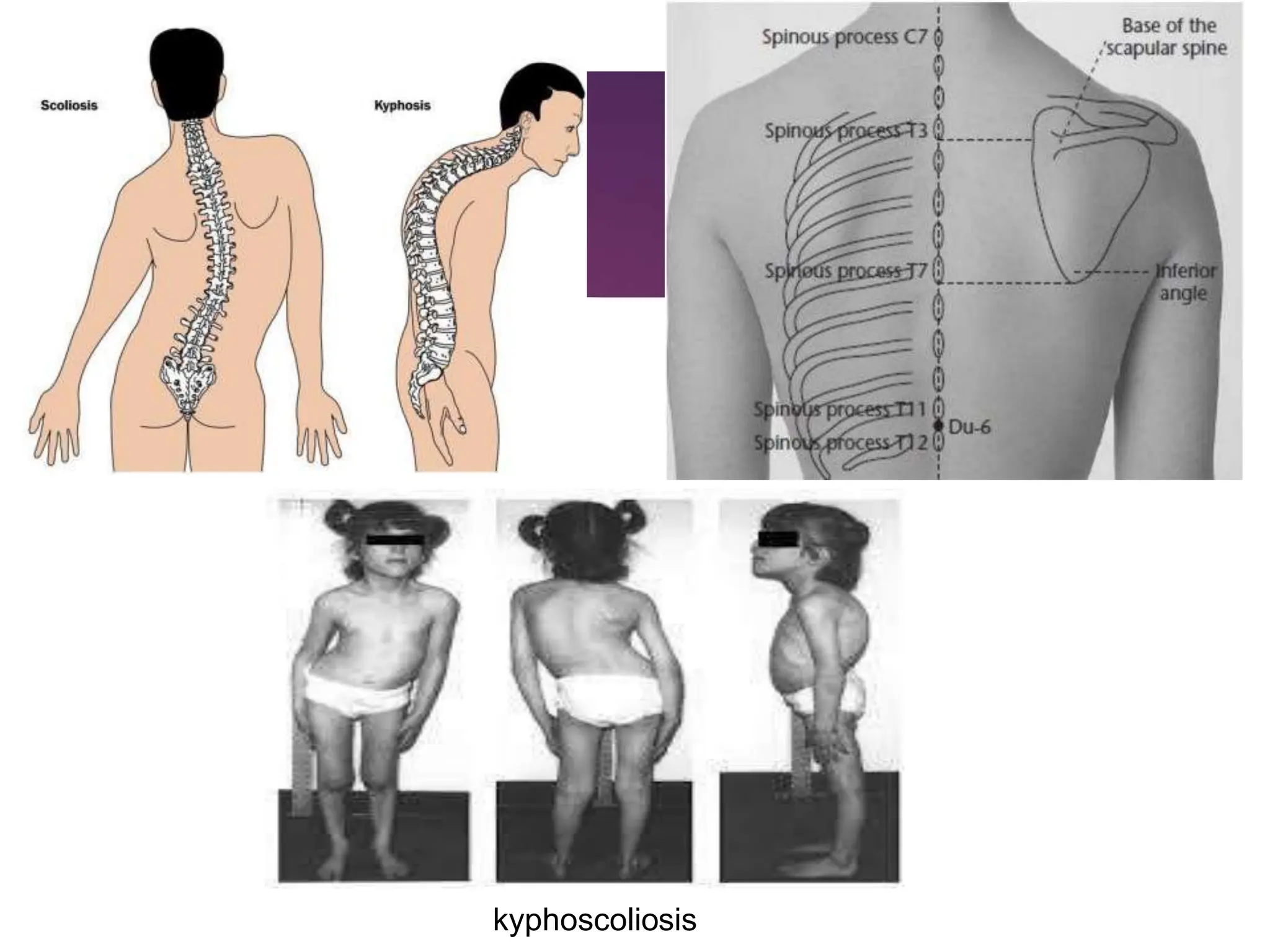
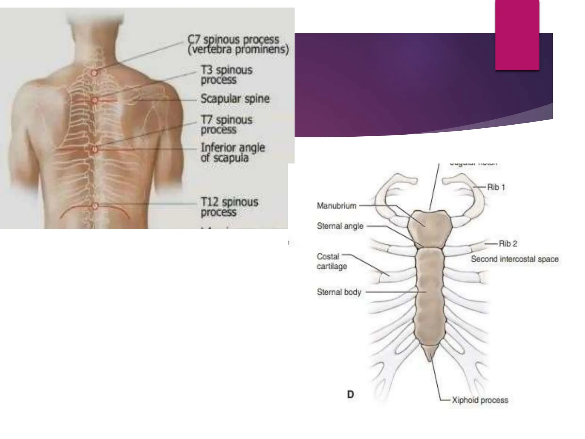
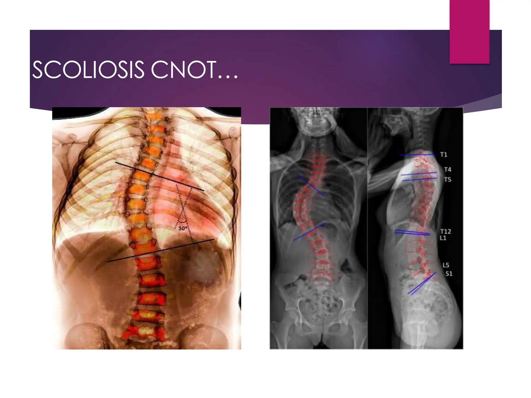
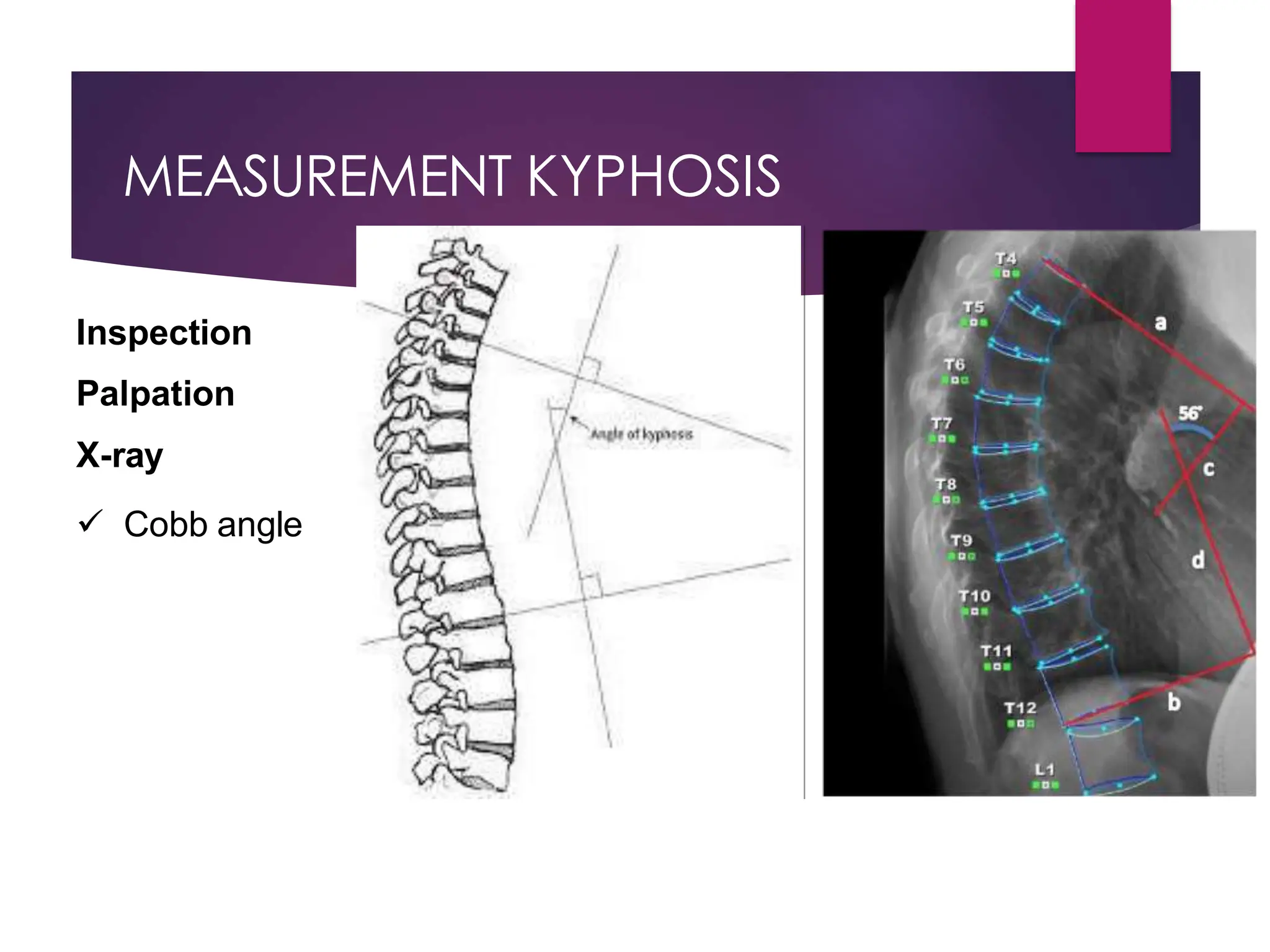
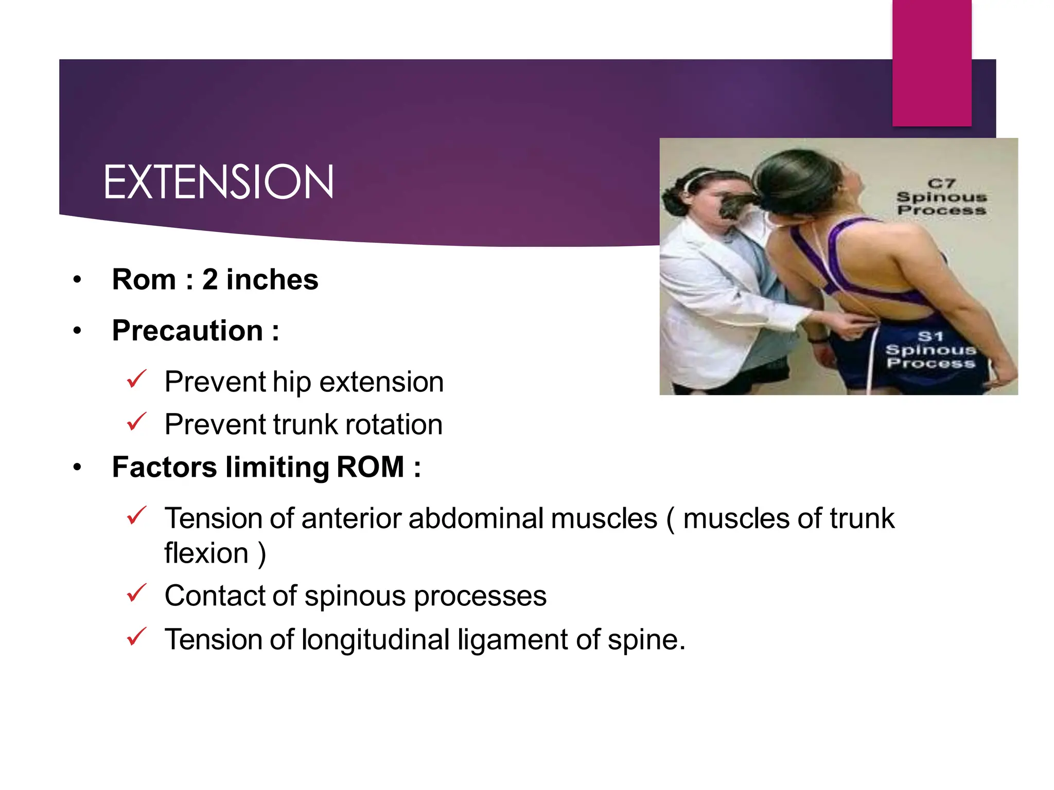
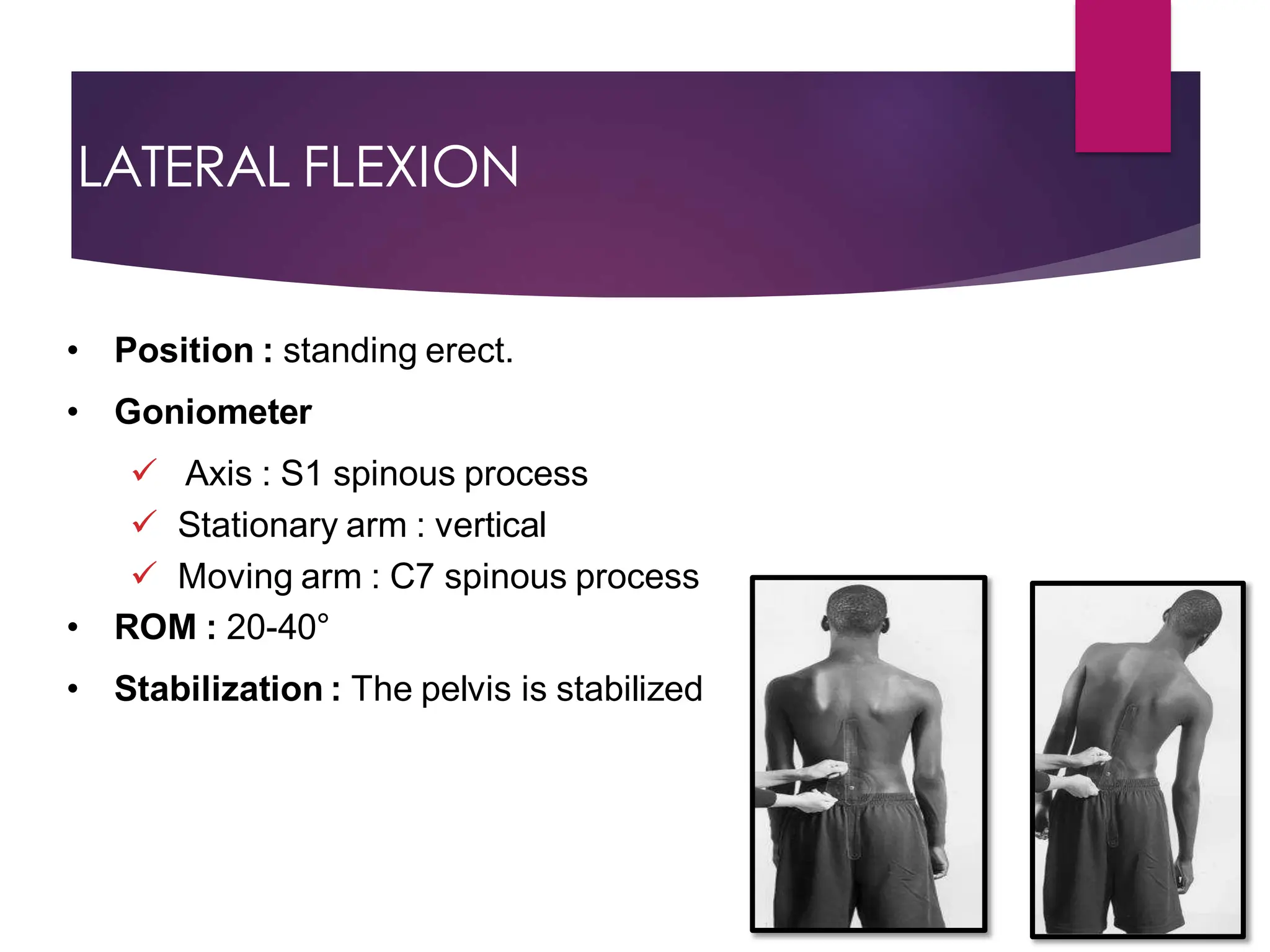
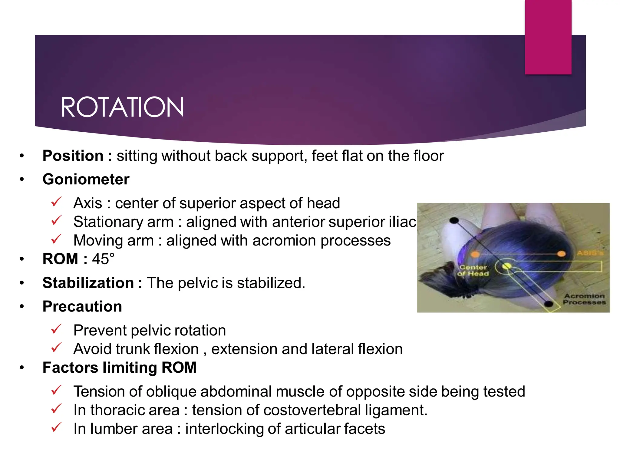
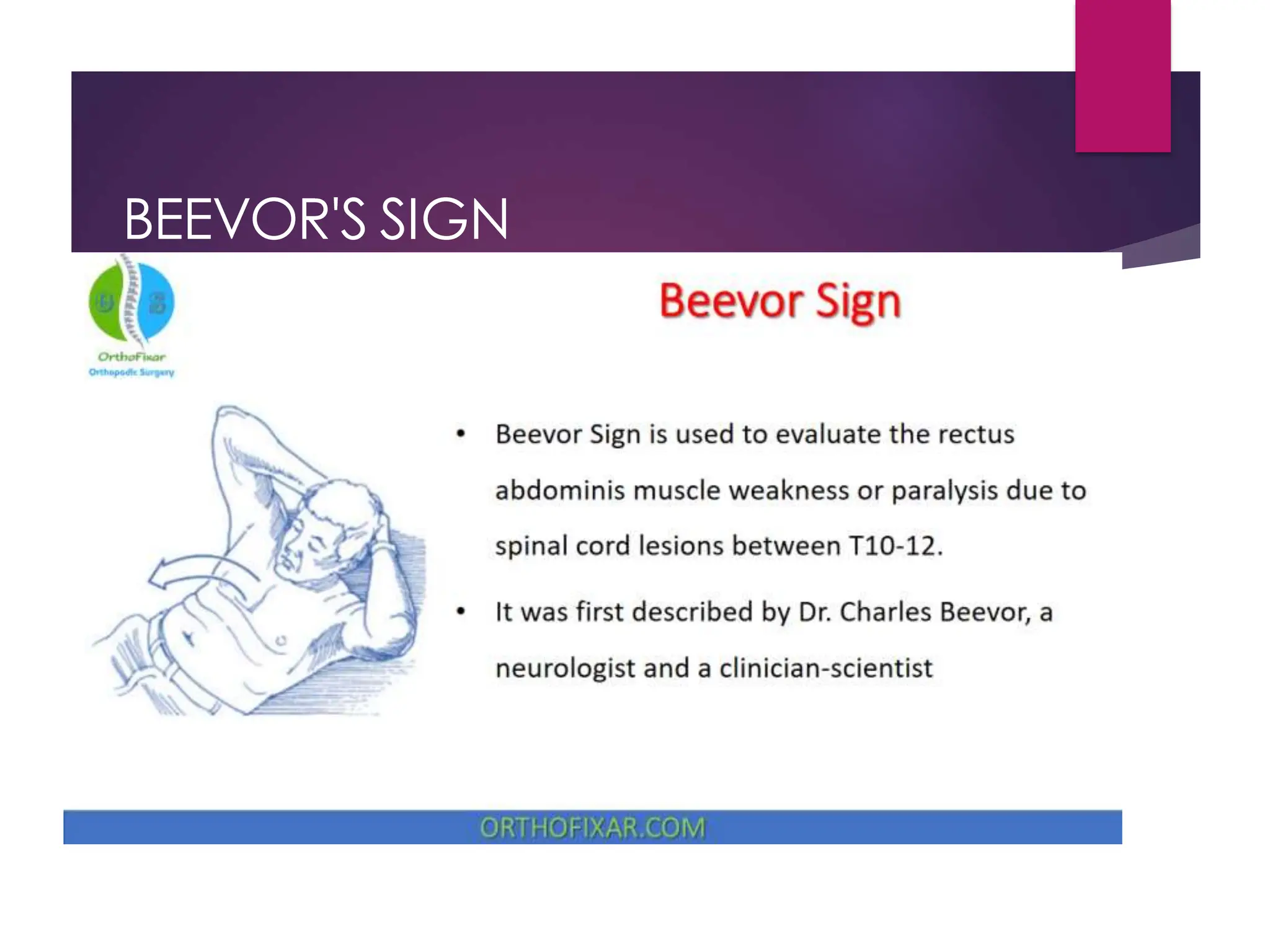
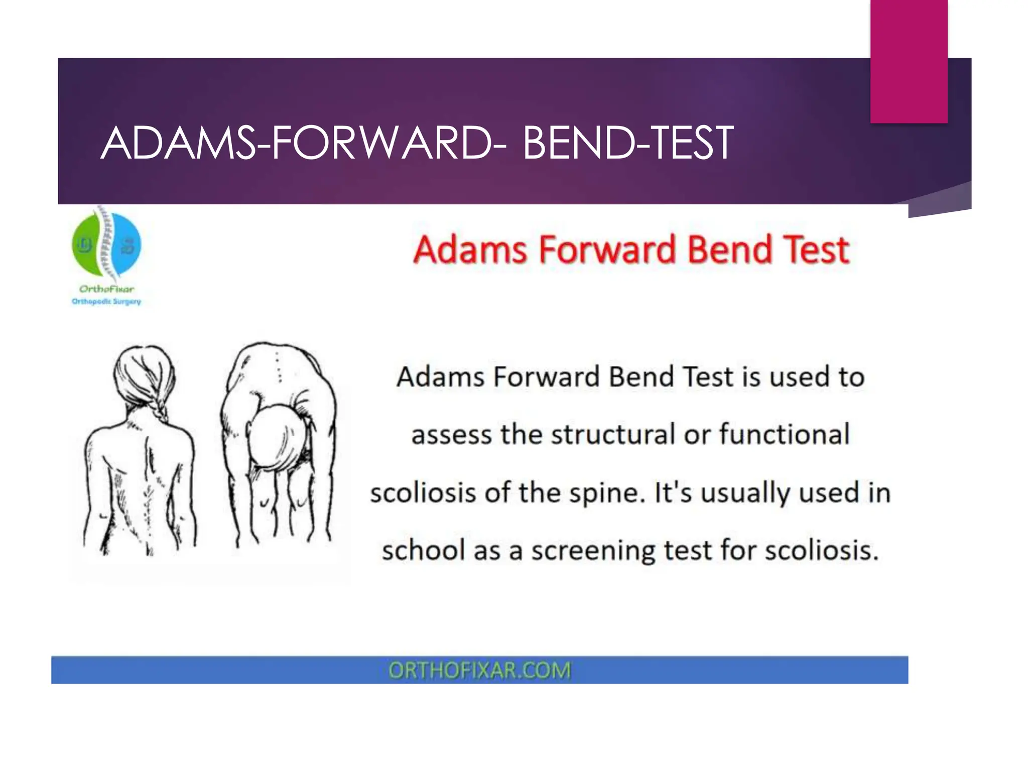
THORACIC
SPINE
by Dr. Khaled AL-Sayani
THORACIC SPAIN
 It is the second segment
 Located between the cervical and lumbar vertebral
segment
 Consist of 12 vertebrae , separated by
intervertebral disc
 Also from part of thoracic cage
 It lies in the upper back and provide attachment for
the ribs
 Natural , slight kyphotic curve exist in T-spain
OBSERVATION
From the side
 Kyphosis (posterior convexity of the spine)
• Senile kyphosis ( with osteoporosis , osteomalacia or
pathological fracture)
• scheuermann’s disease ( osteochondritis involving one or
more of the vertebrae)
• Gibbus (angular kyphosis )
• Fracture
• Tuberculosis of the spine
• Congenital abnormality
.
OBSERVATION
From the behind
 Listing of trunk (due to muscle spasm )
 Scoliosis ( lateral curvature of spine )
• Postural : scoliosis disappears with forward flexion of the
spine
• Structural : scoliosis persists with forward flexion of the
spine and a rib hump presents
 Scapula ( spina scapula T3 , end of scapula T7-T9 ,Medial
border of the scapula and spinous processes 5 cm).
 Soft tissues (Skin color , Swelling and scars)
kyphoscoliosis
PALPATION
• Skin temperature
• Soft tissues
• Tenderness
• Increased or decreased prominence of bones, the position of
vertebrae and ribs
• Supine- sternum, ribs, clavicle, sternocostal and
costochondral joints
• Prone- spinous process, costotransverse and costovertebral
MEASUREMENT SCOLIOSIS
Inspection :
 One shoulder blade being more prominent than the other
 Leaning to one side
Palpation :
 Uneven shoulders, hips, waist, legs or rib cage
Special Tests :
 Adam’s forward bend test
X.ray :
 Cobb angle
 ‘significant’ 25-30 degrees
 ‘severe’ 45-50 degrees.
SCOLIOSIS CNOT…
MEASUREMENT KYPHOSIS
Inspection
Palpation
X-ray
 Cobb angle
FLEXION
• Starting position : The tape is placed proximally on the
spinous process of C7 and distal to S1.
• Ending position : following flexion of the vertebral
using the same land mark
• ROM : Approximately 4 inches
• Precaution : The pelvic and hip joints are stabilized
• Factors limiting ROM :
 Tension of spinal extensor muscles
 Contact of last ribs with abdomen
 Bone blockage : contact of lips of vertebral bodies
with subjacent vertebrae anterriorly
 Tension of ligaments: Posterior longitudinal,
Inter spinal ligament , supra spinal ligament.
EXTENSION
• Rom : 2 inches
• Precaution :
 Prevent hip extension
 Prevent trunk rotation
• Factors limiting ROM :
 Tension of anterior abdominal muscles ( muscles of trunk
flexion )
 Contact of spinous processes
 Tension of longitudinal ligament of spine.
LATERAL FLEXION
• Position : standing erect.
• Goniometer
 Axis : S1 spinous process
 Stationary arm : vertical
 Moving arm : C7 spinous process
• ROM : 20-40°
• Stabilization : The pelvis is stabilized
ROTATION
• Position : sitting without back support, feet flat on the floor
• Goniometer
 Axis : center of superior aspect of head
 Stationary arm : aligned with anterior superior iliac spines
 Moving arm : aligned with acromion processes
• ROM : 45°
• Stabilization : The pelvic is stabilized.
• Precaution
 Prevent pelvic rotation
 Avoid trunk flexion , extension and lateral flexion
• Factors limiting ROM
 Tension of oblique abdominal muscle of opposite side being tested
 In thoracic area : tension of costovertebral ligament.
 In lumber area : interlocking of articular facets
BEEVOR'S SIGN
ADAMS-FORWARD- BEND-TEST
 The patient takes off his/her t-shirt so that the spine is
visible.
 The patient needs to bend forward, starting at the waist
until the back comes in the horizontal plane, with the
feet together, arms hanging and the knees in
extension. The palms are held together.
 The examiner stands at the back of the patient and
looks along the horizontal plane of the spine, searching
for abnormalities of the spinal curve, like increased or
decreased lordosis/ kyphosis, and an asymmetry of the
trunk.
 A positive test is indicated if asymmetry is observed
(one side of the spine is higher than the other).
thoracic assessment.pdf  طبعي تقيم الصدر علاج

thoracic assessment.pdf طبعي تقيم الصدر علاج

  • 1.
  • 2.
    THORACIC SPAIN  Itis the second segment  Located between the cervical and lumbar vertebral segment  Consist of 12 vertebrae , separated by intervertebral disc  Also from part of thoracic cage  It lies in the upper back and provide attachment for the ribs  Natural , slight kyphotic curve exist in T-spain
  • 4.
    OBSERVATION From the side Kyphosis (posterior convexity of the spine) • Senile kyphosis ( with osteoporosis , osteomalacia or pathological fracture) • scheuermann’s disease ( osteochondritis involving one or more of the vertebrae) • Gibbus (angular kyphosis ) • Fracture • Tuberculosis of the spine • Congenital abnormality .
  • 5.
    OBSERVATION From the behind Listing of trunk (due to muscle spasm )  Scoliosis ( lateral curvature of spine ) • Postural : scoliosis disappears with forward flexion of the spine • Structural : scoliosis persists with forward flexion of the spine and a rib hump presents  Scapula ( spina scapula T3 , end of scapula T7-T9 ,Medial border of the scapula and spinous processes 5 cm).  Soft tissues (Skin color , Swelling and scars)
  • 6.
  • 7.
    PALPATION • Skin temperature •Soft tissues • Tenderness • Increased or decreased prominence of bones, the position of vertebrae and ribs • Supine- sternum, ribs, clavicle, sternocostal and costochondral joints • Prone- spinous process, costotransverse and costovertebral
  • 9.
    MEASUREMENT SCOLIOSIS Inspection : One shoulder blade being more prominent than the other  Leaning to one side Palpation :  Uneven shoulders, hips, waist, legs or rib cage Special Tests :  Adam’s forward bend test X.ray :  Cobb angle  ‘significant’ 25-30 degrees  ‘severe’ 45-50 degrees.
  • 10.
  • 11.
  • 12.
    FLEXION • Starting position: The tape is placed proximally on the spinous process of C7 and distal to S1. • Ending position : following flexion of the vertebral using the same land mark • ROM : Approximately 4 inches • Precaution : The pelvic and hip joints are stabilized • Factors limiting ROM :  Tension of spinal extensor muscles  Contact of last ribs with abdomen  Bone blockage : contact of lips of vertebral bodies with subjacent vertebrae anterriorly  Tension of ligaments: Posterior longitudinal, Inter spinal ligament , supra spinal ligament.
  • 14.
    EXTENSION • Rom :2 inches • Precaution :  Prevent hip extension  Prevent trunk rotation • Factors limiting ROM :  Tension of anterior abdominal muscles ( muscles of trunk flexion )  Contact of spinous processes  Tension of longitudinal ligament of spine.
  • 15.
    LATERAL FLEXION • Position: standing erect. • Goniometer  Axis : S1 spinous process  Stationary arm : vertical  Moving arm : C7 spinous process • ROM : 20-40° • Stabilization : The pelvis is stabilized
  • 16.
    ROTATION • Position :sitting without back support, feet flat on the floor • Goniometer  Axis : center of superior aspect of head  Stationary arm : aligned with anterior superior iliac spines  Moving arm : aligned with acromion processes • ROM : 45° • Stabilization : The pelvic is stabilized. • Precaution  Prevent pelvic rotation  Avoid trunk flexion , extension and lateral flexion • Factors limiting ROM  Tension of oblique abdominal muscle of opposite side being tested  In thoracic area : tension of costovertebral ligament.  In lumber area : interlocking of articular facets
  • 17.
  • 18.
  • 19.
     The patienttakes off his/her t-shirt so that the spine is visible.  The patient needs to bend forward, starting at the waist until the back comes in the horizontal plane, with the feet together, arms hanging and the knees in extension. The palms are held together.  The examiner stands at the back of the patient and looks along the horizontal plane of the spine, searching for abnormalities of the spinal curve, like increased or decreased lordosis/ kyphosis, and an asymmetry of the trunk.  A positive test is indicated if asymmetry is observed (one side of the spine is higher than the other).