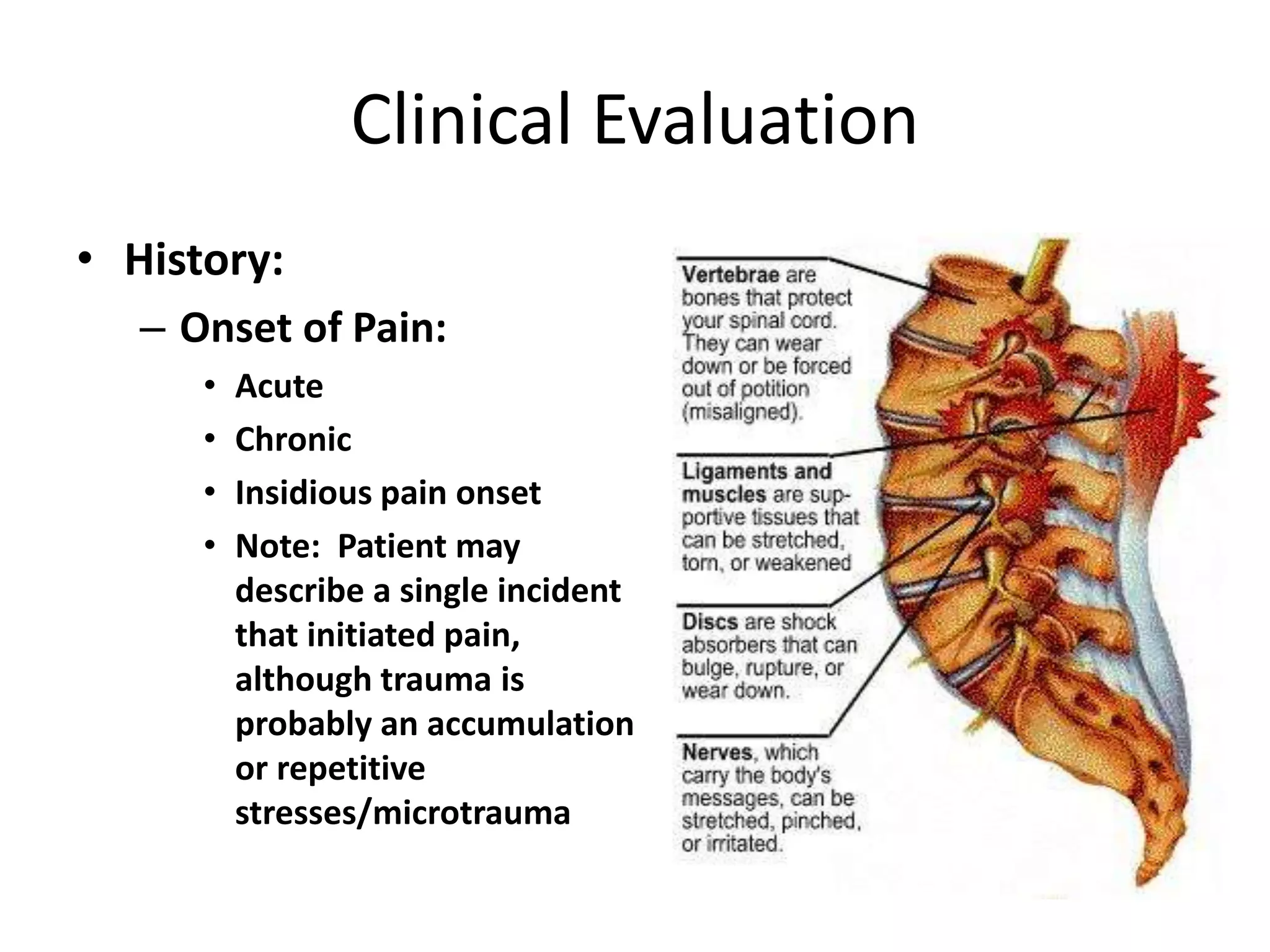
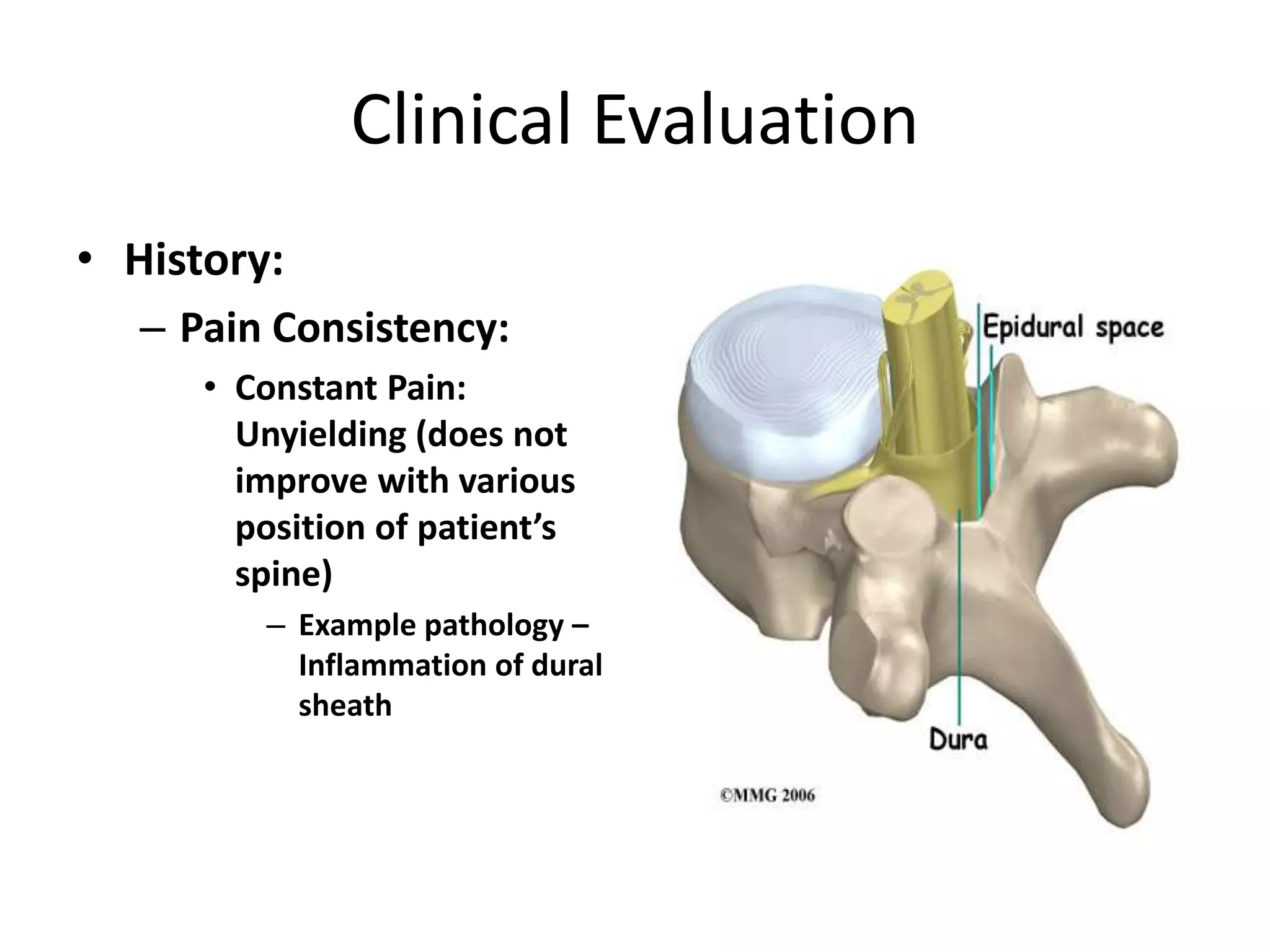
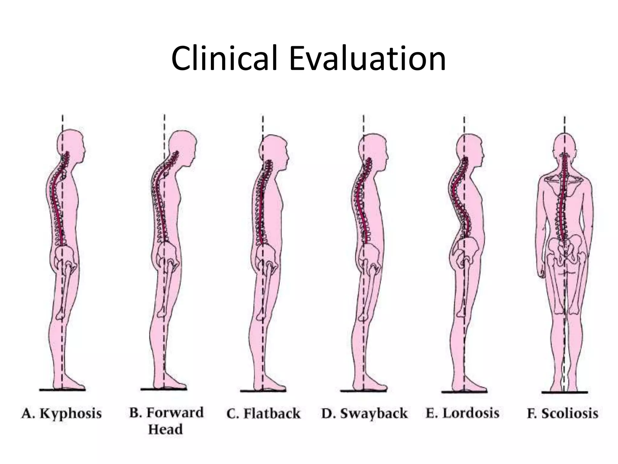
Thoracic and Lumbar Spine
Clinical Evaluation
Clinical Evaluation
• History:
– Location of Pain:
• Pain radiating into extremities
• Peripheral paresthesia or numbness:
– Result of impingement or pressure on nerve root exiting
intervertebral foramen or dural irritation proximal to pain
site
• Pain Locations:
– Lumbar pain – possible ambiguous cause
– Sacroiliac pathology – pain around PSIS or radiating pain in
hip/groin
– Piriformis spasm – symptoms of sciatic nerve dysfunction
Clinical Evaluation
Clinical Evaluation
• History:
– Onset of Pain:
• Acute
• Chronic
• Insidious pain onset
• Note: Patient may
describe a single incident
that initiated pain,
although trauma is
probably an accumulation
or repetitive
stresses/microtrauma
Clinical Evaluation
• History:
– Mechanism of Injury:
• Movement: Flexion,
Extension, Lateral
Bending, Rotation
• Blunt Trauma: Direct
blow to lumbar/thoracic
area
– Contusions
• Compressive Stress:
– Hyperextension of
spine
Clinical Evaluation
• History:
– Pain Consistency:
• Constant Pain:
Unyielding (does not
improve with various
position of patient’s
spine)
– Example pathology –
Inflammation of dural
sheath
Clinical Evaluation
• History:
– Pain Consistency:
• Intermittent Pain:
– Mechanical Origin – certain spinal positions may ↑ or ↓
pain symptoms
» Compression/stretching of nerve root – Increase pain
» Positioning (flexion, traction) – lessen the pressure on
involved structure
Clinical Evaluation
• History:
– Bowel or bladder signs:
• Does the patient have any bowel or bladder
problems?
• Incontinence: Loss of bowel or bladder control
– May indicate lower nerve root lesions (cauda equina
syndrome), or spinal cord injury
– Description: urinary incontinence may range from
occasionally leaking urine (during cough/sneeze) to having
sudden episodes of strong urinary urgency
• History:
– Bowel or Bladder Signs:
• Cauda Equina Syndrome:
– Nerves within the spinal canal have been damaged
– Result: nerves supplying the muscles of the legs, bladder, bowel
and genitals do not function properly
» Patients experience numbness, loss of sensation and pain in the
legs, buttocks and pelvic region (damage usually permanent)
– Causes:
» Spina bifida (abnormality in closure of spinal canal)
» Tumors
» Injury (spinal fractures)
» Intravertebral disc herniation
» Vascular (blood vessel) problems or infections of the cauda equina
Clinical Evaluation
• History:
– History of spinal injury:
• Previous injuries:
– Structural degeneration
– Predisposition to injury
– Changes in activity:
• Exercise habits (intensity
levels, duration,
frequency)
• Footwear, running
surfaces
• New bed
Clinical Evaluation
• General Inspection:
– Frontal Curvature:
• Alignment of lumbar,
thoracic, cervical vertebrae
with patient lying prone or
standing
– Normal alignment –
straight
– Abnormal alignment:
» Scoliosis – lateral
curvature (lumbar
and/or thoracic spine)
Clinical Evaluation
• General Inspection: Scoliosis
– Signs and symptoms:
• Uneven shoulders
• One shoulder blade appears more
prominent
• Uneven waist / 1 hip higher vs.
other
• Leaning to one side
• Back pain and difficulty breathing
(severe scoliosis)
– Causes:
• Idiopathic (85% of cases)
• Underlying neuromuscular disease,
leg-length discrepancy, birth defect,
fetal development (congenital)
• Not caused by poor posture, diet,
exercise, or the use of backpacks
Clinical Evaluation
• Diagnosis:
– Angle: X-ray
• Normal Spine (0
degrees)
• Scoliosis: (> 10
degrees)
– Complications: (severe
scoliosis)
• Lung and heart
damage: compression
of rib cage against
heart, lungs
– > 70 degrees
• Back problems
Clinical Evaluation
• General Inspection:
– Scoliosis Test: Adam’s Forward Bend Test
• Patient Position: Standing with hands held in front (arms straight)
• Evaluation Procedure: Patient bends forward, sliding hands down
the front of each leg
• Positive Test:
– Asymmetrical hump along lateral aspect of thoracolumbar spine
– One shoulder blade appears more prominent
– Uneven hips
• Implications:
– Functional scoliosis: scoliosis present when patient stands straight,
disappears during flexion
– Structural scoliosis: present during both standing and with flexion
Clinical Evaluation
Clinical Evaluation
• General Inspection:
– Sagital Curvature:
• Normal Alignment:
– Lordotic cervical
– Kyphotic thoracic
– Lordotic lumbar
– Kyphotic sacral
Clinical Evaluation
Clinical Evaluation
• General Inspection:
– Observation of GAIT:
• Spinal pain –
influence on walking
and running gait
– Slouching
– Shuffling
– Shortened gait
 Walking on tip toe – S1 weakness
 Walking on heels – L5 weakness
Clinical Evaluation
• General Inspection:
– Skin Markings:
• presence of darkened
areas of skin
pigmentation
– Normal
– Collagen disease
Clinical Evaluation
• General Inspection:
– Breathing patterns:
• Irregular breathing (i.e. shallow respirations, pain)
– Injury to thoracic vertebrae
– Pressure on thoracic nerves
– Trauma to ribs, costal cartilage
Clinical Evaluation
• General Inspection:
– Kyphosis:
• Abnormal forward rounding
of the upper back (> 40 to 45
degrees)
• Round back or hunchback
• Causes:
– Developmental problems,
degenerative diseases
(arthritis), osteoporosis with
compression fractures,
trauma
– Severe cases:
» Can affect lungs, nerves,
causing pain and other
problems
Clinical Evaluation
• General Inspection:
– Kyphosis Test: Forward
bend test
• Patient bends forward
from the waist views the
spine from the side
– With kyphosis, the rounding
of the upper back may
become more obvious in this
position
– Postural kyphosis – the
deformity corrects itself when
patient lies on their back
Clinical Evaluation
• Postural kyphosis:
– May improve on its own
• Exercises to strengthen back muscles, correct posture, and
sleeping on a firm bed
• Structural kyphosis:
– Caused by spinal abnormalities
– Scheuermann's disease:
• Developmental disorder that causes a stooped forward or bent-
over posture
• Affects between 0.5% and 8% of the general population
• Osteoporosis-related kyphosis:
– Multiple compression fractures
• Low bone density
Clinical Evaluation
Clinical Evaluation
• General Inspection:
– Movement and Posture:
• Poor posture (standing,
sitting, bending)
– Lordotic Curve:
• Reduction:
– Muscle spasm
– Hamstring tightness
• Increased:
– Hip flexor tightness
– Abdominal weakness
Clinical Evaluation
Clinical Evaluation
• General Inspection:
– Standing Posture:
• Lateral shift in trunk and pelvis
– Nerve root impingement (lateral shift ↓ pressure)
– Erector Spinae Muscle Tone:
• Unilateral hypertrophy or atrophy
Clinical Evaluation
• Palpation: Thoracic Spine
– Spinous Processes
– Supraspinous Ligaments:
• Fills space between the spinous processes
– Costovertebral Junction:
• Articulation between ribs and thoracic vertebrae
– Trapezius:
• Origin to insertion
• Rhomboids and levator scapulae lie deep to middle/upper traps
– Paravertebral Muscles
– Scapular Muscles
• 1 – Spinous Processes
• 2 – Supraspinous
Ligaments
• 3 – Costovertebral
Junction
• 4 – Trapezius
• 5 – Paravertebral
Muscles
• 6 – Scapular Muscles
Structure Landmark
Cervical vertebral bodies Same level as spinous processes
C1 transverse process One finger’s breadth inferior to mastoid process
C3-C4 vertebrae Posterior to hyoid bone
C4-C5 vertebrae Posterior to thyroid cartilage
C6 vertebrae Posterior to cricoid cartilage; moves during flexion and
extension of cervical spine
C7 vertebrae Prominent posterior spinous process
T1 vertebrae Prominent protrusion inferior to cervical spine
T2 vertebrae Posterior from jugular notch of the sternum
T3 vertebrae Even with the medial border of the scapular spine
T7 vertebrae Even with the inferior angle of the scapula
L3 vertebrae Posterior from the umbilicus
L4 vertebrae Level with the iliac crest
L5 vertebrae Typically demarcated by bilateral dimples, but variable
from person to person
S2 At level of the posterior superior iliac spine
Clinical Evaluation
C7
T1
T2
T3
T4
T5
• 1 – Spinous Processes
• 2 – Step-off Deformity
• 3 – Paravertebral Muscles
Clinical Evaluation
• Spondylolisthesis:
– Forward slippage of a vertebrae on the one below it
• L4 and L5 / L5 and S1
– Affects 5-6% of males, 2-3% of females
– Causes:
• Strenuous physical activity (weightlifting, gymnastics, football)
– Types:
• Developmental:
– May exist at birth, or may develop during childhood (generally not
noticed until later in childhood/adult life)
• Acquired:
– Degeneration: caused by the daily stresses that are put on spine
(i.e. carrying heavy items, physical sports)
» Connections between the vertebrae weaken
– Single or repeated force
Clinical Evaluation
• Spondylolisthesis:
– Grade 1:
• 25% of vertebral body has
slipped forward
– Grade 2:
• 50%
– Grade 3:
• 75%
– Grade 4:
• 100%
– Grade 5:
• Vertebral body completely
fallen off
(i.e.,spondyloptosis)
Clinical Evaluation
• Symptoms:
– May be asymptomatic
– Low back pain (especially
after exercise)
– ↑ lordosis
– Pain/weakness in one or
both legs
– ↓ ability to control bowel/
bladder functions
– Tight hamstrings
– Advanced spondylolisthesis:
changes may occur in the
way patient stands/walks

thoracic and lumbar spine-1.pptx

  • 1.
    Thoracic and LumbarSpine Clinical Evaluation
  • 2.
    Clinical Evaluation • History: –Location of Pain: • Pain radiating into extremities • Peripheral paresthesia or numbness: – Result of impingement or pressure on nerve root exiting intervertebral foramen or dural irritation proximal to pain site • Pain Locations: – Lumbar pain – possible ambiguous cause – Sacroiliac pathology – pain around PSIS or radiating pain in hip/groin – Piriformis spasm – symptoms of sciatic nerve dysfunction
  • 3.
  • 4.
    Clinical Evaluation • History: –Onset of Pain: • Acute • Chronic • Insidious pain onset • Note: Patient may describe a single incident that initiated pain, although trauma is probably an accumulation or repetitive stresses/microtrauma
  • 5.
    Clinical Evaluation • History: –Mechanism of Injury: • Movement: Flexion, Extension, Lateral Bending, Rotation • Blunt Trauma: Direct blow to lumbar/thoracic area – Contusions • Compressive Stress: – Hyperextension of spine
  • 7.
    Clinical Evaluation • History: –Pain Consistency: • Constant Pain: Unyielding (does not improve with various position of patient’s spine) – Example pathology – Inflammation of dural sheath
  • 8.
    Clinical Evaluation • History: –Pain Consistency: • Intermittent Pain: – Mechanical Origin – certain spinal positions may ↑ or ↓ pain symptoms » Compression/stretching of nerve root – Increase pain » Positioning (flexion, traction) – lessen the pressure on involved structure
  • 9.
    Clinical Evaluation • History: –Bowel or bladder signs: • Does the patient have any bowel or bladder problems? • Incontinence: Loss of bowel or bladder control – May indicate lower nerve root lesions (cauda equina syndrome), or spinal cord injury – Description: urinary incontinence may range from occasionally leaking urine (during cough/sneeze) to having sudden episodes of strong urinary urgency
  • 10.
    • History: – Bowelor Bladder Signs: • Cauda Equina Syndrome: – Nerves within the spinal canal have been damaged – Result: nerves supplying the muscles of the legs, bladder, bowel and genitals do not function properly » Patients experience numbness, loss of sensation and pain in the legs, buttocks and pelvic region (damage usually permanent) – Causes: » Spina bifida (abnormality in closure of spinal canal) » Tumors » Injury (spinal fractures) » Intravertebral disc herniation » Vascular (blood vessel) problems or infections of the cauda equina
  • 11.
    Clinical Evaluation • History: –History of spinal injury: • Previous injuries: – Structural degeneration – Predisposition to injury – Changes in activity: • Exercise habits (intensity levels, duration, frequency) • Footwear, running surfaces • New bed
  • 12.
    Clinical Evaluation • GeneralInspection: – Frontal Curvature: • Alignment of lumbar, thoracic, cervical vertebrae with patient lying prone or standing – Normal alignment – straight – Abnormal alignment: » Scoliosis – lateral curvature (lumbar and/or thoracic spine)
  • 13.
    Clinical Evaluation • GeneralInspection: Scoliosis – Signs and symptoms: • Uneven shoulders • One shoulder blade appears more prominent • Uneven waist / 1 hip higher vs. other • Leaning to one side • Back pain and difficulty breathing (severe scoliosis) – Causes: • Idiopathic (85% of cases) • Underlying neuromuscular disease, leg-length discrepancy, birth defect, fetal development (congenital) • Not caused by poor posture, diet, exercise, or the use of backpacks
  • 14.
    Clinical Evaluation • Diagnosis: –Angle: X-ray • Normal Spine (0 degrees) • Scoliosis: (> 10 degrees) – Complications: (severe scoliosis) • Lung and heart damage: compression of rib cage against heart, lungs – > 70 degrees • Back problems
  • 15.
    Clinical Evaluation • GeneralInspection: – Scoliosis Test: Adam’s Forward Bend Test • Patient Position: Standing with hands held in front (arms straight) • Evaluation Procedure: Patient bends forward, sliding hands down the front of each leg • Positive Test: – Asymmetrical hump along lateral aspect of thoracolumbar spine – One shoulder blade appears more prominent – Uneven hips • Implications: – Functional scoliosis: scoliosis present when patient stands straight, disappears during flexion – Structural scoliosis: present during both standing and with flexion
  • 16.
  • 17.
    Clinical Evaluation • GeneralInspection: – Sagital Curvature: • Normal Alignment: – Lordotic cervical – Kyphotic thoracic – Lordotic lumbar – Kyphotic sacral
  • 18.
  • 19.
    Clinical Evaluation • GeneralInspection: – Observation of GAIT: • Spinal pain – influence on walking and running gait – Slouching – Shuffling – Shortened gait  Walking on tip toe – S1 weakness  Walking on heels – L5 weakness
  • 20.
    Clinical Evaluation • GeneralInspection: – Skin Markings: • presence of darkened areas of skin pigmentation – Normal – Collagen disease
  • 21.
    Clinical Evaluation • GeneralInspection: – Breathing patterns: • Irregular breathing (i.e. shallow respirations, pain) – Injury to thoracic vertebrae – Pressure on thoracic nerves – Trauma to ribs, costal cartilage
  • 22.
    Clinical Evaluation • GeneralInspection: – Kyphosis: • Abnormal forward rounding of the upper back (> 40 to 45 degrees) • Round back or hunchback • Causes: – Developmental problems, degenerative diseases (arthritis), osteoporosis with compression fractures, trauma – Severe cases: » Can affect lungs, nerves, causing pain and other problems
  • 23.
    Clinical Evaluation • GeneralInspection: – Kyphosis Test: Forward bend test • Patient bends forward from the waist views the spine from the side – With kyphosis, the rounding of the upper back may become more obvious in this position – Postural kyphosis – the deformity corrects itself when patient lies on their back
  • 24.
    Clinical Evaluation • Posturalkyphosis: – May improve on its own • Exercises to strengthen back muscles, correct posture, and sleeping on a firm bed • Structural kyphosis: – Caused by spinal abnormalities – Scheuermann's disease: • Developmental disorder that causes a stooped forward or bent- over posture • Affects between 0.5% and 8% of the general population • Osteoporosis-related kyphosis: – Multiple compression fractures • Low bone density
  • 25.
  • 26.
    Clinical Evaluation • GeneralInspection: – Movement and Posture: • Poor posture (standing, sitting, bending) – Lordotic Curve: • Reduction: – Muscle spasm – Hamstring tightness • Increased: – Hip flexor tightness – Abdominal weakness
  • 27.
  • 28.
    Clinical Evaluation • GeneralInspection: – Standing Posture: • Lateral shift in trunk and pelvis – Nerve root impingement (lateral shift ↓ pressure) – Erector Spinae Muscle Tone: • Unilateral hypertrophy or atrophy
  • 29.
    Clinical Evaluation • Palpation:Thoracic Spine – Spinous Processes – Supraspinous Ligaments: • Fills space between the spinous processes – Costovertebral Junction: • Articulation between ribs and thoracic vertebrae – Trapezius: • Origin to insertion • Rhomboids and levator scapulae lie deep to middle/upper traps – Paravertebral Muscles – Scapular Muscles
  • 30.
    • 1 –Spinous Processes • 2 – Supraspinous Ligaments • 3 – Costovertebral Junction • 4 – Trapezius • 5 – Paravertebral Muscles • 6 – Scapular Muscles
  • 31.
    Structure Landmark Cervical vertebralbodies Same level as spinous processes C1 transverse process One finger’s breadth inferior to mastoid process C3-C4 vertebrae Posterior to hyoid bone C4-C5 vertebrae Posterior to thyroid cartilage C6 vertebrae Posterior to cricoid cartilage; moves during flexion and extension of cervical spine C7 vertebrae Prominent posterior spinous process T1 vertebrae Prominent protrusion inferior to cervical spine T2 vertebrae Posterior from jugular notch of the sternum T3 vertebrae Even with the medial border of the scapular spine T7 vertebrae Even with the inferior angle of the scapula L3 vertebrae Posterior from the umbilicus L4 vertebrae Level with the iliac crest L5 vertebrae Typically demarcated by bilateral dimples, but variable from person to person S2 At level of the posterior superior iliac spine
  • 32.
  • 33.
    • 1 –Spinous Processes • 2 – Step-off Deformity • 3 – Paravertebral Muscles
  • 34.
    Clinical Evaluation • Spondylolisthesis: –Forward slippage of a vertebrae on the one below it • L4 and L5 / L5 and S1 – Affects 5-6% of males, 2-3% of females – Causes: • Strenuous physical activity (weightlifting, gymnastics, football) – Types: • Developmental: – May exist at birth, or may develop during childhood (generally not noticed until later in childhood/adult life) • Acquired: – Degeneration: caused by the daily stresses that are put on spine (i.e. carrying heavy items, physical sports) » Connections between the vertebrae weaken – Single or repeated force
  • 35.
    Clinical Evaluation • Spondylolisthesis: –Grade 1: • 25% of vertebral body has slipped forward – Grade 2: • 50% – Grade 3: • 75% – Grade 4: • 100% – Grade 5: • Vertebral body completely fallen off (i.e.,spondyloptosis)
  • 36.
    Clinical Evaluation • Symptoms: –May be asymptomatic – Low back pain (especially after exercise) – ↑ lordosis – Pain/weakness in one or both legs – ↓ ability to control bowel/ bladder functions – Tight hamstrings – Advanced spondylolisthesis: changes may occur in the way patient stands/walks