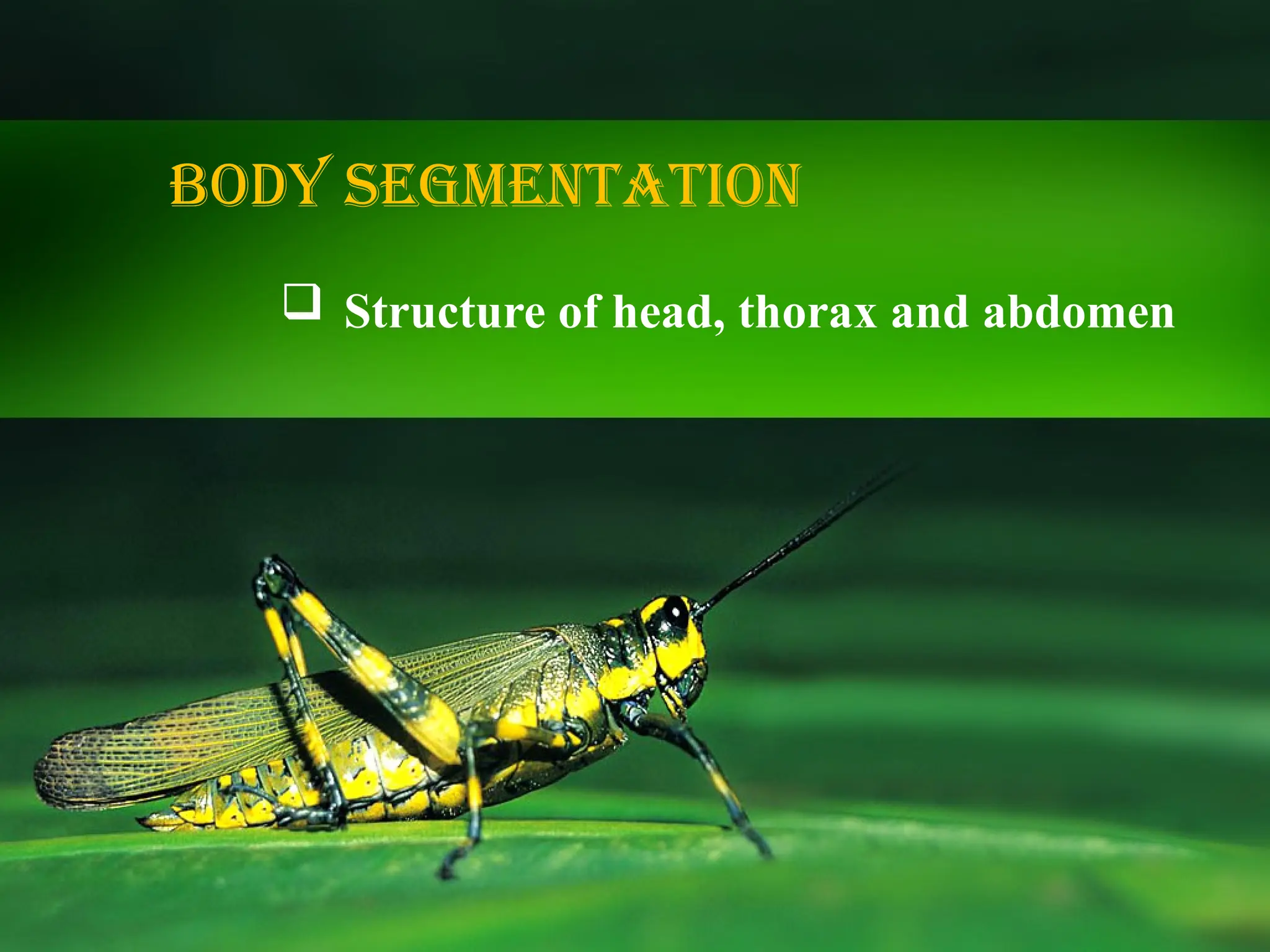
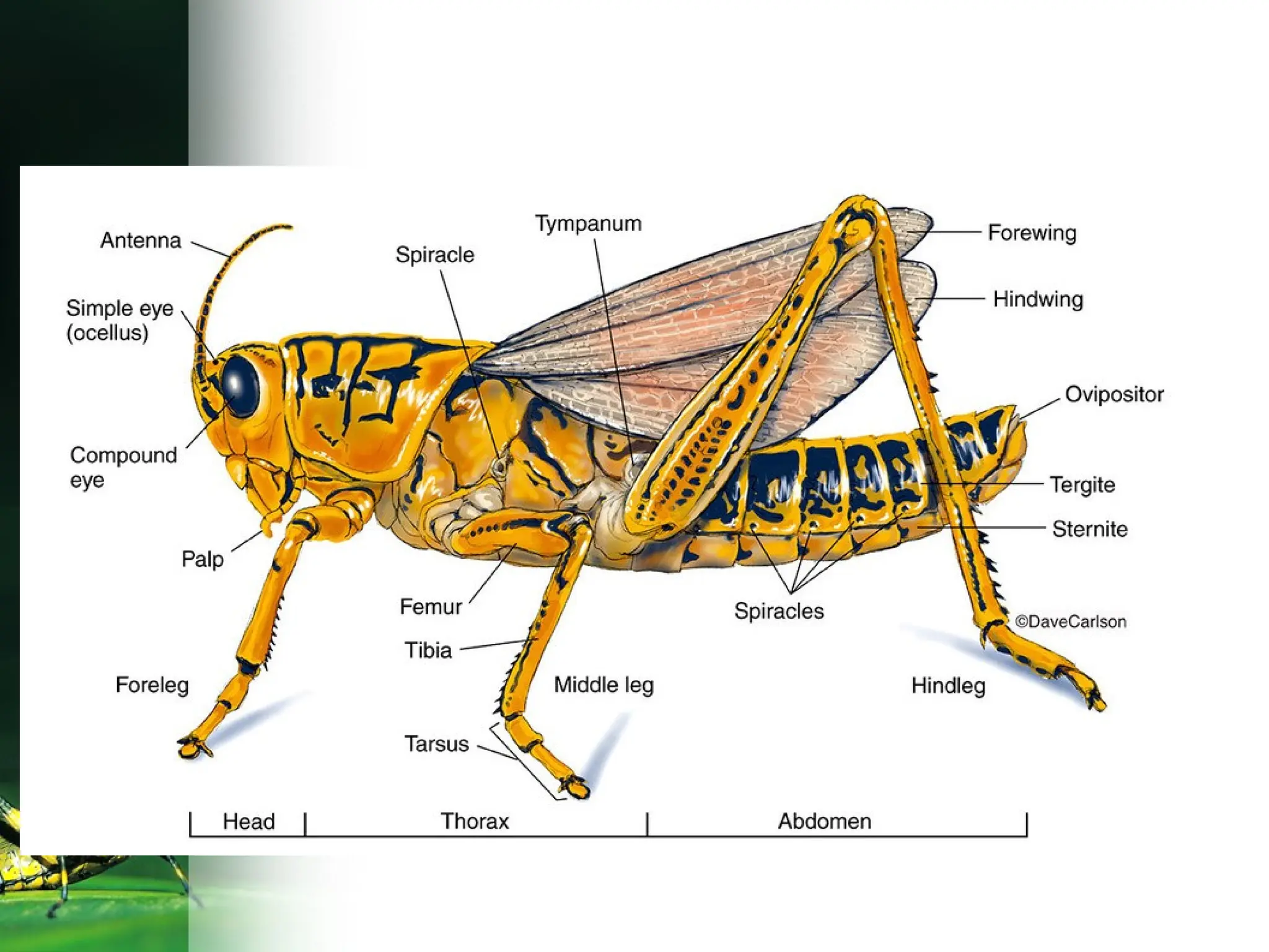
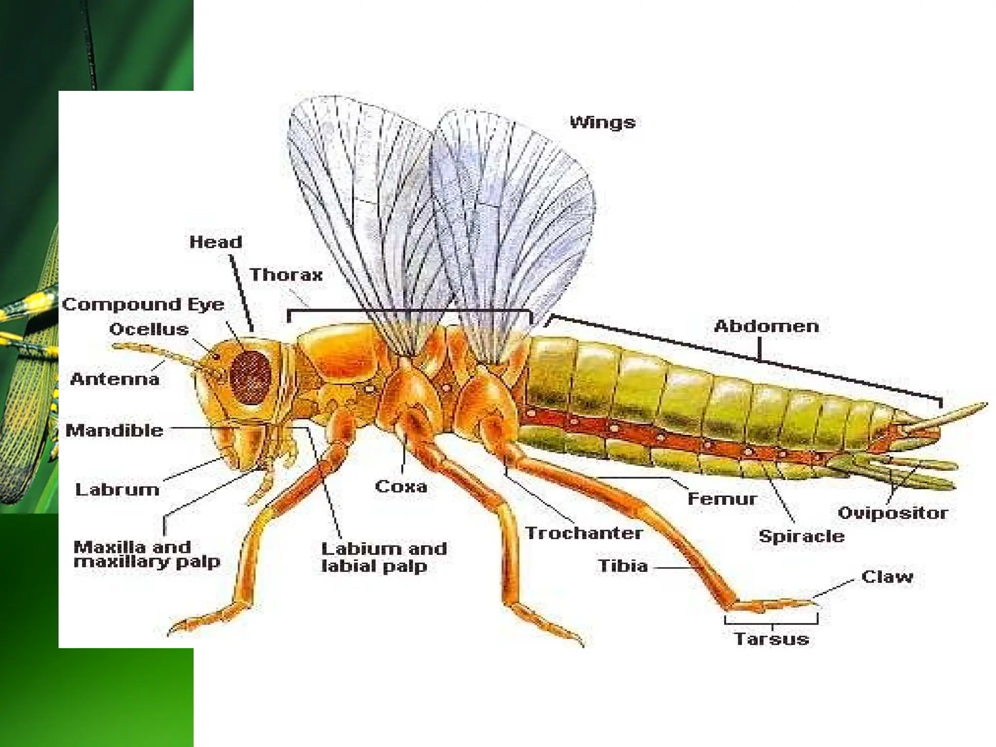
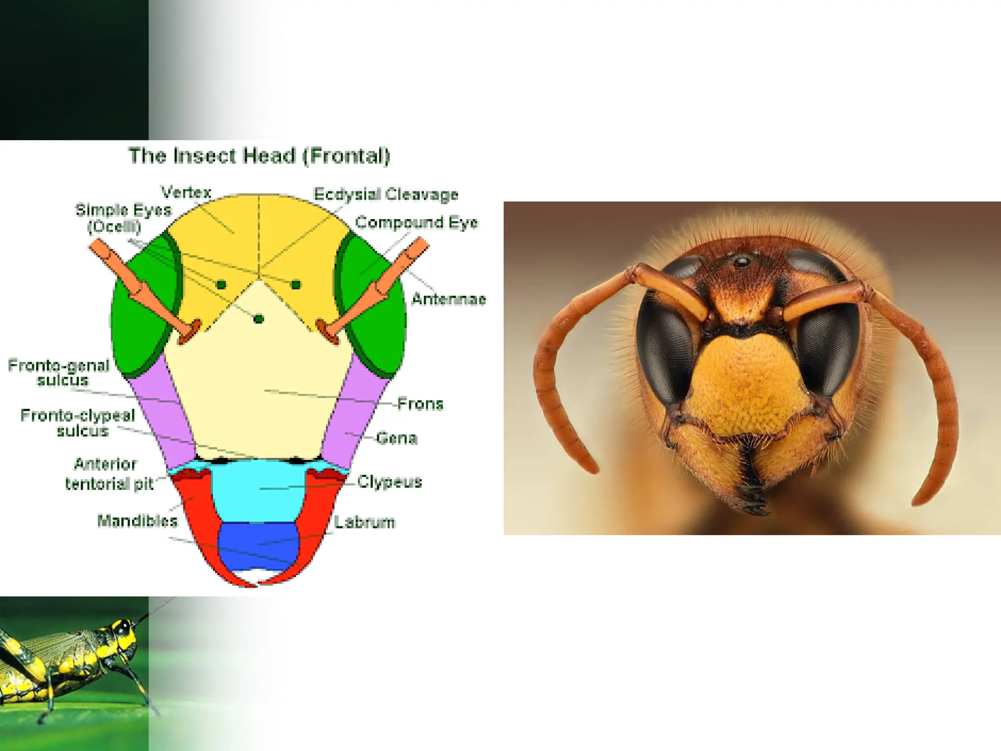
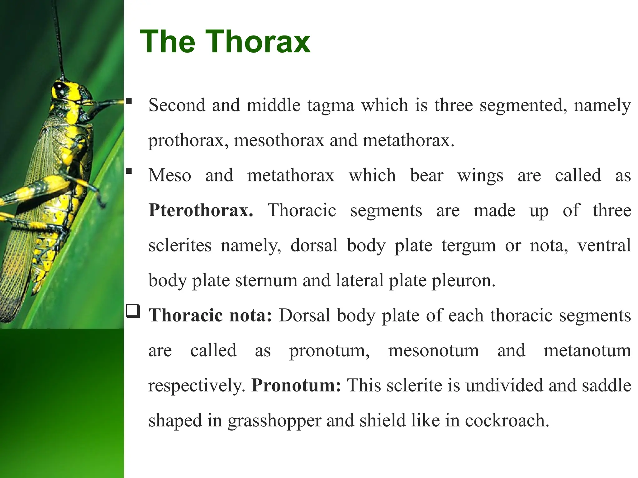
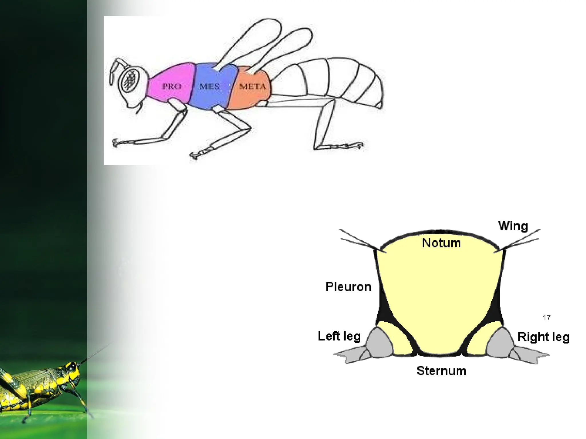
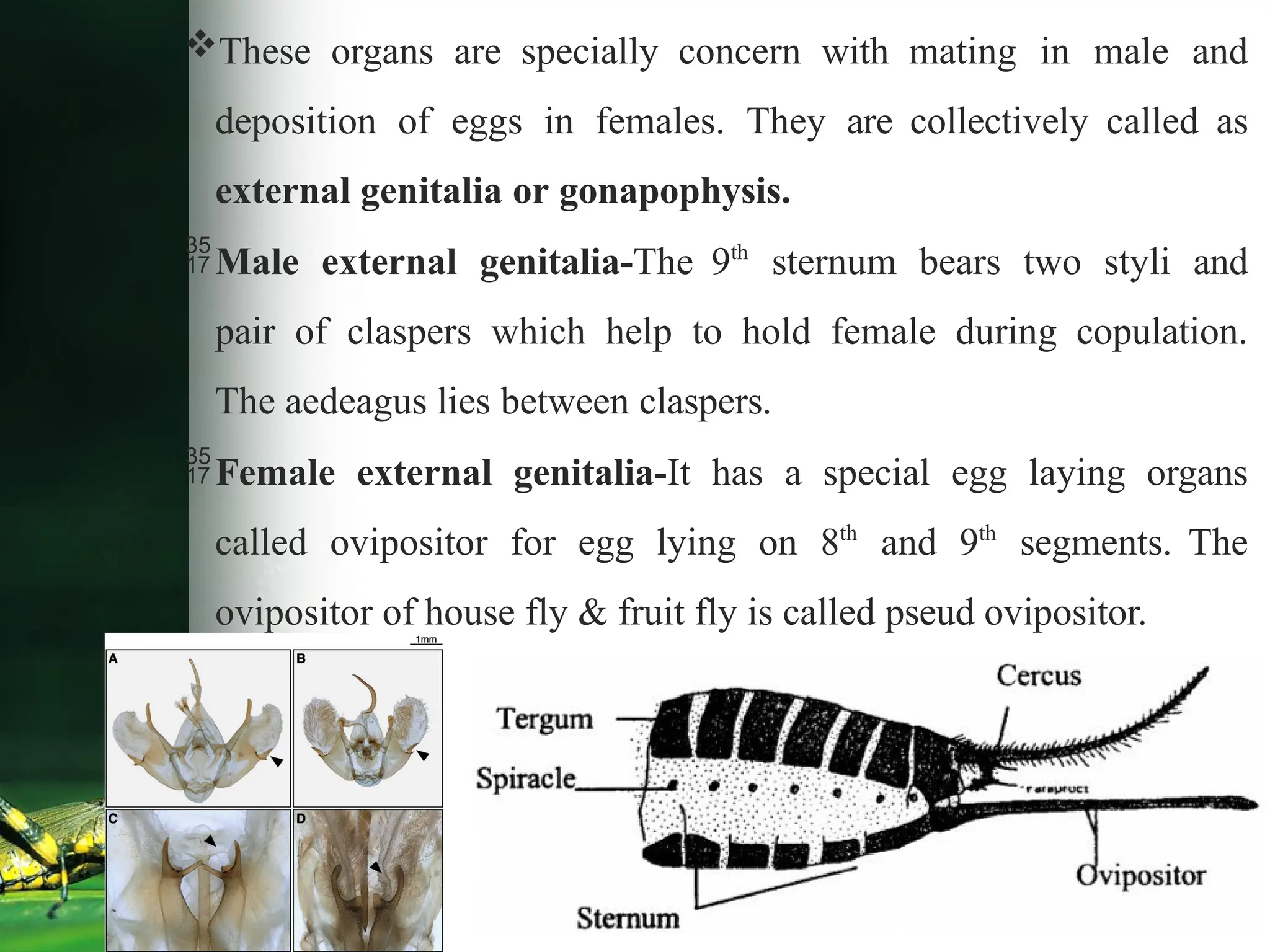
Body Segmentation
 Structure of head, thorax and abdomen
Agricultural Entomology
(Sixth Deans’Committee Recommended Syllabus)
Course No: ENTO: 121
Course Title: Fundamentals of Entomology
By,
Mr. Arshad K. Shaikh
Assistant Professor
Institute:
Sharadchandraji Pawar Institute of Agricultural
Sciences,
Kharavate- Dahiwali, Tal. Chiplun, Dist. Ratnagiri.
Dr. Balasaheb Sawant Konkan Krishi Vidyapeeth, Dapoli
mrarshadshaikh23@gmail.com
 Insect body is divided in to three regions or tagmata namely
head, thorax and abdomen. This grouping of body segments in
to regions is known as tagmosis.
 The cuticle forms the external surface of the body wall and
contains chitin. The cuticle hardens (Sclerotizes) at localized areas
to form sclerites. The sclerites are separated from each other by
means of the impressed line is known as sutures ( Border).
 The body wall is divisible into three regions
a) The dorsal (upper) region is called dorsum or tergum.
b) The ventral (lower) region is called venter or sternum.
c) The two lateral regions are known as plurae.
 Head consists of 6 segments possesses mouthparts,
compound eyes, simple eyes (ocelli) and a pair of
antennae.
 Thorax consists of 3 segments i.e. prothorax,
mesothorax and metathorax, Meso and metathorax are
together known as pterothorax. All the three thoracic
segments possess a pair of legs and meso and meta
thorax possess one pair of wings.
 Abdomen has 11 segments with genital appendages
on 8th and 9th
segments.
 The insect body generally consists of 20 segments.
1. Head:
 First anterior tagma formed by the fusion of six segments
along with appendages. It is concerned with feeding
mechanism and carries special sensory organs like antennae
and eyes. Head is formed by following segment.
Sr. No. Name of segments Name of appendage
1 Pre- antennary Pair of compound eyes
2 Antennary Pair of antennae
3 Intercalary Single labrum (Upper lip)
4 Mandibular Pair of mandibles (Teeth)
5 Maxillary Pair of maxillae (Tounge)
6 Labial Labium ( Lower lip)
 Head is attached or articulated to the thorax through neck or
cervix.
 Head capsule is sclerotized and the head capsule excluding
appendages formed by the fusion of several sclerites is known as
cranium.
 Sclerites of Head
i. Vertex: Summit of the head between compound eyes.
ii. Frons: Facial area below the vertex and above clypeus.
iii. Gena: Lateral cranial area behind the compound eyes.
iv. Clypeus: Cranial area below the frons to which labrum is
attached.
v. Occiput : Cranial area between occipital and post occipital
suture.
Sutures of Head: The linear invaginations of the exoskeleton
between two sclerites are called as suture (some times referred
as sulcus).
i. Epicranial suture/ ecdysial cleavage: Inverted `Y' shaped
suture found medially on the top of head, with a median
suture (coronal suture) and lateral sutures (frontal suture).
ii. Epistomal suture/ Fronto clypeal suture: Found between
frons and clypeus. (epi –above; stoma- mouth parts)
iii. Clypeo-labral suture: Found between clypeus and labrum
(upper lip).
iv. Postoccipital suture: Groove bordering occipital foramen..
 Endoskeleton of insect cuticle provides space for
attachment of muscles of antenna and mouthparts, called
as tentorium.
 The appendages like a pair of compound eyes, 0-3 ocelli, a
pair of antenna and mouth parts are called as cephalic
appendages.
 Functions of Head
 i. Food ingestion
 ii. Sensory perception
 iii. Coordination of bodily activities
 iv. Protection of the coordinating centers
Types of head position
According to the position or projection of mouth parts the head of the
insect can be classified as;
(a) Hypognathous (Hypo – Below: Gnathous – Jaw )
The head remain vertical and is at right angle to the long axis of the
body and mouth parts are ventrally placed and projected downwards. This
is also known as Orthopteroid type.
Eg: Grass hopper, Cockroach.
(b) Prognathous : (Pro – infront: Gnathous – Jaw )
The head remains in the same axis to body and mouth parts are
projected forward.
This is also known as Coleopteroid type. Eg: beetles
(c) Opisthognathous : (Opistho – behind: Gnathous Jaw )
It is same as prognathous but mouthparts are directed backward
and held in between the fore legs. This is also known as
Hemipteroid or Opisthorhynchous type. Eg: bugs
 Second and middle tagma which is three segmented, namely
prothorax, mesothorax and metathorax.
 Meso and metathorax which bear wings are called as
Pterothorax. Thoracic segments are made up of three
sclerites namely, dorsal body plate tergum or nota, ventral
body plate sternum and lateral plate pleuron.
 Thoracic nota: Dorsal body plate of each thoracic segments
are called as pronotum, mesonotum and metanotum
respectively. Pronotum: This sclerite is undivided and saddle
shaped in grasshopper and shield like in cockroach.
The Thorax
17
18
 Pterothoracic notum: Have 3 transverse sutures (antecostal,
prescutal and scuto-scutellar) and 5 tergites (acrotergite,
prescutum, scutum, scutellum and post-scutellum).
 Thoracic sterna: Vental body plate of each thoracic segments
are called as prosternum, mesosternum and metasternum.
Thoracic sterna is made up of a segmental plate called
eusternun and an intersternite called spinasternum. Eusternum
is made up of three sternites viz., presternum, basisternum and
sternellum.
 Thoracic pleura: Lateral body wall of thoracic segment
between notum and sternum. Sclerites of pleuron is called as
pleurite and they fuse to form pleural plate.
Pleural plate is divided into anterior episternum and posterior
epimeron by pleural suture. Pterothoracic pleuron provides space
for articulation of wings and legs.
Thoracic appendages are three pairs of legs and two pairs of
wings. Two pairs of spiracles are also present in the mesopleuron
and metapleuron.
Functions of thorax: Mainly concerned with locomotion.
The Abdomen
The abdomen in the embryo usually consists 11 segments.
The abdominal segments are sometimes designated as
uromeres.
The terminal region of abdomen is called telson which bears
anus.
The 1st abdominal segment gets fused to metathorax
forming propodeum. (In ants, bees and wasps).
The first eight abdominal segments carry a pair of spiracles
each.
Abdomen generally concerned with reproduction and
metabolic activity.
 Appendages (Attachments) of abdomen–
1. Non reproductive appendages –
a)Cerci –They are present on 11th
segment in most of the insects.
It is present in male cockroach, silverfish, grasshopper Cerci
usually act as tactile organ or sound receptors in grasshopper.
They become a part of male genitalia in caddis fly. In
earwigs, cerci are modified into defensive organ.
b)Prolegs in insect larvae- The larvae of Lepidoptera bear five
pairs of abdominal legs called Prolegs on 3rd
4th
5th
6th
and
10th
segments. These Prolegs bear spines like structures called
crochets, on terminal ends to grip the plant surfaces. In case of
larvae of sawfly there are eight pairs of Prolegs but are without
crochets.
c)Abdominal gills-
It is present in aquatic insects for respiration. eg. Nymph of
odonata.
d)Cornicles: Aphids have a pair of short tubes known as
cornicles or siphonculi projecting from dorsum of fifth or sixth
abdominal segment. They permit the escape of waxy fluid
which perhaps serves for protection against predators.
2. Reproductive appendages –
It includes abdominal Segments from 1 to 7 are pregenital
segments, 8th
and 9th
are known as genital segments as they
form genital appendages i.e. ovipositor in females and aedeagus
or penis in males. 10th
and 11 th
segments are known as
postgenital segments.
These organs are specially concern with mating in male and
deposition of eggs in females. They are collectively called as
external genitalia or gonapophysis.
Male external genitalia-The 9th
sternum bears two styli and
pair of claspers which help to hold female during copulation.
The aedeagus lies between claspers.
Female external genitalia-It has a special egg laying organs
called ovipositor for egg lying on 8th
and 9th
segments. The
ovipositor of house fly & fruit fly is called pseud ovipositor.
*"The Segmented Blueprint: Unlocking Insect Body Architecture"*.pptx

*"The Segmented Blueprint: Unlocking Insect Body Architecture"*.pptx

  • 1.
    Body Segmentation  Structureof head, thorax and abdomen
  • 2.
    Agricultural Entomology (Sixth Deans’CommitteeRecommended Syllabus) Course No: ENTO: 121 Course Title: Fundamentals of Entomology By, Mr. Arshad K. Shaikh Assistant Professor Institute: Sharadchandraji Pawar Institute of Agricultural Sciences, Kharavate- Dahiwali, Tal. Chiplun, Dist. Ratnagiri. Dr. Balasaheb Sawant Konkan Krishi Vidyapeeth, Dapoli mrarshadshaikh23@gmail.com
  • 3.
     Insect bodyis divided in to three regions or tagmata namely head, thorax and abdomen. This grouping of body segments in to regions is known as tagmosis.  The cuticle forms the external surface of the body wall and contains chitin. The cuticle hardens (Sclerotizes) at localized areas to form sclerites. The sclerites are separated from each other by means of the impressed line is known as sutures ( Border).  The body wall is divisible into three regions a) The dorsal (upper) region is called dorsum or tergum. b) The ventral (lower) region is called venter or sternum. c) The two lateral regions are known as plurae.
  • 4.
     Head consistsof 6 segments possesses mouthparts, compound eyes, simple eyes (ocelli) and a pair of antennae.  Thorax consists of 3 segments i.e. prothorax, mesothorax and metathorax, Meso and metathorax are together known as pterothorax. All the three thoracic segments possess a pair of legs and meso and meta thorax possess one pair of wings.  Abdomen has 11 segments with genital appendages on 8th and 9th segments.  The insect body generally consists of 20 segments.
  • 7.
    1. Head:  Firstanterior tagma formed by the fusion of six segments along with appendages. It is concerned with feeding mechanism and carries special sensory organs like antennae and eyes. Head is formed by following segment. Sr. No. Name of segments Name of appendage 1 Pre- antennary Pair of compound eyes 2 Antennary Pair of antennae 3 Intercalary Single labrum (Upper lip) 4 Mandibular Pair of mandibles (Teeth) 5 Maxillary Pair of maxillae (Tounge) 6 Labial Labium ( Lower lip)
  • 8.
     Head isattached or articulated to the thorax through neck or cervix.  Head capsule is sclerotized and the head capsule excluding appendages formed by the fusion of several sclerites is known as cranium.  Sclerites of Head i. Vertex: Summit of the head between compound eyes. ii. Frons: Facial area below the vertex and above clypeus. iii. Gena: Lateral cranial area behind the compound eyes. iv. Clypeus: Cranial area below the frons to which labrum is attached. v. Occiput : Cranial area between occipital and post occipital suture.
  • 10.
    Sutures of Head:The linear invaginations of the exoskeleton between two sclerites are called as suture (some times referred as sulcus). i. Epicranial suture/ ecdysial cleavage: Inverted `Y' shaped suture found medially on the top of head, with a median suture (coronal suture) and lateral sutures (frontal suture). ii. Epistomal suture/ Fronto clypeal suture: Found between frons and clypeus. (epi –above; stoma- mouth parts) iii. Clypeo-labral suture: Found between clypeus and labrum (upper lip).
  • 11.
    iv. Postoccipital suture:Groove bordering occipital foramen..  Endoskeleton of insect cuticle provides space for attachment of muscles of antenna and mouthparts, called as tentorium.  The appendages like a pair of compound eyes, 0-3 ocelli, a pair of antenna and mouth parts are called as cephalic appendages.  Functions of Head  i. Food ingestion  ii. Sensory perception  iii. Coordination of bodily activities  iv. Protection of the coordinating centers
  • 12.
    Types of headposition According to the position or projection of mouth parts the head of the insect can be classified as; (a) Hypognathous (Hypo – Below: Gnathous – Jaw ) The head remain vertical and is at right angle to the long axis of the body and mouth parts are ventrally placed and projected downwards. This is also known as Orthopteroid type. Eg: Grass hopper, Cockroach.
  • 13.
    (b) Prognathous :(Pro – infront: Gnathous – Jaw ) The head remains in the same axis to body and mouth parts are projected forward. This is also known as Coleopteroid type. Eg: beetles
  • 14.
    (c) Opisthognathous :(Opistho – behind: Gnathous Jaw ) It is same as prognathous but mouthparts are directed backward and held in between the fore legs. This is also known as Hemipteroid or Opisthorhynchous type. Eg: bugs
  • 16.
     Second andmiddle tagma which is three segmented, namely prothorax, mesothorax and metathorax.  Meso and metathorax which bear wings are called as Pterothorax. Thoracic segments are made up of three sclerites namely, dorsal body plate tergum or nota, ventral body plate sternum and lateral plate pleuron.  Thoracic nota: Dorsal body plate of each thoracic segments are called as pronotum, mesonotum and metanotum respectively. Pronotum: This sclerite is undivided and saddle shaped in grasshopper and shield like in cockroach. The Thorax
  • 17.
  • 18.
    18  Pterothoracic notum:Have 3 transverse sutures (antecostal, prescutal and scuto-scutellar) and 5 tergites (acrotergite, prescutum, scutum, scutellum and post-scutellum).  Thoracic sterna: Vental body plate of each thoracic segments are called as prosternum, mesosternum and metasternum. Thoracic sterna is made up of a segmental plate called eusternun and an intersternite called spinasternum. Eusternum is made up of three sternites viz., presternum, basisternum and sternellum.  Thoracic pleura: Lateral body wall of thoracic segment between notum and sternum. Sclerites of pleuron is called as pleurite and they fuse to form pleural plate.
  • 19.
    Pleural plate isdivided into anterior episternum and posterior epimeron by pleural suture. Pterothoracic pleuron provides space for articulation of wings and legs. Thoracic appendages are three pairs of legs and two pairs of wings. Two pairs of spiracles are also present in the mesopleuron and metapleuron. Functions of thorax: Mainly concerned with locomotion.
  • 20.
    The Abdomen The abdomenin the embryo usually consists 11 segments. The abdominal segments are sometimes designated as uromeres. The terminal region of abdomen is called telson which bears anus. The 1st abdominal segment gets fused to metathorax forming propodeum. (In ants, bees and wasps). The first eight abdominal segments carry a pair of spiracles each. Abdomen generally concerned with reproduction and metabolic activity.
  • 21.
     Appendages (Attachments)of abdomen– 1. Non reproductive appendages – a)Cerci –They are present on 11th segment in most of the insects. It is present in male cockroach, silverfish, grasshopper Cerci usually act as tactile organ or sound receptors in grasshopper. They become a part of male genitalia in caddis fly. In earwigs, cerci are modified into defensive organ. b)Prolegs in insect larvae- The larvae of Lepidoptera bear five pairs of abdominal legs called Prolegs on 3rd 4th 5th 6th and 10th segments. These Prolegs bear spines like structures called crochets, on terminal ends to grip the plant surfaces. In case of larvae of sawfly there are eight pairs of Prolegs but are without crochets.
  • 23.
    c)Abdominal gills- It ispresent in aquatic insects for respiration. eg. Nymph of odonata. d)Cornicles: Aphids have a pair of short tubes known as cornicles or siphonculi projecting from dorsum of fifth or sixth abdominal segment. They permit the escape of waxy fluid which perhaps serves for protection against predators. 2. Reproductive appendages – It includes abdominal Segments from 1 to 7 are pregenital segments, 8th and 9th are known as genital segments as they form genital appendages i.e. ovipositor in females and aedeagus or penis in males. 10th and 11 th segments are known as postgenital segments.
  • 25.
    These organs arespecially concern with mating in male and deposition of eggs in females. They are collectively called as external genitalia or gonapophysis. Male external genitalia-The 9th sternum bears two styli and pair of claspers which help to hold female during copulation. The aedeagus lies between claspers. Female external genitalia-It has a special egg laying organs called ovipositor for egg lying on 8th and 9th segments. The ovipositor of house fly & fruit fly is called pseud ovipositor.

Editor's Notes

  • #3 Head sutures- border lines or boundaries between different parts of insect head.
  • #8 Sclerites-Harden plates that provide protection and support to head. Cranium- hard outer covering of insect head. It protects the brain eyes and other vital organs. Think of it like a helmet that shields the insect head. Occiput- Backside of head
  • #10 Head sutures- border lines or boundaries between different parts of insect head.
  • #20 Uromeres- Tail segment, insects tail, propodeum- rear thorax of insect
  • #21 Uromeres- Tail segment, insects tail, propodeum- rear thorax of insect