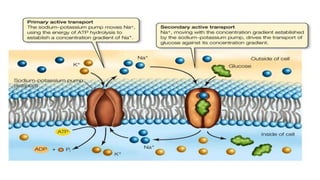
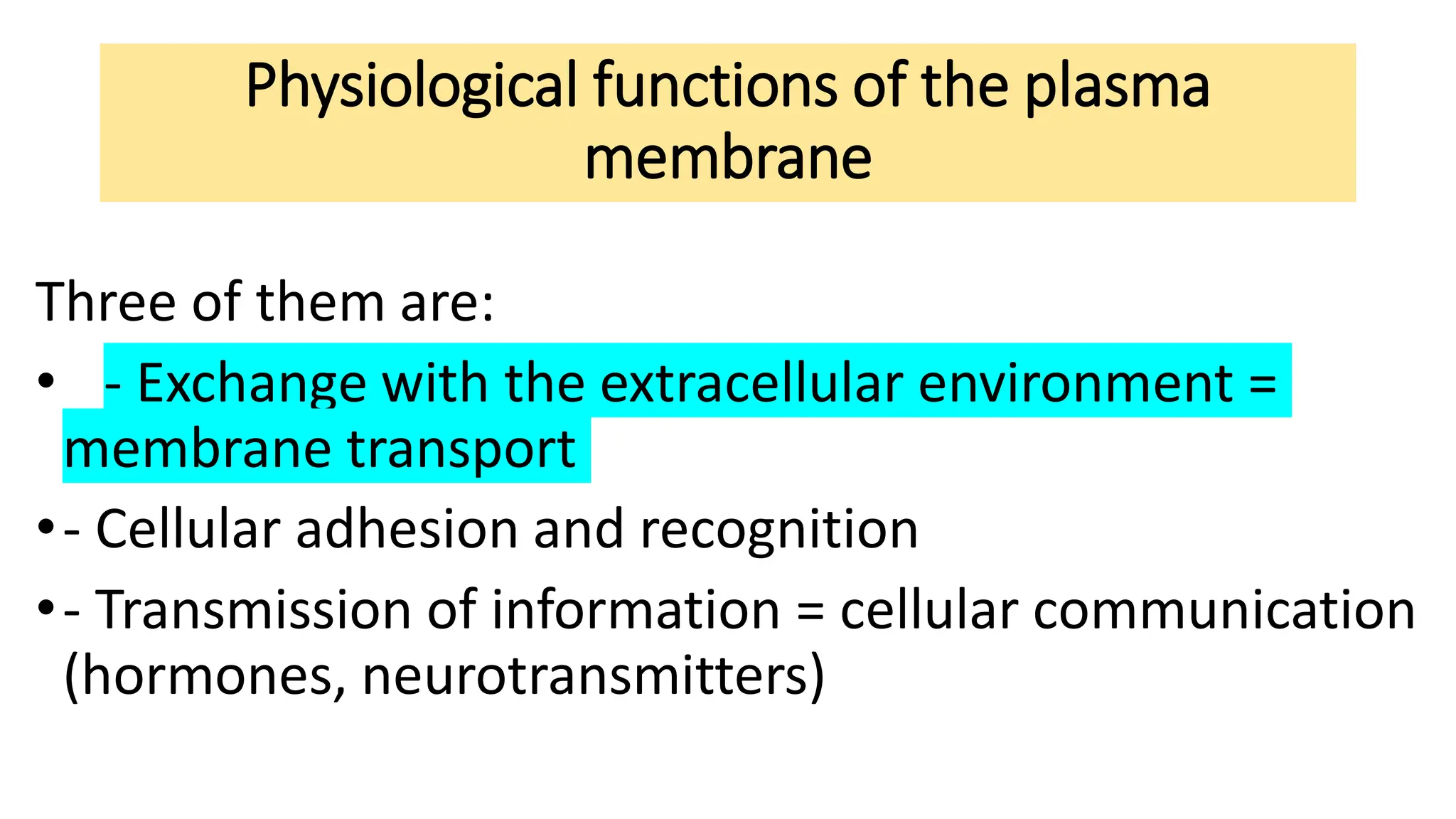
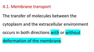
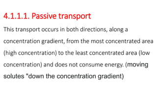
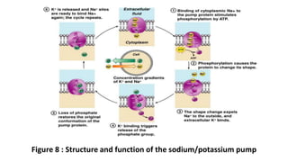
Physiological functions of the plasma
membrane
Three of them are:
• - Exchange with the extracellular environment =
membrane transport
•- Cellular adhesion and recognition
•- Transmission of information = cellular communication
(hormones, neurotransmitters)
4.1. Membrane transport
The transfer of molecules between the
cytoplasm and the extracellular environment
occurs in both directions with or without
deformation of the membrane.
• 4.1.1. Membrane exchanges without deformation of the plasma
membrane
• It is done through the membrane constituents (lipids, proteins) and
without intervention of the cytoskeleton. There are 2 criteria for
classifying membrane transport.
* Energy consumption or not
• Transport without energy Passive transport
• Transport with energy Active transport
* Presence or absence of a permease: Intrinsic protein of the plasma
membrane or group of proteins responsible for transport.
• Transport without permease Simple diffusion
• Transport with permease Facilitated diffusion
4.1.1.1. Passive transport
This transport occurs in both directions, along a
concentration gradient, from the most concentrated area
(high concentration) to the least concentrated area (low
concentration) and does not consume energy. (moving
solutes "down the concentration gradient)
* Simple diffusion
It concerns uncharged molecules (non-electrolytes)
that are sufficiently small. Simple diffusion is
general for water (osmosis phenomenon) whose
permeability coefficient is one of the highest.(figure
1)
Figure 1 : diffusion and osmosis
• Osmosis is water moving along its concentration gradient.
• Solute molecules decrease the number of water molecules that are free to move
around. Water moves from where free molecules are abundant to where there are
fewer.
• Example of red blood cells placed in 3 types of areas at different concentrations :
• - Isotonic area (Na Cl = 0.9%). Constant volume
• - Hypotonic environment (Na Cl < 0.9%) Increases in volume (hemolysis)
• - Hypertonic environment (Na Cl >0.9%) Decreases in volume (plasmolysis)
•Simple diffusion exists in all cells but is amplified in
cells specialized in the absorption function (Figure 2):
•- the renal cell has invaginations at its base
•- the intestinal cell has microvilli on its surface
Figure 2 : cell membrane specialized in absorption function
•Simple diffusion depends on several factors (figure 3):
•-Liposolubility, fat-soluble substances (alcohols, aldehydes,
ketones, anesthetics) penetrate more easily.
•-Solubility in water (hydrosolubility), water-soluble
substances (methanol, ethanol, urea, O2, Co2 ...etc.) pass
through the membrane at the same time as the water.
Figure 3 : simple diffusion
Facilitated diffusion
• It involves the intervention of a membrane transport protein which
can be a carrier protein or a protein channel. These channels are
generally specific: only one very specific substance can pass through
them and no other. So, not just any substance can pass through the
membrane = selective permeability.
Example 1: Transport of water
•Example 1: Transport of water
•Water can pass through aquaporins (Table I), it is a
temporary pore system allowing the entry or exit of
water according to the laws of osmosis.
Table I: Some types of aquaporins
Protein Activity Main location
AQP0 water channel Eye
AQP1 water channel Erythrocytes, kidney,eye, brain,
heart, lung
AQP2 water channel Kidney (collecting duct)
AQP3 water channel Erythrocytes, kidney,
gastrointestinal tract, lung, bladder
AQP4 water channel Brain, eye, kidney, lung, intestin
AQP5 water channel Lacrimal and salivary glands
Alpha-TIP water channel Seed vacuoles
• The control of certain aquaporins is done through hormones, we have noticed in
the bladder:
• - in the resting state, before stimulation by ADH (vasopressin): urinary
reabsorption is low.
• in the active state (after stimulation by ADH): the reabsorption of urinary water is
important.
• Indeed, cryodecapping shows that at rest, the integrated proteins of the plasma
membrane are dispersed and that in activity the proteins are gathered together
and seem to form a channel or a pore which allows the transport of water.
Example 2: Transport of non-electrolytic molecules
• It concerns uncharged molecules, such as sugars and aa
• Glucose transport in the RBC
• This substance is poorly lipid soluble, however it penetrates very quickly into
the cell in the direction of the concentration gradient, in fact glucose crosses
the plasma membrane combined with a specific membrane protein. This
transport exhibits a saturation phenomenon for high substrate concentrations
and is characterized by high substrate specificity. Only a few molecules of
similar structure can behave as inhibitors through competition.
• It is generally accepted that facilitated diffusion involves the
following 4 steps (Figure 4).
• 1 – Fixation of glucose on the external side and formation of a
glucose-permease complex.
• 2 – Translocation of the complex into the lipid bilayer.
• 3 – Dissociation of the complex and release of glucose into the
cytoplasm.
• 4 – Return to initial state
Figure 4 : glucose transport in the red blood cells
• The nature of the complex formed is poorly known, it could be:
• -A covalent bond between the substrate and its transporter which would
imply an enzymatic type activity hence the name permease given to the
transporter.
• -The translocation could result from a rotation or a simple diffusion of the
integrated protein – molecule complex transported through the lipid phase.
• -Or on the contrary be due to a modification of the structure of the
integrated protein which would cause the movement of the molecule
transported across the membrane.
Glucose transport in adipocytes
• It is subject to hormonal control, the steps are as follows
(Figure 5):
• 1 – Binding of insulin to membrane receptors
• 2 – Transmission of the hormonal message inside the cell
• 3 – Translocation of glucose transporters to the plasma
membrane (by exocytosis)
• 4 – Fixation of glucose on its receptor
• 5 – Release of glucose into the cytosol
• 6 – The transporters are recovered by endocytosis at the end of
stimulation
Figure 5 : glucose transport in adipocytes
Example 3: Transport of electrolytes
• In all cells, the intracellular ionic concentration is different
from that of the extracellular environment. Indeed the
extracellular environment is very rich in Na+, Cl- and poor in
K+, on the other hand the intracellular fluid is very rich in K+
and organic anions (which do not cross the plasma
membrane) and it is poor in Na+, Cl-
• There is an equilibrium called “Donnan equilibrium”, this equilibrium is achieved when:
• - the product of the ion concentrations is the same on both sides of the plasma membrane,
• [Na+]1 [cl-]1= [Na+]2 [Cl-]2
• - the electrical neutrality of each compartment is maintained
• As much charge + and charge – in each compartment
• There are two types of ion channels allowing very rapid exchanges depending on the
concentration gradient:
• - potential-gated or voltage-gated ion channels (a class of transmembrane proteins that form ion channels
that are activated by changes in the electrical membrane potential near the channel.)
• - ligand-gated ion channels
4.1.1.2. Active transport
• Particularly studied in the case of the movements of Na+ and k+; Nearly
40% of the energy spent each day is used for active transportation.
• - Does not deform the plasma membrane
• - Is carried out against the concentration gradient
• - Requires energy expenditure
• - Allows cells to maintain an internal environment different from the
external environment (Figure 6):
Example 1: Na+/K+ pump
• Properties of the pump: It is a protein which ensures the coupled
transport of sodium and potassium ions, the export of 3 Na+
corresponding to the import of 2 K+. This Na/K ATPase is an intrinsic
protein of the plasma membrane, formed of 2 large  subunits (S/U
) and 2 small S/U . These are the S/U  which contain the specific
binding sites for Na+, K+, and ATP.
• Operation (Figure 7 and 8):
Figure 6 : active transport
• 1. Phosphorylation of the protein from ATP. This step depends on
the presence of a Mg2+ cofactor.
• Cofactor (Figure 22) is a small molecule that binds to the active site.
Without the cofactor, the enzyme site is inactive. Many cofactors
are metal ions (Cu, Zn, Mn, etc.). Some are small organic molecules
(relative to the size of the enzyme); they are then called coenzymes.
Many vitamins are coenzymes (or are coenzyme precursors).
Figure 7: Activation of the enzyme binding site by the cofactor
•2. This phosphorylation results in the transfer of
Na+ outside the cell.
•3. The phosphorylated enzyme subsequently fixes
2 molecules of K+
•4. Dephosphorylation results in the release of K+
inside the cell.
Figure 8 : Structure and function of the sodium/potassium pump
•There are also K, Na, Ca+2 and H pumps,
which also operate against the concentration
gradient.
Example 2: Cotransport
• This transport concerns molecules (aa or sugar) / ions or ions/ions
which move in the same direction (symport) or in the opposite
direction (antiport). It is done thanks to a membrane protein which
has binding sites for the molecules and ions to be transported. This
transport requires the energy provided by the hydrolysis of ATP
(example Na+/K+ pump). It is directly coupled with a flow of ions
which directly follows an electrochemical gradient.( figure 9 )
Figure 9 : cotransport
Transport of glucose and Na+: Symport
cotransport
In kidney or intestinal cells, D glucose and Na+ are transported together
• inside the cell thanks to a protein called sugar/ion cotransporter
(Figure 10), which has a binding site for D glucose and another for
Na+, the glucose then leaves the cell according to a diffusion process
ease. The output of Na+ is coupled to the input of K+ through an
active transport process.
Figure 10: Symport cotransport Glucose/Na+
Transport of Ca++: Antiport cotransport
An ion (Na in general) diffuses following its concentration gradient.
• This displacement allows a substance to cross in the opposite direction against its
concentration gradient.
• Example of the Na+ / Mg++ Pump: the diffusion of Na in the cell allows the
expulsion of Mg++ against its gradient.
• In cardiac cells the entry of Na+ is coupled to the exit of Ca++ (Figure 11). This
system reduces the intracellular concentration of Ca++, which leads to a reduction
in cardiac contractions.
Figure 11 : Calcium/Sodium Antiport cotransport in the cardiac cells
• Among the inhibitors of active transport, we note glucosides,
for example ouabain which compete at the K+ binding sites of
the Na+/K+ pump. These drugs are of great clinical interest
since they are used in the treatment of heart failure, by
increasing cardiac contractions.
• ATPase Na+/K+ inhibited intracellular [Na+]; inhibition of
intracellular Na+/ Ca+2 antiport cotransport of muscle
contractions
The plasma membrane course cytophysiology.pdf

The plasma membrane course cytophysiology.pdf

  • 1.
    Physiological functions ofthe plasma membrane Three of them are: • - Exchange with the extracellular environment = membrane transport •- Cellular adhesion and recognition •- Transmission of information = cellular communication (hormones, neurotransmitters)
  • 2.
    4.1. Membrane transport Thetransfer of molecules between the cytoplasm and the extracellular environment occurs in both directions with or without deformation of the membrane.
  • 3.
    • 4.1.1. Membraneexchanges without deformation of the plasma membrane • It is done through the membrane constituents (lipids, proteins) and without intervention of the cytoskeleton. There are 2 criteria for classifying membrane transport. * Energy consumption or not • Transport without energy Passive transport • Transport with energy Active transport * Presence or absence of a permease: Intrinsic protein of the plasma membrane or group of proteins responsible for transport. • Transport without permease Simple diffusion • Transport with permease Facilitated diffusion
  • 5.
    4.1.1.1. Passive transport Thistransport occurs in both directions, along a concentration gradient, from the most concentrated area (high concentration) to the least concentrated area (low concentration) and does not consume energy. (moving solutes "down the concentration gradient)
  • 7.
    * Simple diffusion Itconcerns uncharged molecules (non-electrolytes) that are sufficiently small. Simple diffusion is general for water (osmosis phenomenon) whose permeability coefficient is one of the highest.(figure 1)
  • 8.
    Figure 1 :diffusion and osmosis
  • 9.
    • Osmosis iswater moving along its concentration gradient. • Solute molecules decrease the number of water molecules that are free to move around. Water moves from where free molecules are abundant to where there are fewer. • Example of red blood cells placed in 3 types of areas at different concentrations : • - Isotonic area (Na Cl = 0.9%). Constant volume • - Hypotonic environment (Na Cl < 0.9%) Increases in volume (hemolysis) • - Hypertonic environment (Na Cl >0.9%) Decreases in volume (plasmolysis)
  • 11.
    •Simple diffusion existsin all cells but is amplified in cells specialized in the absorption function (Figure 2): •- the renal cell has invaginations at its base •- the intestinal cell has microvilli on its surface
  • 12.
    Figure 2 :cell membrane specialized in absorption function
  • 13.
    •Simple diffusion dependson several factors (figure 3): •-Liposolubility, fat-soluble substances (alcohols, aldehydes, ketones, anesthetics) penetrate more easily. •-Solubility in water (hydrosolubility), water-soluble substances (methanol, ethanol, urea, O2, Co2 ...etc.) pass through the membrane at the same time as the water.
  • 14.
    Figure 3 :simple diffusion
  • 15.
    Facilitated diffusion Facilitated diffusion •It involves the intervention of a membrane transport protein which can be a carrier protein or a protein channel. These channels are generally specific: only one very specific substance can pass through them and no other. So, not just any substance can pass through the membrane = selective permeability.
  • 16.
    Example 1: Transportof water •Example 1: Transport of water •Water can pass through aquaporins (Table I), it is a temporary pore system allowing the entry or exit of water according to the laws of osmosis.
  • 17.
    Table I: Sometypes of aquaporins Protein Activity Main location AQP0 water channel Eye AQP1 water channel Erythrocytes, kidney,eye, brain, heart, lung AQP2 water channel Kidney (collecting duct) AQP3 water channel Erythrocytes, kidney, gastrointestinal tract, lung, bladder AQP4 water channel Brain, eye, kidney, lung, intestin AQP5 water channel Lacrimal and salivary glands Alpha-TIP water channel Seed vacuoles
  • 18.
    • The controlof certain aquaporins is done through hormones, we have noticed in the bladder: • - in the resting state, before stimulation by ADH (vasopressin): urinary reabsorption is low. • in the active state (after stimulation by ADH): the reabsorption of urinary water is important. • Indeed, cryodecapping shows that at rest, the integrated proteins of the plasma membrane are dispersed and that in activity the proteins are gathered together and seem to form a channel or a pore which allows the transport of water.
  • 19.
    Example 2: Transportof non-electrolytic molecules • It concerns uncharged molecules, such as sugars and aa • Glucose transport in the RBC • This substance is poorly lipid soluble, however it penetrates very quickly into the cell in the direction of the concentration gradient, in fact glucose crosses the plasma membrane combined with a specific membrane protein. This transport exhibits a saturation phenomenon for high substrate concentrations and is characterized by high substrate specificity. Only a few molecules of similar structure can behave as inhibitors through competition.
  • 20.
    • It isgenerally accepted that facilitated diffusion involves the following 4 steps (Figure 4). • 1 – Fixation of glucose on the external side and formation of a glucose-permease complex. • 2 – Translocation of the complex into the lipid bilayer. • 3 – Dissociation of the complex and release of glucose into the cytoplasm. • 4 – Return to initial state
  • 21.
    Figure 4 :glucose transport in the red blood cells
  • 22.
    • The natureof the complex formed is poorly known, it could be: • -A covalent bond between the substrate and its transporter which would imply an enzymatic type activity hence the name permease given to the transporter. • -The translocation could result from a rotation or a simple diffusion of the integrated protein – molecule complex transported through the lipid phase. • -Or on the contrary be due to a modification of the structure of the integrated protein which would cause the movement of the molecule transported across the membrane.
  • 23.
    Glucose transport inadipocytes • It is subject to hormonal control, the steps are as follows (Figure 5): • 1 – Binding of insulin to membrane receptors • 2 – Transmission of the hormonal message inside the cell • 3 – Translocation of glucose transporters to the plasma membrane (by exocytosis) • 4 – Fixation of glucose on its receptor • 5 – Release of glucose into the cytosol • 6 – The transporters are recovered by endocytosis at the end of stimulation
  • 24.
    Figure 5 :glucose transport in adipocytes
  • 25.
    Example 3: Transportof electrolytes • In all cells, the intracellular ionic concentration is different from that of the extracellular environment. Indeed the extracellular environment is very rich in Na+, Cl- and poor in K+, on the other hand the intracellular fluid is very rich in K+ and organic anions (which do not cross the plasma membrane) and it is poor in Na+, Cl-
  • 26.
    • There isan equilibrium called “Donnan equilibrium”, this equilibrium is achieved when: • - the product of the ion concentrations is the same on both sides of the plasma membrane, • [Na+]1 [cl-]1= [Na+]2 [Cl-]2 • - the electrical neutrality of each compartment is maintained • As much charge + and charge – in each compartment • There are two types of ion channels allowing very rapid exchanges depending on the concentration gradient: • - potential-gated or voltage-gated ion channels (a class of transmembrane proteins that form ion channels that are activated by changes in the electrical membrane potential near the channel.) • - ligand-gated ion channels
  • 27.
    4.1.1.2. Active transport •Particularly studied in the case of the movements of Na+ and k+; Nearly 40% of the energy spent each day is used for active transportation. • - Does not deform the plasma membrane • - Is carried out against the concentration gradient • - Requires energy expenditure • - Allows cells to maintain an internal environment different from the external environment (Figure 6):
  • 28.
    Example 1: Na+/K+pump • Properties of the pump: It is a protein which ensures the coupled transport of sodium and potassium ions, the export of 3 Na+ corresponding to the import of 2 K+. This Na/K ATPase is an intrinsic protein of the plasma membrane, formed of 2 large  subunits (S/U ) and 2 small S/U . These are the S/U  which contain the specific binding sites for Na+, K+, and ATP. • Operation (Figure 7 and 8):
  • 29.
    Figure 6 :active transport
  • 30.
    • 1. Phosphorylationof the protein from ATP. This step depends on the presence of a Mg2+ cofactor. • Cofactor (Figure 22) is a small molecule that binds to the active site. Without the cofactor, the enzyme site is inactive. Many cofactors are metal ions (Cu, Zn, Mn, etc.). Some are small organic molecules (relative to the size of the enzyme); they are then called coenzymes. Many vitamins are coenzymes (or are coenzyme precursors).
  • 31.
    Figure 7: Activationof the enzyme binding site by the cofactor
  • 32.
    •2. This phosphorylationresults in the transfer of Na+ outside the cell. •3. The phosphorylated enzyme subsequently fixes 2 molecules of K+ •4. Dephosphorylation results in the release of K+ inside the cell.
  • 33.
    Figure 8 :Structure and function of the sodium/potassium pump
  • 34.
    •There are alsoK, Na, Ca+2 and H pumps, which also operate against the concentration gradient.
  • 35.
    Example 2: Cotransport •This transport concerns molecules (aa or sugar) / ions or ions/ions which move in the same direction (symport) or in the opposite direction (antiport). It is done thanks to a membrane protein which has binding sites for the molecules and ions to be transported. This transport requires the energy provided by the hydrolysis of ATP (example Na+/K+ pump). It is directly coupled with a flow of ions which directly follows an electrochemical gradient.( figure 9 )
  • 36.
    Figure 9 :cotransport
  • 37.
    Transport of glucoseand Na+: Symport cotransport In kidney or intestinal cells, D glucose and Na+ are transported together • inside the cell thanks to a protein called sugar/ion cotransporter (Figure 10), which has a binding site for D glucose and another for Na+, the glucose then leaves the cell according to a diffusion process ease. The output of Na+ is coupled to the input of K+ through an active transport process.
  • 38.
    Figure 10: Symportcotransport Glucose/Na+
  • 39.
    Transport of Ca++:Antiport cotransport An ion (Na in general) diffuses following its concentration gradient. • This displacement allows a substance to cross in the opposite direction against its concentration gradient. • Example of the Na+ / Mg++ Pump: the diffusion of Na in the cell allows the expulsion of Mg++ against its gradient. • In cardiac cells the entry of Na+ is coupled to the exit of Ca++ (Figure 11). This system reduces the intracellular concentration of Ca++, which leads to a reduction in cardiac contractions.
  • 40.
    Figure 11 :Calcium/Sodium Antiport cotransport in the cardiac cells
  • 41.
    • Among theinhibitors of active transport, we note glucosides, for example ouabain which compete at the K+ binding sites of the Na+/K+ pump. These drugs are of great clinical interest since they are used in the treatment of heart failure, by increasing cardiac contractions. • ATPase Na+/K+ inhibited intracellular [Na+]; inhibition of intracellular Na+/ Ca+2 antiport cotransport of muscle contractions