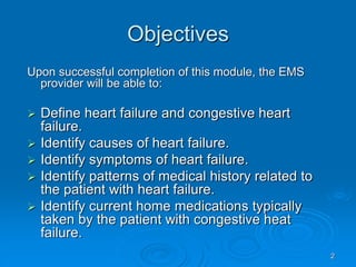
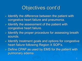
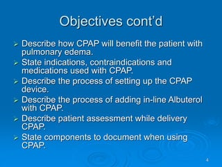
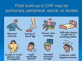
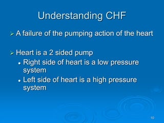
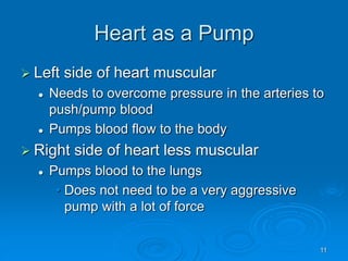
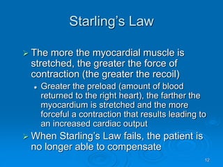
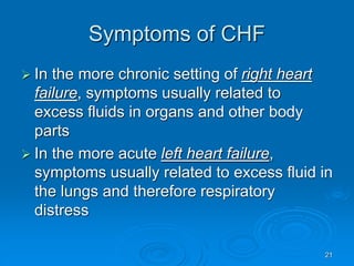
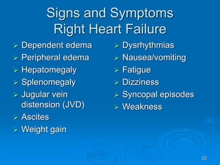
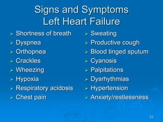
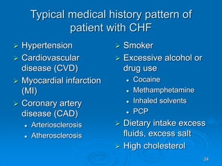
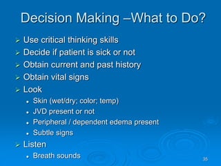
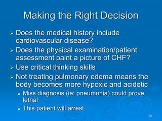
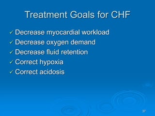
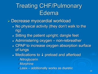
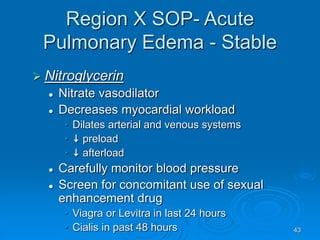
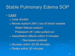
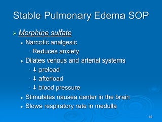
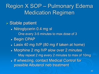
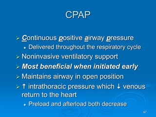
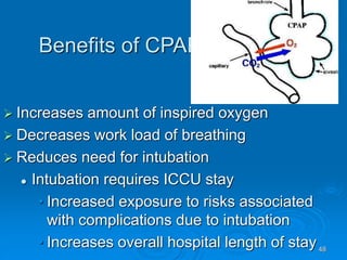
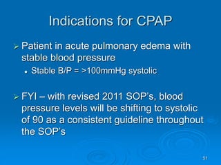
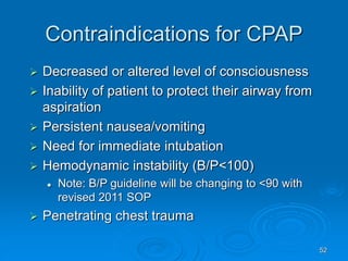
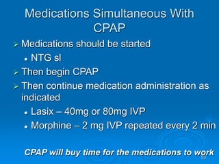
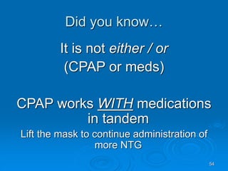
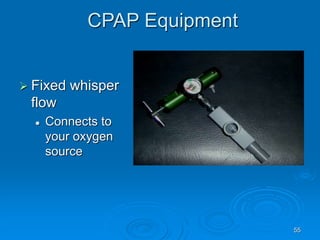
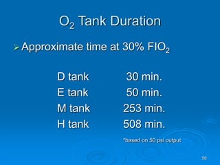
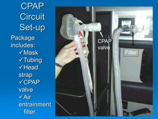
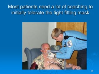
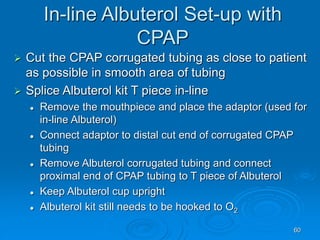
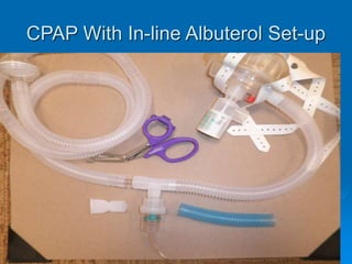
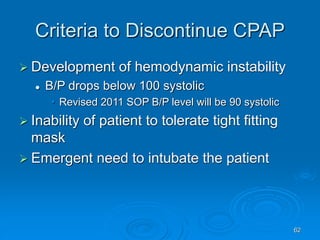
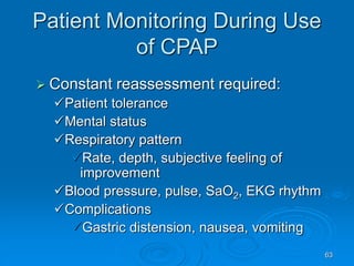
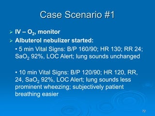
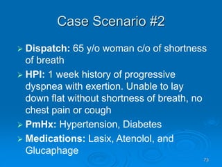
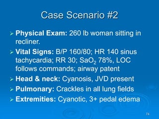
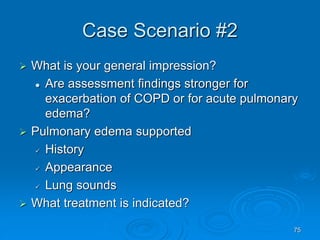
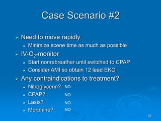
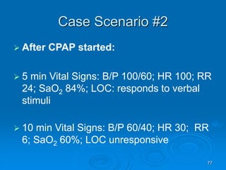
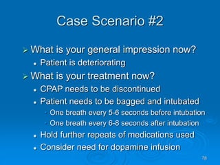
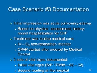
1
The Patient with Heart
Failure
CPAP as an Intervention
2
Objectives
Upon successful completion of this module, the EMS
provider will be able to:
 Define heart failure and congestive heart
failure.
 Identify causes of heart failure.
 Identify symptoms of heart failure.
 Identify patterns of medical history related to
the patient with heart failure.
 Identify current home medications typically
taken by the patient with congestive heat
failure.
3
Objectives cont’d
 Identify the difference between the patient with
congestive heart failure and pneumonia.
 Identify the assessment of the patient with
congestive heart failure.
 Identify the proper procedure for assessing breath
sounds.
 Identify treatment goals and options for congestive
heart failure following Region X SOP’s.
 Define CPAP as used by EMS for the patient with
pulmonary edema.
4
Objectives cont’d
 Describe how CPAP will benefit the patient with
pulmonary edema.
 State indications, contraindications and
medications used with CPAP.
 Describe the process of setting up the CPAP
device.
 Describe the process of adding in-line Albuterol
with CPAP.
 Describe patient assessment while delivery
CPAP.
 State components to document when using
CPAP.
5
Objectives cont’d
 Demonstrate the set up of CPAP.
 Demonstrate the set-up of regular and
in-line Albuterol.
 Demonstrate adding in-line Albuterol
with CPAP.
 Actively participate in case scenario
discussion.
 Successfully complete the post quiz with a
score of 80% or better.
6
What is Heart Failure?
 A clinical syndrome
 Heart’s mechanical performance (ie:
pumping action) is compromised
 Cardiac output unable to meet the demands
of the body’s needs
 Generally divided into backward
ventricular failure (right heart failure) and
forward ventricular failure (left heart
failure)
 Can be of a chronic or acute nature
7
Heart Failure
 Variety of causes
 Valve disease
 Heart disease
 Contributing factors to heart failure
 Diet - excess fluid or salt intake
 Hypertension
 Pulmonary embolism
 Excessive alcohol or drug usage
 Progression of an underlying disease
8
What is CHF?
 Congestive heart failure = CHF
 Condition of excess build-up of fluid in the
lungs and/or other body parts/organs
 Fluid build-up causes congestion in the
organs seen as edema
 May be brought on by diseased heart
valves, hypertension, or some form of
obstructive pulmonary disease
 Often a complication of AMI
9
Fluid build-up in CHF may be
pulmonary, peripheral, sacral, or ascites
10
Understanding CHF
 A failure of the pumping action of the heart
 Heart is a 2 sided pump
 Right side of heart is a low pressure
system
 Left side of heart is a high pressure
system
11
Heart as a Pump
 Left side of heart muscular
 Needs to overcome pressure in the arteries to
push/pump blood
 Pumps blood flow to the body
 Right side of heart less muscular
 Pumps blood to the lungs
• Does not need to be a very aggressive
pump with a lot of force
12
Starling’s Law
 The more the myocardial muscle is
stretched, the greater the force of
contraction (the greater the recoil)
 Greater the preload (amount of blood
returned to the right heart), the farther the
myocardium is stretched and the more
forceful a contraction that results leading to
an increased cardiac output
 When Starling’s Law fails, the patient is
no longer able to compensate
13
Hypertension
 B/P is a measurement
of force against the wall of the arteries
 When vessels stiffen due to calcium build-
up (arteriosclerosis) and plaque develops
(atherosclerosis), vessels are less
compliant
 Higher pressures are needed to pump
blood through stiffer vessels
14
Right Ventricular Failure
 Failure of right ventricle as a forward
pump
 Back pressure of blood into systemic
venous circulation system
 Common causes
 Left ventricular failure (AMI)
 Systemic hypertension
 Pulmonary hypertension
 Cor pulmonale – heart
disease due to pulmonary
disease
(ie; effects of COPD)
15
Progression of Right Heart Failure
 Right ventricle cannot eject all of the blood
out
 Fluid/pressure builds up
• In right atrium
Backs up into the venous system
Results in pedal/dependent
edema
 Visible as JVD
16
Right
Sided
Heart
Failure -
A
Systemic
Picture
17
Left Ventricular Failure
 Failure of left ventricle to function as a forward
pump
 Back pressure of blood into pulmonary circulation
 Often causes pulmonary edema
 Common causes
 Various types of heart disease
• Ischemia / acute MI
• Coronary artery disease (CAD)-
arteriosclerosis/atherosclerosis
• Valve disease
• Chronic hypertension -  afterload
• Dysrhythmias
18
Progression of
Left Ventricular Failure
 Left ventricle cannot eject all the blood
delivered from the right heart via the
lungs
 Left atrial pressure rises and transmitted
to pulmonary veins and capillaries
 These high pressures force blood plasma
into alveoli (ie: pulmonary edema)
 Oxygen capacity of lungs reduced
 Hypoxia develops
 Acidosis develops
19
Pulmonary
Edema
 Severest form
of congestive
heart failure
 Left ventricular forward failure
 Think left/lungs
 Patient develops respiratory distress due to
fluid in the lungs
 Note: extremely rare to have unilateral pulmonary
edema; then related to unusual pathology/med hx
20
Pathophysiological Changes in
Pulmonary Edema
 Left ventricle cannot empty effectively
 Fluid moves from capillary beds into
surrounding interstitial tissue  alveoli
 Fluid in alveoli impedes oxygen exchange
 Surfactant lining alveoli washes out
 Alveoli stiffen
 Alveoli collapse after each breath and are harder to
open
 Lungs develop  compliance,
airflow obstruction, hyperinflation
  to workload of breathing
21
Symptoms of CHF
 In the more chronic setting of right heart
failure, symptoms usually related to
excess fluids in organs and other body
parts
 In the more acute left heart failure,
symptoms usually related to excess fluid in
the lungs and therefore respiratory
distress
22
Signs and Symptoms
Right Heart Failure
 Dependent edema
 Peripheral edema
 Hepatomegaly
 Splenomegaly
 Jugular vein
distension (JVD)
 Ascites
 Weight gain
 Dysrhythmias
 Nausea/vomiting
 Fatigue
 Dizziness
 Syncopal episodes
 Weakness
23
Signs and Symptoms
Left Heart Failure
 Shortness of breath
 Dyspnea
 Orthopnea
 Crackles
 Wheezing
 Hypoxia
 Respiratory acidosis
 Chest pain
 Sweating
 Productive cough
 Blood tinged sputum
 Cyanosis
 Palpitations
 Dysrhythmias
 Hypertension
 Anxiety/restlessness
24
Typical medical history pattern of
patient with CHF
 Hypertension
 Cardiovascular
disease (CVD)
 Myocardial infarction
(MI)
 Coronary artery
disease (CAD)
 Arteriosclerosis
 Atherosclerosis
 Smoker
 Excessive alcohol or
drug use
 Cocaine
 Methamphetamine
 Inhaled solvents
 PCP
 Dietary intake excess
fluids, excess salt
 High cholesterol
25
Typical home medication history
pattern of patient with CHF
 Diuretic
 Digoxin
  contractility force of
the heart (inotropic)
 Home oxygen therapy
 Anti-hypertensive
 ACE inhibitors (end in “pril”)
 Beta blockers
•  heart rate & force
of contractions  B/P
• Often end in “olol”
 Calcium channel
inhibitors
• Slows movement of
calcium into small
muscles wrapped
around blood
vessels relaxing
blood vessels
•  peripheral
vascular resistance
relaxing blood
vessels
26
Herbal remedies that may be harmful
when mixed with heart failure
 St. John’s wort
 Ephedra
 Gingko biloba
 Kava
 Licorice
 Ginseng
 Aconite
 Alisma plantago
 Bearberry buchu
 Couch grass
 Dandelion
 Horsetail rush
 Juniper
27
Evaluation CHF/PE Pneumonia COPD
History HTN, heart
problems
n/a Lung problems
Dyspnea Orthopnea,
PND
Orthopnea
possible
Chronic;
pursed lips
Recent hx Acute weight
gain, dependent
edema
Fever, malaise Gradual
weight loss
Cough Frothy
sputum
Productive thick
green
Chronic;
productive
Onset Rapid Gradual Gradual
B/P High Normal Normal
Meds Dig, anti-HTN,
diuretic
Antibiotic, cold prep Bronchodilators,
steroids
Tx O2, NTG,
lasix, MS
O2, neb, fluids O2, neb
28
Separating Signs/Symptoms
Symptom CHF/PE Pneumonia COPD
SOB Yes Yes Yes
Cough Maybe Yes Early a.m.
Sputum Frothy pink Yellow/green Thick brown
Fever No Yes No
Skin Cold/clammy Hot/dry Normal or dusky
Chest pain Possible Maybe No
Smoking hx Possible Possible Usually
Wheezing Maybe;
bilateral
Maybe; same
side as disease
Usually,
bilateral
Crackles Yes; bilateral Maybe; same
side as disease
No
29
A Note…
“Old geezers don’t become new
wheezers!”
 COPD develops over a long period of time. If
an elderly person does not have a history of
COPD and they are suddenly wheezing, think
a cardiac problem or pulmonary edema.
Assume the worst,
hope for the best
30
Patient Assessment - CHF
 Acute findings
 Recent trouble sleeping
•  trips to the bathroom at night
• Orthopnea with  number of pillows
• Sleeping in the recliner
• New episodes of paroxysmal nocturnal
dyspnea (PND)
•  use of nitroglycerin to stop chest pain
•  use of oxygen
31
Patient Assessment - CHF
 General impression
 Labored respirations
 Audible noisy respirations
 Tripod positioning
 Frothy sputum production
  work of breathing – retractions, tachypnea
 Wheezing/crackles bilaterally
 Diaphoretic
 Change in skin color from norm
 Severe anxiety/restlessness
 Severe hypertension may be present
32
Patient Assessment - CHF
 Signs and symptoms pulmonary edema
 Tachypnea
 Orthopnea
 PND
 Noisy labored respirations
 Fine crackles/rales
 Wheezing – “cardiac asthma”
 Coarse crackles/rhonchi larger airways
 Coughing with frothy blood tinged sputum
33
Obtaining Breath Sounds
 Use flat diaphragm surface of stethoscope
 Rub stethoscope head between hands to
warm it up before placing on patient’s skin
 If audible sounds are heard, ask patient to
cough gently to clear upper airway
 Auscultate side to side and top to bottom
 Anterior: Posterior:
34
Adventitious (Extra) Breath
Sounds
 Check for asymmetry
 Crackles: high pitched, continuous sounds
like rubbing hair between fingers
 Wheezes: generally high pitched, of musical
quality
 Stridor: Harsh inspiratory wheeze indicating
upper airway obstruction
 Rhonchi: snoring or gurgling quality
 Any extra sound not a crackle or wheeze
is usually rhonchi
35
Decision Making –What to Do?
 Use critical thinking skills
 Decide if patient is sick or not
 Obtain current and past history
 Obtain vital signs
 Look
 Skin (wet/dry; color; temp)
 JVD present or not
 Peripheral / dependent edema present
 Subtle signs
 Listen
 Breath sounds
36
Making the Right Decision
 Does the medical history include
cardiovascular disease?
 Does the physical examination/patient
assessment paint a picture of CHF?
 Use critical thinking skills
 Not treating pulmonary edema means the
body becomes more hypoxic and acidotic
 Miss diagnosis (ie: pneumonia) could prove
lethal
 This patient will arrest
37
Treatment Goals for CHF
 Decrease myocardial workload
 Decrease oxygen demand
 Decrease fluid retention
 Correct hypoxia
 Correct acidosis
38
Treating CHF/Pulmonary
Edema
 Decrease myocardial workload
 No physical activity (they don’t walk to the
rig)
 Sitting the patient upright; dangle feet
 Administering oxygen – non-rebreather
 CPAP to increase oxygen absorption surface
of lungs
 Medications to  preload and afterload
Nitroglycerin
Morphine
Lasix – additionally works as diuretic
39
Treatment Goals for Pneumonia
 Supply supplemental oxygen as needed
 Treat the bacterial infection
 Hydrate the patient
• Usually found in the elderly
• Often vague symptoms; use to feeling ill
• Immune system often already weakened
so mortality rate is high with this diagnosis
40
Region X SOP- Acute
Pulmonary Edema
 Begin Routine Medical Care
 Take standard precautions
 Perform assessments
 Identify priority patient and make transport
decisions
• Stay and play?
• Load N go?
 Perform routine tasks
• IV-O2-monitor
41
What About the IV and
Nitroglycerin?
 Region X Medical Directors discussion:
 Majority of patients in pulmonary edema will be
hypertensive
 Nitroglycerin will help reduce preload which will
lower blood pressure (beneficial)
 Do not delay NTG dose, if no contraindications,
to start the IV
• If patient deteriorates before IV established,
can always place an IO
42
Region X SOP- Acute
Pulmonary Edema
 Determine if the patient is stable or
unstable
 Stability guided by status of perfusion
B/P and level of consciousness
 If stable, the patient can receive more
aggressive care including medications and
procedures (ie: CPAP)
 If unstable, Medical Control needs to
coordinate degree of care provided in the
field (ie: meds and CPAP)
43
Region X SOP- Acute
Pulmonary Edema - Stable
 Nitroglycerin
 Nitrate vasodilator
 Decreases myocardial workload
• Dilates arterial and venous systems
•  preload
•  afterload
 Carefully monitor blood pressure
 Screen for concomitant use of sexual
enhancement drug
• Viagra or Levitra in last 24 hours
• Cialis in past 48 hours
44
Stable Pulmonary Edema SOP
 Lasix
 Loop diuretic
 Moves sodium (NA+) out of blood vessels
• Water follows sodium
• Potassium (K+) also pulled out
 Vasodilation effects within 5 minutes
• Decreases preload
 Diuresis within 20-30 minutes
 Peaks within 30 minutes
45
Stable Pulmonary Edema SOP
 Morphine sulfate
 Narcotic analgesic
• Reduces anxiety
 Dilates venous and arterial systems
•  preload
•  afterload
•  blood pressure
 Stimulates nausea center in the brain
 Slows respiratory rate in medulla
46
Region X SOP – Pulmonary Edema
Medication Regimen
 Stable patient
 Nitroglycerin 0.4 mg sl
• One every 3-5 minutes to max dose of 3
 Begin CPAP
 Lasix 40 mg IVP (80 mg if taken at home)
 Morphine 2 mg IVP slow over 2 minutes
• May repeat 2 mg every 2 minutes to max of 10mg
 If wheezing, contact Medical Control for
possible Albuterol neb treatment
47
CPAP
 Continuous positive airway pressure
 Delivered throughout the respiratory cycle
 Noninvasive ventilatory support
 Most beneficial when initiated early
 Maintains airway in open position
  intrathoracic pressure which  venous
return to the heart
 Preload and afterload both decrease
48
Benefits of CPAP
 Increases amount of inspired oxygen
 Decreases work load of breathing
 Reduces need for intubation
 Intubation requires ICCU stay
• Increased exposure to risks associated
with complications due to intubation
• Increases overall hospital length of stay
49
Redistribution of extravascular lung
water during use of CPAP
Without CPAP With CPAP
50
51
Indications for CPAP
 Patient in acute pulmonary edema with
stable blood pressure
 Stable B/P = >100mmHg systolic
 FYI – with revised 2011 SOP’s, blood
pressure levels will be shifting to systolic
of 90 as a consistent guideline throughout
the SOP’s
52
Contraindications for CPAP
 Decreased or altered level of consciousness
 Inability of patient to protect their airway from
aspiration
 Persistent nausea/vomiting
 Need for immediate intubation
 Hemodynamic instability (B/P<100)
 Note: B/P guideline will be changing to <90 with
revised 2011 SOP
 Penetrating chest trauma
53
Medications Simultaneous With
CPAP
 Medications should be started
 NTG sl
 Then begin CPAP
 Then continue medication administration as
indicated
 Lasix – 40mg or 80mg IVP
 Morphine – 2 mg IVP repeated every 2 min
CPAP will buy time for the medications to work
54
Did you know…
It is not either / or
(CPAP or meds)
CPAP works WITH medications
in tandem
Lift the mask to continue administration of
more NTG
55
CPAP Equipment
 Fixed whisper
flow
 Connects to
your oxygen
source
56
O2 Tank Duration
Approximate time at 30% FIO2
D tank 30 min.
E tank 50 min.
M tank 253 min.
H tank 508 min.
*based on 50 psi output
57
CPAP
Circuit
Set-up
Package
includes:
Mask
Tubing
Head
strap
CPAP
valve
Air
entrainment
filter
Filter
CPAP
valve
58
Most patients need a lot of coaching to
initially tolerate the tight fitting mask
59
If The Patient is Wheezing
 Contact Medical Control to consider an
order for Albuterol via nebulizer
 Medical Control needs to give this
physician’s order
 Contact ECRN on radio
• Needs to give the ED MD a report
• Obtains MD’s order
• Relays the response to EMS
 If Albuterol is given, monitor for cardiac
side effects (ie: tachycardia)
60
In-line Albuterol Set-up with
CPAP
 Cut the CPAP corrugated tubing as close to patient
as possible in smooth area of tubing
 Splice Albuterol kit T piece in-line
 Remove the mouthpiece and place the adaptor (used for
in-line Albuterol)
 Connect adaptor to distal cut end of corrugated CPAP
tubing
 Remove Albuterol corrugated tubing and connect
proximal end of CPAP tubing to T piece of Albuterol
 Keep Albuterol cup upright
 Albuterol kit still needs to be hooked to O2
61
CPAP With In-line Albuterol Set-up
62
Criteria to Discontinue CPAP
 Development of hemodynamic instability
 B/P drops below 100 systolic
• Revised 2011 SOP B/P level will be 90 systolic
 Inability of patient to tolerate tight fitting
mask
 Emergent need to intubate the patient
63
Patient Monitoring During Use
of CPAP
 Constant reassessment required:
Patient tolerance
Mental status
Respiratory pattern
Rate, depth, subjective feeling of
improvement
Blood pressure, pulse, SaO2, EKG rhythm
Complications
Gastric distension, nausea, vomiting
64
Monitoring Improvement With
CPAP
 It’s working when:
 Level of distress decreases
 Respiratory rate is returning toward normal
 Pulse oximetry (SaO2) increasing
 Pulse rate decreasing toward normal
 Decrease in use of accessory muscles
 Ability to speak in fuller sentences returning
65
Contacting Medical Control
 Remember:
 Early communication with receiving
hospital
 Hospital needs to get their regulator for
oxygen source connection
• Usually not kept in each room
66
Documentation With CPAP
 Assessment leading your general
impression to a diagnosis of pulmonary
edema
 CPAP level provided (10cmH2O)
 FiO2 provided (100%)
 SaO2 serial levels
 Vital signs over time
 Response to treatment
 Any adverse reactions noted
67
So, What’s Different About BiPAP?
 Bi-level positive airway pressure
 Uses 2 levels of pressure
 Helps move more air into lungs without need
to exhale against higher pressures
 CPAP is a larger & noisier machine
 Uses extra effort to exhale and can be tiring
 Both can be used for sleep apnea
 BiPAP easier on those with COPD and
neuromuscular diseases
68
Case Scenarios
Small Group and Large Group
Discussions
 Read the presentation
 Form a general impression
 Discuss treatment options
 Discuss what/how/when to reassess the
patient
 Decide what treatment to continue or what
adjustments need to be made
 Note: Additional questions are asked on ppt that can be
discussed during group presentations.
69
Case Scenario #1
 Dispatch: You are called to a 70 y/o man
c/o breathing problems
 HPI: Increasing shortness of breath for
1 day despite the use of inhalers
 PmHx: COPD, Hypertension, and
Diabetes
 Medications: Albuterol Inhaler, Lasix, and
Aspirin
 Allergies: Penicillin
70
Case Scenario #1
 Physical Exam: Thin white man on home
oxygen breathing through pursed lips sitting in a
tripod position
 Vital Signs: B/P 180/90; HR 120 sinus
tachycardia; RR 30; SaO2 88%; LOC alert;
airway patent
 Head & neck: Perioral cyanosis, no JVD
 Pulmonary: Lung auscultation reveals
inspiratory and expiratory wheezes
 Extremities: Cyanotic, no pedal edema
71
Case Scenario #1
 What is your general impression?
 Are assessment findings stronger for
exacerbation of COPD or for acute
pulmonary edema?
 COPD supported
 History
 Appearance
 Lung sounds
 What treatment is indicated?
72
Case Scenario #1
 IV – O2, monitor
 Albuterol nebulizer started:
• 5 min Vital Signs: B/P 160/90; HR 130; RR 24;
SaO2 92%, LOC Alert; lung sounds unchanged
• 10 min Vital Signs: B/P 120/90; HR 120, RR,
24, SaO2 92%, LOC Alert; lung sounds less
prominent wheezing; subjectively patient
breathing easier
73
Case Scenario #2
 Dispatch: 65 y/o woman c/o of shortness
of breath
 HPI: 1 week history of progressive
dyspnea with exertion. Unable to lay
down flat without shortness of breath, no
chest pain or cough
 PmHx: Hypertension, Diabetes
 Medications: Lasix, Atenolol, and
Glucaphage
74
Case Scenario #2
 Physical Exam: 260 lb woman sitting in
recliner.
 Vital Signs: B/P 160/80; HR 140 sinus
tachycardia; RR 30; SaO2 78%, LOC
follows commands; airway patent
 Head & neck: Cyanosis, JVD present
 Pulmonary: Crackles in all lung fields
 Extremities: Cyanotic, 3+ pedal edema
75
Case Scenario #2
 What is your general impression?
 Are assessment findings stronger for
exacerbation of COPD or for acute pulmonary
edema?
 Pulmonary edema supported
 History
 Appearance
 Lung sounds
 What treatment is indicated?
76
Case Scenario #2
 Need to move rapidly
 Minimize scene time as much as possible
 IV-O2-monitor
 Start nonrebreather until switched to CPAP
 Consider AMI so obtain 12 lead EKG
 Any contraindications to treatment?
 Nitroglycerin?
 CPAP?
 Lasix?
 Morphine?
NO
NO
NO
NO
77
Case Scenario #2
 After CPAP started:
 5 min Vital Signs: B/P 100/60; HR 100; RR
24; SaO2 84%; LOC: responds to verbal
stimuli
 10 min Vital Signs: B/P 60/40; HR 30; RR
6; SaO2 60%; LOC unresponsive
78
Case Scenario #2
 What is your general impression now?
 Patient is deteriorating
 What is your treatment now?
 CPAP needs to be discontinued
 Patient needs to be bagged and intubated
• One breath every 5-6 seconds before intubation
• One breath every 6-8 seconds after intubation
 Hold further repeats of medications used
 Consider need for dopamine infusion
79
Case Scenario #3 Documentation
 Initial impression was acute pulmonary edema
 Based on physical assessment; history;
recent hospitalization for CHF
 Treatment was routine medical care
 IV – O2 non-rebreather- monitor
 CPAP started after ordered by Medical
Control
 2 sets of vital signs documented
 Initial vital signs (B/P 170/98 – 92 – 32)
 Second reading at the hospital
80
Case Scenario #3 Comments
Documented
 Upon arrival patient found sitting upright,
agitated, complaining of chest pain and
difficulty breathing. Audible congested
breathing standing next to patient. Unable to
complete a full sentence. Bilateral pedal
edema noted. Began oxygen via
nonrebreather. IV started. Moved patient to
ambulance. Medical Control contacted and
ordered CPAP to be started. Patient becoming
more agitated. After 5 minutes, SaO2
increasing. Patient stated breathing was
becoming easier.
81
Case Scenario #3 Documentation
cont’d
 Patient transported sitting upright.
Continued CPAP during entire call.
Transported patient into ED on portable O2
with CPAP continued.
82
Case Scenario #3 Documentation
cont’d
 Pt contact: 0954
 Depart scene: 1025
 “Drugs”
 0959 - Oxygen - 15 l – non-rebreather
 1001 – 0.9 NS 1000ml – TKO – IV
 1005 – CPAP /oxygen – 15l – CPAP mask
 “`Cardiac rhythm”
 0958 – sinus
 1035 - sinus
83
Case Scenario #3 Documentation
Discussion
What went well?
 Recognized pulmonary edema
 CPAP used with positive patient response
84
Case Scenario #3 Documentation
Discussion
 What could be improved upon?
 Long on-scene time (0954 – 1025 -31 mins)
 Delay in initiating O2 therapy – 5 minutes
 Waited for MC to order CPAP – 11 min delay
• No Medical Control direction needed to initiate
 No other meds given for pulmonary edema
 Only 2 sets of vital signs taken on a critical
patient
85
Case Scenario #4
 Dispatch: You are called to a 84 year-old
female c/o breathing problems
 HPI: Running low grade fevers, not feeling
well for 4 days
 PmHx: MI, Hypertension, TIA’s
 Medications: Plavix, Lasix, Lisinopril
 Allergies: Iodine, shellfish
86
Case Scenario #4
 Physical Exam:
 Vital Signs: B/P 142/80; HR 96 sinus
rhythm; RR 28; SaO2 92%, LOC follows
commands; airway patent
 Head & neck: Pale, no JVD
 Pulmonary: Crackles in right lower lung
field
 Extremities: Pale, pedal pulses palpable
87
Case Scenario #4
 What is your general impression?
 Are assessment findings stronger for
acute pulmonary edema or pneumonia?
 Pneumonia supported?
 History
 Appearance
 Lung sounds not so helpful
 What treatment is indicated?
88
Case Scenario #4
 What is your treatment now?
 IV-O2-monitor
 Fluids
• Faster than keep open but not a fluid
challenge
 Diagnosis confirmed at the hospital with
chest x-ray and labs
89
Case Scenario #4
 Patients with pneumonia need fluids
 Patients with congestive heart failure need
fluid restrictions
 A wrong diagnosis and therefore wrong
treatment approach could be harmful for
both patients
90
Case Scenario #5
 Dispatch: You are called to a home for a 78
year-old male with severe SOB
 HPI: Has been getting progressively SOB past 2
days; slept in recliner last night
 PmHx: MI x3; hypertension, diverticulitis,
seizures
 Medications: Aspirin, Hydrodiuril, Verapamil,
NTG PRN, Coumadin, Phenobarbital
 Allergies: none
91
Case Scenario #5
 Physical Exam:
 Vital Signs: B/P 172/96; HR 110 sinus
tachycardia; RR 36; SaO2 88%, LOC follows
commands; extremely anxious; airway patent
 Head & neck: JVD
 Pulmonary: Crackles mid way up lung fields
bilaterally
 Extremities: Cyanotic, pedal edema palpable
92
Case Scenario #5
 What is your general impression?
 What is your treatment plan?
 Write a run report
 Include initial assessment
 Document treatment interventions indicated
 Document reassessment performed
 Discuss as a group what needs to be
included
93
94
95
Bibliography
 Bledsoe, B., Porter, R., Cherry, R. Paramedic
Care: Principles and Practices. Brady. 2009.
 Limmer, D., O’Keefe, M. Emergency Care, 10th
Edition. Brady. 2005.
 Region X SOP’s March 2007; Amended
January 1, 2008.
 http://whisperflow.respironics.com/
 www.emsworld.com
 Variety internet websites for CPAP and
pulmonary edema

The Cardiac Failure by CPAP intervention

  • 1.
    1 The Patient withHeart Failure CPAP as an Intervention
  • 2.
    2 Objectives Upon successful completionof this module, the EMS provider will be able to:  Define heart failure and congestive heart failure.  Identify causes of heart failure.  Identify symptoms of heart failure.  Identify patterns of medical history related to the patient with heart failure.  Identify current home medications typically taken by the patient with congestive heat failure.
  • 3.
    3 Objectives cont’d  Identifythe difference between the patient with congestive heart failure and pneumonia.  Identify the assessment of the patient with congestive heart failure.  Identify the proper procedure for assessing breath sounds.  Identify treatment goals and options for congestive heart failure following Region X SOP’s.  Define CPAP as used by EMS for the patient with pulmonary edema.
  • 4.
    4 Objectives cont’d  Describehow CPAP will benefit the patient with pulmonary edema.  State indications, contraindications and medications used with CPAP.  Describe the process of setting up the CPAP device.  Describe the process of adding in-line Albuterol with CPAP.  Describe patient assessment while delivery CPAP.  State components to document when using CPAP.
  • 5.
    5 Objectives cont’d  Demonstratethe set up of CPAP.  Demonstrate the set-up of regular and in-line Albuterol.  Demonstrate adding in-line Albuterol with CPAP.  Actively participate in case scenario discussion.  Successfully complete the post quiz with a score of 80% or better.
  • 6.
    6 What is HeartFailure?  A clinical syndrome  Heart’s mechanical performance (ie: pumping action) is compromised  Cardiac output unable to meet the demands of the body’s needs  Generally divided into backward ventricular failure (right heart failure) and forward ventricular failure (left heart failure)  Can be of a chronic or acute nature
  • 7.
    7 Heart Failure  Varietyof causes  Valve disease  Heart disease  Contributing factors to heart failure  Diet - excess fluid or salt intake  Hypertension  Pulmonary embolism  Excessive alcohol or drug usage  Progression of an underlying disease
  • 8.
    8 What is CHF? Congestive heart failure = CHF  Condition of excess build-up of fluid in the lungs and/or other body parts/organs  Fluid build-up causes congestion in the organs seen as edema  May be brought on by diseased heart valves, hypertension, or some form of obstructive pulmonary disease  Often a complication of AMI
  • 9.
    9 Fluid build-up inCHF may be pulmonary, peripheral, sacral, or ascites
  • 10.
    10 Understanding CHF  Afailure of the pumping action of the heart  Heart is a 2 sided pump  Right side of heart is a low pressure system  Left side of heart is a high pressure system
  • 11.
    11 Heart as aPump  Left side of heart muscular  Needs to overcome pressure in the arteries to push/pump blood  Pumps blood flow to the body  Right side of heart less muscular  Pumps blood to the lungs • Does not need to be a very aggressive pump with a lot of force
  • 12.
    12 Starling’s Law  Themore the myocardial muscle is stretched, the greater the force of contraction (the greater the recoil)  Greater the preload (amount of blood returned to the right heart), the farther the myocardium is stretched and the more forceful a contraction that results leading to an increased cardiac output  When Starling’s Law fails, the patient is no longer able to compensate
  • 13.
    13 Hypertension  B/P isa measurement of force against the wall of the arteries  When vessels stiffen due to calcium build- up (arteriosclerosis) and plaque develops (atherosclerosis), vessels are less compliant  Higher pressures are needed to pump blood through stiffer vessels
  • 14.
    14 Right Ventricular Failure Failure of right ventricle as a forward pump  Back pressure of blood into systemic venous circulation system  Common causes  Left ventricular failure (AMI)  Systemic hypertension  Pulmonary hypertension  Cor pulmonale – heart disease due to pulmonary disease (ie; effects of COPD)
  • 15.
    15 Progression of RightHeart Failure  Right ventricle cannot eject all of the blood out  Fluid/pressure builds up • In right atrium Backs up into the venous system Results in pedal/dependent edema  Visible as JVD
  • 16.
  • 17.
    17 Left Ventricular Failure Failure of left ventricle to function as a forward pump  Back pressure of blood into pulmonary circulation  Often causes pulmonary edema  Common causes  Various types of heart disease • Ischemia / acute MI • Coronary artery disease (CAD)- arteriosclerosis/atherosclerosis • Valve disease • Chronic hypertension -  afterload • Dysrhythmias
  • 18.
    18 Progression of Left VentricularFailure  Left ventricle cannot eject all the blood delivered from the right heart via the lungs  Left atrial pressure rises and transmitted to pulmonary veins and capillaries  These high pressures force blood plasma into alveoli (ie: pulmonary edema)  Oxygen capacity of lungs reduced  Hypoxia develops  Acidosis develops
  • 19.
    19 Pulmonary Edema  Severest form ofcongestive heart failure  Left ventricular forward failure  Think left/lungs  Patient develops respiratory distress due to fluid in the lungs  Note: extremely rare to have unilateral pulmonary edema; then related to unusual pathology/med hx
  • 20.
    20 Pathophysiological Changes in PulmonaryEdema  Left ventricle cannot empty effectively  Fluid moves from capillary beds into surrounding interstitial tissue  alveoli  Fluid in alveoli impedes oxygen exchange  Surfactant lining alveoli washes out  Alveoli stiffen  Alveoli collapse after each breath and are harder to open  Lungs develop  compliance, airflow obstruction, hyperinflation   to workload of breathing
  • 21.
    21 Symptoms of CHF In the more chronic setting of right heart failure, symptoms usually related to excess fluids in organs and other body parts  In the more acute left heart failure, symptoms usually related to excess fluid in the lungs and therefore respiratory distress
  • 22.
    22 Signs and Symptoms RightHeart Failure  Dependent edema  Peripheral edema  Hepatomegaly  Splenomegaly  Jugular vein distension (JVD)  Ascites  Weight gain  Dysrhythmias  Nausea/vomiting  Fatigue  Dizziness  Syncopal episodes  Weakness
  • 23.
    23 Signs and Symptoms LeftHeart Failure  Shortness of breath  Dyspnea  Orthopnea  Crackles  Wheezing  Hypoxia  Respiratory acidosis  Chest pain  Sweating  Productive cough  Blood tinged sputum  Cyanosis  Palpitations  Dysrhythmias  Hypertension  Anxiety/restlessness
  • 24.
    24 Typical medical historypattern of patient with CHF  Hypertension  Cardiovascular disease (CVD)  Myocardial infarction (MI)  Coronary artery disease (CAD)  Arteriosclerosis  Atherosclerosis  Smoker  Excessive alcohol or drug use  Cocaine  Methamphetamine  Inhaled solvents  PCP  Dietary intake excess fluids, excess salt  High cholesterol
  • 25.
    25 Typical home medicationhistory pattern of patient with CHF  Diuretic  Digoxin   contractility force of the heart (inotropic)  Home oxygen therapy  Anti-hypertensive  ACE inhibitors (end in “pril”)  Beta blockers •  heart rate & force of contractions  B/P • Often end in “olol”  Calcium channel inhibitors • Slows movement of calcium into small muscles wrapped around blood vessels relaxing blood vessels •  peripheral vascular resistance relaxing blood vessels
  • 26.
    26 Herbal remedies thatmay be harmful when mixed with heart failure  St. John’s wort  Ephedra  Gingko biloba  Kava  Licorice  Ginseng  Aconite  Alisma plantago  Bearberry buchu  Couch grass  Dandelion  Horsetail rush  Juniper
  • 27.
    27 Evaluation CHF/PE PneumoniaCOPD History HTN, heart problems n/a Lung problems Dyspnea Orthopnea, PND Orthopnea possible Chronic; pursed lips Recent hx Acute weight gain, dependent edema Fever, malaise Gradual weight loss Cough Frothy sputum Productive thick green Chronic; productive Onset Rapid Gradual Gradual B/P High Normal Normal Meds Dig, anti-HTN, diuretic Antibiotic, cold prep Bronchodilators, steroids Tx O2, NTG, lasix, MS O2, neb, fluids O2, neb
  • 28.
    28 Separating Signs/Symptoms Symptom CHF/PEPneumonia COPD SOB Yes Yes Yes Cough Maybe Yes Early a.m. Sputum Frothy pink Yellow/green Thick brown Fever No Yes No Skin Cold/clammy Hot/dry Normal or dusky Chest pain Possible Maybe No Smoking hx Possible Possible Usually Wheezing Maybe; bilateral Maybe; same side as disease Usually, bilateral Crackles Yes; bilateral Maybe; same side as disease No
  • 29.
    29 A Note… “Old geezersdon’t become new wheezers!”  COPD develops over a long period of time. If an elderly person does not have a history of COPD and they are suddenly wheezing, think a cardiac problem or pulmonary edema. Assume the worst, hope for the best
  • 30.
    30 Patient Assessment -CHF  Acute findings  Recent trouble sleeping •  trips to the bathroom at night • Orthopnea with  number of pillows • Sleeping in the recliner • New episodes of paroxysmal nocturnal dyspnea (PND) •  use of nitroglycerin to stop chest pain •  use of oxygen
  • 31.
    31 Patient Assessment -CHF  General impression  Labored respirations  Audible noisy respirations  Tripod positioning  Frothy sputum production   work of breathing – retractions, tachypnea  Wheezing/crackles bilaterally  Diaphoretic  Change in skin color from norm  Severe anxiety/restlessness  Severe hypertension may be present
  • 32.
    32 Patient Assessment -CHF  Signs and symptoms pulmonary edema  Tachypnea  Orthopnea  PND  Noisy labored respirations  Fine crackles/rales  Wheezing – “cardiac asthma”  Coarse crackles/rhonchi larger airways  Coughing with frothy blood tinged sputum
  • 33.
    33 Obtaining Breath Sounds Use flat diaphragm surface of stethoscope  Rub stethoscope head between hands to warm it up before placing on patient’s skin  If audible sounds are heard, ask patient to cough gently to clear upper airway  Auscultate side to side and top to bottom  Anterior: Posterior:
  • 34.
    34 Adventitious (Extra) Breath Sounds Check for asymmetry  Crackles: high pitched, continuous sounds like rubbing hair between fingers  Wheezes: generally high pitched, of musical quality  Stridor: Harsh inspiratory wheeze indicating upper airway obstruction  Rhonchi: snoring or gurgling quality  Any extra sound not a crackle or wheeze is usually rhonchi
  • 35.
    35 Decision Making –Whatto Do?  Use critical thinking skills  Decide if patient is sick or not  Obtain current and past history  Obtain vital signs  Look  Skin (wet/dry; color; temp)  JVD present or not  Peripheral / dependent edema present  Subtle signs  Listen  Breath sounds
  • 36.
    36 Making the RightDecision  Does the medical history include cardiovascular disease?  Does the physical examination/patient assessment paint a picture of CHF?  Use critical thinking skills  Not treating pulmonary edema means the body becomes more hypoxic and acidotic  Miss diagnosis (ie: pneumonia) could prove lethal  This patient will arrest
  • 37.
    37 Treatment Goals forCHF  Decrease myocardial workload  Decrease oxygen demand  Decrease fluid retention  Correct hypoxia  Correct acidosis
  • 38.
    38 Treating CHF/Pulmonary Edema  Decreasemyocardial workload  No physical activity (they don’t walk to the rig)  Sitting the patient upright; dangle feet  Administering oxygen – non-rebreather  CPAP to increase oxygen absorption surface of lungs  Medications to  preload and afterload Nitroglycerin Morphine Lasix – additionally works as diuretic
  • 39.
    39 Treatment Goals forPneumonia  Supply supplemental oxygen as needed  Treat the bacterial infection  Hydrate the patient • Usually found in the elderly • Often vague symptoms; use to feeling ill • Immune system often already weakened so mortality rate is high with this diagnosis
  • 40.
    40 Region X SOP-Acute Pulmonary Edema  Begin Routine Medical Care  Take standard precautions  Perform assessments  Identify priority patient and make transport decisions • Stay and play? • Load N go?  Perform routine tasks • IV-O2-monitor
  • 41.
    41 What About theIV and Nitroglycerin?  Region X Medical Directors discussion:  Majority of patients in pulmonary edema will be hypertensive  Nitroglycerin will help reduce preload which will lower blood pressure (beneficial)  Do not delay NTG dose, if no contraindications, to start the IV • If patient deteriorates before IV established, can always place an IO
  • 42.
    42 Region X SOP-Acute Pulmonary Edema  Determine if the patient is stable or unstable  Stability guided by status of perfusion B/P and level of consciousness  If stable, the patient can receive more aggressive care including medications and procedures (ie: CPAP)  If unstable, Medical Control needs to coordinate degree of care provided in the field (ie: meds and CPAP)
  • 43.
    43 Region X SOP-Acute Pulmonary Edema - Stable  Nitroglycerin  Nitrate vasodilator  Decreases myocardial workload • Dilates arterial and venous systems •  preload •  afterload  Carefully monitor blood pressure  Screen for concomitant use of sexual enhancement drug • Viagra or Levitra in last 24 hours • Cialis in past 48 hours
  • 44.
    44 Stable Pulmonary EdemaSOP  Lasix  Loop diuretic  Moves sodium (NA+) out of blood vessels • Water follows sodium • Potassium (K+) also pulled out  Vasodilation effects within 5 minutes • Decreases preload  Diuresis within 20-30 minutes  Peaks within 30 minutes
  • 45.
    45 Stable Pulmonary EdemaSOP  Morphine sulfate  Narcotic analgesic • Reduces anxiety  Dilates venous and arterial systems •  preload •  afterload •  blood pressure  Stimulates nausea center in the brain  Slows respiratory rate in medulla
  • 46.
    46 Region X SOP– Pulmonary Edema Medication Regimen  Stable patient  Nitroglycerin 0.4 mg sl • One every 3-5 minutes to max dose of 3  Begin CPAP  Lasix 40 mg IVP (80 mg if taken at home)  Morphine 2 mg IVP slow over 2 minutes • May repeat 2 mg every 2 minutes to max of 10mg  If wheezing, contact Medical Control for possible Albuterol neb treatment
  • 47.
    47 CPAP  Continuous positiveairway pressure  Delivered throughout the respiratory cycle  Noninvasive ventilatory support  Most beneficial when initiated early  Maintains airway in open position   intrathoracic pressure which  venous return to the heart  Preload and afterload both decrease
  • 48.
    48 Benefits of CPAP Increases amount of inspired oxygen  Decreases work load of breathing  Reduces need for intubation  Intubation requires ICCU stay • Increased exposure to risks associated with complications due to intubation • Increases overall hospital length of stay
  • 49.
    49 Redistribution of extravascularlung water during use of CPAP Without CPAP With CPAP
  • 50.
  • 51.
    51 Indications for CPAP Patient in acute pulmonary edema with stable blood pressure  Stable B/P = >100mmHg systolic  FYI – with revised 2011 SOP’s, blood pressure levels will be shifting to systolic of 90 as a consistent guideline throughout the SOP’s
  • 52.
    52 Contraindications for CPAP Decreased or altered level of consciousness  Inability of patient to protect their airway from aspiration  Persistent nausea/vomiting  Need for immediate intubation  Hemodynamic instability (B/P<100)  Note: B/P guideline will be changing to <90 with revised 2011 SOP  Penetrating chest trauma
  • 53.
    53 Medications Simultaneous With CPAP Medications should be started  NTG sl  Then begin CPAP  Then continue medication administration as indicated  Lasix – 40mg or 80mg IVP  Morphine – 2 mg IVP repeated every 2 min CPAP will buy time for the medications to work
  • 54.
    54 Did you know… Itis not either / or (CPAP or meds) CPAP works WITH medications in tandem Lift the mask to continue administration of more NTG
  • 55.
    55 CPAP Equipment  Fixedwhisper flow  Connects to your oxygen source
  • 56.
    56 O2 Tank Duration Approximatetime at 30% FIO2 D tank 30 min. E tank 50 min. M tank 253 min. H tank 508 min. *based on 50 psi output
  • 57.
  • 58.
    58 Most patients needa lot of coaching to initially tolerate the tight fitting mask
  • 59.
    59 If The Patientis Wheezing  Contact Medical Control to consider an order for Albuterol via nebulizer  Medical Control needs to give this physician’s order  Contact ECRN on radio • Needs to give the ED MD a report • Obtains MD’s order • Relays the response to EMS  If Albuterol is given, monitor for cardiac side effects (ie: tachycardia)
  • 60.
    60 In-line Albuterol Set-upwith CPAP  Cut the CPAP corrugated tubing as close to patient as possible in smooth area of tubing  Splice Albuterol kit T piece in-line  Remove the mouthpiece and place the adaptor (used for in-line Albuterol)  Connect adaptor to distal cut end of corrugated CPAP tubing  Remove Albuterol corrugated tubing and connect proximal end of CPAP tubing to T piece of Albuterol  Keep Albuterol cup upright  Albuterol kit still needs to be hooked to O2
  • 61.
    61 CPAP With In-lineAlbuterol Set-up
  • 62.
    62 Criteria to DiscontinueCPAP  Development of hemodynamic instability  B/P drops below 100 systolic • Revised 2011 SOP B/P level will be 90 systolic  Inability of patient to tolerate tight fitting mask  Emergent need to intubate the patient
  • 63.
    63 Patient Monitoring DuringUse of CPAP  Constant reassessment required: Patient tolerance Mental status Respiratory pattern Rate, depth, subjective feeling of improvement Blood pressure, pulse, SaO2, EKG rhythm Complications Gastric distension, nausea, vomiting
  • 64.
    64 Monitoring Improvement With CPAP It’s working when:  Level of distress decreases  Respiratory rate is returning toward normal  Pulse oximetry (SaO2) increasing  Pulse rate decreasing toward normal  Decrease in use of accessory muscles  Ability to speak in fuller sentences returning
  • 65.
    65 Contacting Medical Control Remember:  Early communication with receiving hospital  Hospital needs to get their regulator for oxygen source connection • Usually not kept in each room
  • 66.
    66 Documentation With CPAP Assessment leading your general impression to a diagnosis of pulmonary edema  CPAP level provided (10cmH2O)  FiO2 provided (100%)  SaO2 serial levels  Vital signs over time  Response to treatment  Any adverse reactions noted
  • 67.
    67 So, What’s DifferentAbout BiPAP?  Bi-level positive airway pressure  Uses 2 levels of pressure  Helps move more air into lungs without need to exhale against higher pressures  CPAP is a larger & noisier machine  Uses extra effort to exhale and can be tiring  Both can be used for sleep apnea  BiPAP easier on those with COPD and neuromuscular diseases
  • 68.
    68 Case Scenarios Small Groupand Large Group Discussions  Read the presentation  Form a general impression  Discuss treatment options  Discuss what/how/when to reassess the patient  Decide what treatment to continue or what adjustments need to be made  Note: Additional questions are asked on ppt that can be discussed during group presentations.
  • 69.
    69 Case Scenario #1 Dispatch: You are called to a 70 y/o man c/o breathing problems  HPI: Increasing shortness of breath for 1 day despite the use of inhalers  PmHx: COPD, Hypertension, and Diabetes  Medications: Albuterol Inhaler, Lasix, and Aspirin  Allergies: Penicillin
  • 70.
    70 Case Scenario #1 Physical Exam: Thin white man on home oxygen breathing through pursed lips sitting in a tripod position  Vital Signs: B/P 180/90; HR 120 sinus tachycardia; RR 30; SaO2 88%; LOC alert; airway patent  Head & neck: Perioral cyanosis, no JVD  Pulmonary: Lung auscultation reveals inspiratory and expiratory wheezes  Extremities: Cyanotic, no pedal edema
  • 71.
    71 Case Scenario #1 What is your general impression?  Are assessment findings stronger for exacerbation of COPD or for acute pulmonary edema?  COPD supported  History  Appearance  Lung sounds  What treatment is indicated?
  • 72.
    72 Case Scenario #1 IV – O2, monitor  Albuterol nebulizer started: • 5 min Vital Signs: B/P 160/90; HR 130; RR 24; SaO2 92%, LOC Alert; lung sounds unchanged • 10 min Vital Signs: B/P 120/90; HR 120, RR, 24, SaO2 92%, LOC Alert; lung sounds less prominent wheezing; subjectively patient breathing easier
  • 73.
    73 Case Scenario #2 Dispatch: 65 y/o woman c/o of shortness of breath  HPI: 1 week history of progressive dyspnea with exertion. Unable to lay down flat without shortness of breath, no chest pain or cough  PmHx: Hypertension, Diabetes  Medications: Lasix, Atenolol, and Glucaphage
  • 74.
    74 Case Scenario #2 Physical Exam: 260 lb woman sitting in recliner.  Vital Signs: B/P 160/80; HR 140 sinus tachycardia; RR 30; SaO2 78%, LOC follows commands; airway patent  Head & neck: Cyanosis, JVD present  Pulmonary: Crackles in all lung fields  Extremities: Cyanotic, 3+ pedal edema
  • 75.
    75 Case Scenario #2 What is your general impression?  Are assessment findings stronger for exacerbation of COPD or for acute pulmonary edema?  Pulmonary edema supported  History  Appearance  Lung sounds  What treatment is indicated?
  • 76.
    76 Case Scenario #2 Need to move rapidly  Minimize scene time as much as possible  IV-O2-monitor  Start nonrebreather until switched to CPAP  Consider AMI so obtain 12 lead EKG  Any contraindications to treatment?  Nitroglycerin?  CPAP?  Lasix?  Morphine? NO NO NO NO
  • 77.
    77 Case Scenario #2 After CPAP started:  5 min Vital Signs: B/P 100/60; HR 100; RR 24; SaO2 84%; LOC: responds to verbal stimuli  10 min Vital Signs: B/P 60/40; HR 30; RR 6; SaO2 60%; LOC unresponsive
  • 78.
    78 Case Scenario #2 What is your general impression now?  Patient is deteriorating  What is your treatment now?  CPAP needs to be discontinued  Patient needs to be bagged and intubated • One breath every 5-6 seconds before intubation • One breath every 6-8 seconds after intubation  Hold further repeats of medications used  Consider need for dopamine infusion
  • 79.
    79 Case Scenario #3Documentation  Initial impression was acute pulmonary edema  Based on physical assessment; history; recent hospitalization for CHF  Treatment was routine medical care  IV – O2 non-rebreather- monitor  CPAP started after ordered by Medical Control  2 sets of vital signs documented  Initial vital signs (B/P 170/98 – 92 – 32)  Second reading at the hospital
  • 80.
    80 Case Scenario #3Comments Documented  Upon arrival patient found sitting upright, agitated, complaining of chest pain and difficulty breathing. Audible congested breathing standing next to patient. Unable to complete a full sentence. Bilateral pedal edema noted. Began oxygen via nonrebreather. IV started. Moved patient to ambulance. Medical Control contacted and ordered CPAP to be started. Patient becoming more agitated. After 5 minutes, SaO2 increasing. Patient stated breathing was becoming easier.
  • 81.
    81 Case Scenario #3Documentation cont’d  Patient transported sitting upright. Continued CPAP during entire call. Transported patient into ED on portable O2 with CPAP continued.
  • 82.
    82 Case Scenario #3Documentation cont’d  Pt contact: 0954  Depart scene: 1025  “Drugs”  0959 - Oxygen - 15 l – non-rebreather  1001 – 0.9 NS 1000ml – TKO – IV  1005 – CPAP /oxygen – 15l – CPAP mask  “`Cardiac rhythm”  0958 – sinus  1035 - sinus
  • 83.
    83 Case Scenario #3Documentation Discussion What went well?  Recognized pulmonary edema  CPAP used with positive patient response
  • 84.
    84 Case Scenario #3Documentation Discussion  What could be improved upon?  Long on-scene time (0954 – 1025 -31 mins)  Delay in initiating O2 therapy – 5 minutes  Waited for MC to order CPAP – 11 min delay • No Medical Control direction needed to initiate  No other meds given for pulmonary edema  Only 2 sets of vital signs taken on a critical patient
  • 85.
    85 Case Scenario #4 Dispatch: You are called to a 84 year-old female c/o breathing problems  HPI: Running low grade fevers, not feeling well for 4 days  PmHx: MI, Hypertension, TIA’s  Medications: Plavix, Lasix, Lisinopril  Allergies: Iodine, shellfish
  • 86.
    86 Case Scenario #4 Physical Exam:  Vital Signs: B/P 142/80; HR 96 sinus rhythm; RR 28; SaO2 92%, LOC follows commands; airway patent  Head & neck: Pale, no JVD  Pulmonary: Crackles in right lower lung field  Extremities: Pale, pedal pulses palpable
  • 87.
    87 Case Scenario #4 What is your general impression?  Are assessment findings stronger for acute pulmonary edema or pneumonia?  Pneumonia supported?  History  Appearance  Lung sounds not so helpful  What treatment is indicated?
  • 88.
    88 Case Scenario #4 What is your treatment now?  IV-O2-monitor  Fluids • Faster than keep open but not a fluid challenge  Diagnosis confirmed at the hospital with chest x-ray and labs
  • 89.
    89 Case Scenario #4 Patients with pneumonia need fluids  Patients with congestive heart failure need fluid restrictions  A wrong diagnosis and therefore wrong treatment approach could be harmful for both patients
  • 90.
    90 Case Scenario #5 Dispatch: You are called to a home for a 78 year-old male with severe SOB  HPI: Has been getting progressively SOB past 2 days; slept in recliner last night  PmHx: MI x3; hypertension, diverticulitis, seizures  Medications: Aspirin, Hydrodiuril, Verapamil, NTG PRN, Coumadin, Phenobarbital  Allergies: none
  • 91.
    91 Case Scenario #5 Physical Exam:  Vital Signs: B/P 172/96; HR 110 sinus tachycardia; RR 36; SaO2 88%, LOC follows commands; extremely anxious; airway patent  Head & neck: JVD  Pulmonary: Crackles mid way up lung fields bilaterally  Extremities: Cyanotic, pedal edema palpable
  • 92.
    92 Case Scenario #5 What is your general impression?  What is your treatment plan?  Write a run report  Include initial assessment  Document treatment interventions indicated  Document reassessment performed  Discuss as a group what needs to be included
  • 93.
  • 94.
  • 95.
    95 Bibliography  Bledsoe, B.,Porter, R., Cherry, R. Paramedic Care: Principles and Practices. Brady. 2009.  Limmer, D., O’Keefe, M. Emergency Care, 10th Edition. Brady. 2005.  Region X SOP’s March 2007; Amended January 1, 2008.  http://whisperflow.respironics.com/  www.emsworld.com  Variety internet websites for CPAP and pulmonary edema