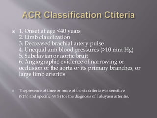
 A chronic, progressive, inflammatory,
occlusive disease of the aorta and its branches.
The inflammatory process results in stenosis,
occlusion, dilation or aneurysm formation in
the arterial wall.
 Aortic arch syndrome
 Pulseless disease
 prevalent in adolescent girls and young women
 1.2–2.6 cases per million in US
 In Asia
 Adolescent girls and young women
 Common age at presentation is 10 to 40 years
 There is panarteritis with inflammatory
mononuclear cell infiltrates and occasionally
giant cells.
 Marked intimal proliferation and fibrosis,
scarring and vascularization of the media, and
disruption and degeneration of the elastic
lamina.
 Narrowing of the lumen occurs with or
without thrombosis.
 circulating immune complexes have been
demonstrated, but their pathogenic significance
is unclear.
 Inflammatory lesions originates in Vasa
vasorum followed by cellular infilteration
maily T cells but NK cells, dentritic cells,
monocytes and neutrophils also invade outer
layer of media.
 Interlukin 6 & 1 are released to maintain
abberrant inflammatory process
 Triggering Antigen is unknown
 Antibodies found in TA are AECA(Antibodies
against endothelial cell)
 AECA may have role but not yet proved.
 CONSTITUNAL:
Dominate the presentation in 1/3rd of patients these include
Asthenia
headache
weight loss
fever
myalgia
arthralgia
Prominent back pain, especially in the thoracic region,
 caused by vascular damage (stenosis, occlusion or
dilataion of vessels)
 Bruit
 Claudication(upper extremity >Lower extremity)
 Hypertension
 Light headedness
 Unequal blood pressure in extremities
 Carotidynia
 Absent pulses
 Bruits at carotid arteries, supraclavicular or
infraclavicular space (subclavian disease)
flexor surface of the upper arm
(axillary artery disease)
abdomen (from renal or mesenteric artery
vasculitis)
 Angina
 Aortic Regurgitation
 Mitral regurgitation (secondary to left
ventricular dilatation due to AR)
 Congestive heart failure ( myocarditis or by
chronic aortic with or without mitral
regurgitation)
 Headache
 Visual Disturbance
 Stroke
 TIA
 Seizures
 Erythema Nodosum
 Ulcerated sabacute nodular lesion
 Pyoderma gangrenosum
 Rheumatic diseases :
Giant cell arteritis
Cogan syndrome
Relapsing polychrondritis,
Ankylosing spondylitis,
Rheumatoid arthritis
Sysstemic lupus erythematosus
Buergr disease and
Behcet s disease
Infectious disease: Syphilis
Other diseases:
Atherosclerosis
inflammatory abdominal aortic aneurysm
ergotism
radiation-induced
damage
retroperitoneal fibrosis
IBD
sarcoidosis, neurofibromatosis,
congenital coarctation, Marfan syndrome
 Measuring blood pressure in both arms,
carefully palpating pulses in all extremities,
and listening
for bruits in the abdomen and chest and along
the carotids
and supraclavicular and axillary areas provide
the best clinical tools in early diagnosis of
Takayasu arteritis.
 1. Onset at age <40 years
2. Limb claudication
3. Decreased brachial artery pulse
4. Unequal arm blood pressures (>10 mm Hg)
5. Subclavian or aortic bruit
6. Angiographic evidence of narrowing or
occlusion of the aorta or its primary branches, or
large limb arteritis
 The presence of three or more of the six criteria was sensitive
(91%) and specific (98%) for the diagnosis of Takayasu arteritis.
 TA does not cause any specific blood test or
urinary abnormalities, however
 ESR and CRP are raised, non specific tests. ESR
may be normal in high disease activity in 30%
patients and high in 40% patients with inactive
disease.
 Pentraxin-3 may more accurately reflect disease
activity than ESR or CRP
 Anemia
 Thrombocytosis
 Raised Creatinine
 Mild proteinuria or hematuria on UCE
 MRI and MRA
 CT and CTA
 Vascular ultrasonography
 Conventional aortography are abnormal in
virtually all patients with TA.
 Conventional angiography is the “gold
standard” for precisely delineating the
stenoses, occlusions and thrombosis.
 Biopsies of the aorta or other actively affected
arteries show a
granulomatous vasculitis with giant cells.
2 or more features :
(1)fever
(2) elevated ESR
(3) symptoms or signs of vascular ischemia or
inflammation (e.g.,claudication, absent pulse,
carotidynia)
(4)typical angiographic features
.
GLUCOCORTICOIDS are very effective in
suppressing vascular inflammation as well as
constitutional symptoms within days to few
weeks; prednisone (1 mg/kg) for 1 to 3 months
and then tapered to 10 mg/d over 4–6 months.
Anemia, thrombocytosis, and elevated ESRs
also usually respond promptly
 Resolution of signs, symptoms, and laboratory
markers of
inflammation, as well as lack of progression of
angiographic
abnormalities, is seen in most patients who
receive glucocorticoid therapy.
Many patients with TA experience relapses of
symptoms or progression of vascular disease that
necessitate restarting high-dose prednisone therapy.
 resistant to steroids/ recurrent disease once
corticosteroids are tapered
cyclophosphamide (1-2 mg/kg/day), not preffered
choice in young females.
azathioprine (1-2mg/kg/day), or
methotrexate (0.3 mg/kg/week)
Mycophenolate mofetil/ anti TNF α agents-
infliximab
 Tricky in patients with extensive Takayasu
arteritis.
 The physician must accept compromises in
blood pressures That sustain perfusion of
critical organs or tissues.
 Pregnancy does not alter disease character
 Complications of HTN mainly affect.
 Outcome usually favourable
 Management of HTN essential
 Measurement of BP in UL impossible/unreliable
more accurate in legs
 HTN in 2nd stage Labour –risk for ICH-shortening
stage .
 pre-eclampsia, CCF, progressive RF,CVA
 Fertility not affected
According to new reseaches 13-fold higher rate of
obstetric complications compared to pregnancies
before TA diagnosis
Obstetric complications: 40% including pree
clampsia/eclampsia , premature delivery and
intrauterine fetal growth restriction or death.
Maternal complications: 39% of pregnancies and
included mainly new-onset or worsening
hypertension.
 Antihypertensive agents that are safely used
with proven success in pregnancy are labetolol,
hydralazine, and alpha-methyldopa
Takayasu Arteritis

Takayasu Arteritis

  • 2.
     A chronic,progressive, inflammatory, occlusive disease of the aorta and its branches. The inflammatory process results in stenosis, occlusion, dilation or aneurysm formation in the arterial wall.  Aortic arch syndrome  Pulseless disease
  • 3.
     prevalent inadolescent girls and young women  1.2–2.6 cases per million in US  In Asia  Adolescent girls and young women  Common age at presentation is 10 to 40 years
  • 4.
     There ispanarteritis with inflammatory mononuclear cell infiltrates and occasionally giant cells.  Marked intimal proliferation and fibrosis, scarring and vascularization of the media, and disruption and degeneration of the elastic lamina.  Narrowing of the lumen occurs with or without thrombosis.
  • 5.
     circulating immunecomplexes have been demonstrated, but their pathogenic significance is unclear.  Inflammatory lesions originates in Vasa vasorum followed by cellular infilteration maily T cells but NK cells, dentritic cells, monocytes and neutrophils also invade outer layer of media.  Interlukin 6 & 1 are released to maintain abberrant inflammatory process
  • 6.
     Triggering Antigenis unknown  Antibodies found in TA are AECA(Antibodies against endothelial cell)  AECA may have role but not yet proved.
  • 7.
     CONSTITUNAL: Dominate thepresentation in 1/3rd of patients these include Asthenia headache weight loss fever myalgia arthralgia Prominent back pain, especially in the thoracic region,
  • 8.
     caused byvascular damage (stenosis, occlusion or dilataion of vessels)  Bruit  Claudication(upper extremity >Lower extremity)  Hypertension  Light headedness  Unequal blood pressure in extremities  Carotidynia  Absent pulses
  • 9.
     Bruits atcarotid arteries, supraclavicular or infraclavicular space (subclavian disease) flexor surface of the upper arm (axillary artery disease) abdomen (from renal or mesenteric artery vasculitis)
  • 10.
     Angina  AorticRegurgitation  Mitral regurgitation (secondary to left ventricular dilatation due to AR)  Congestive heart failure ( myocarditis or by chronic aortic with or without mitral regurgitation)
  • 11.
     Headache  VisualDisturbance  Stroke  TIA  Seizures
  • 12.
     Erythema Nodosum Ulcerated sabacute nodular lesion  Pyoderma gangrenosum
  • 13.
     Rheumatic diseases: Giant cell arteritis Cogan syndrome Relapsing polychrondritis, Ankylosing spondylitis, Rheumatoid arthritis Sysstemic lupus erythematosus Buergr disease and Behcet s disease
  • 14.
    Infectious disease: Syphilis Otherdiseases: Atherosclerosis inflammatory abdominal aortic aneurysm ergotism radiation-induced damage retroperitoneal fibrosis IBD sarcoidosis, neurofibromatosis, congenital coarctation, Marfan syndrome
  • 15.
     Measuring bloodpressure in both arms, carefully palpating pulses in all extremities, and listening for bruits in the abdomen and chest and along the carotids and supraclavicular and axillary areas provide the best clinical tools in early diagnosis of Takayasu arteritis.
  • 16.
     1. Onsetat age <40 years 2. Limb claudication 3. Decreased brachial artery pulse 4. Unequal arm blood pressures (>10 mm Hg) 5. Subclavian or aortic bruit 6. Angiographic evidence of narrowing or occlusion of the aorta or its primary branches, or large limb arteritis  The presence of three or more of the six criteria was sensitive (91%) and specific (98%) for the diagnosis of Takayasu arteritis.
  • 17.
     TA doesnot cause any specific blood test or urinary abnormalities, however  ESR and CRP are raised, non specific tests. ESR may be normal in high disease activity in 30% patients and high in 40% patients with inactive disease.  Pentraxin-3 may more accurately reflect disease activity than ESR or CRP
  • 18.
     Anemia  Thrombocytosis Raised Creatinine  Mild proteinuria or hematuria on UCE
  • 19.
     MRI andMRA  CT and CTA  Vascular ultrasonography  Conventional aortography are abnormal in virtually all patients with TA.  Conventional angiography is the “gold standard” for precisely delineating the stenoses, occlusions and thrombosis.
  • 20.
     Biopsies ofthe aorta or other actively affected arteries show a granulomatous vasculitis with giant cells.
  • 22.
    2 or morefeatures : (1)fever (2) elevated ESR (3) symptoms or signs of vascular ischemia or inflammation (e.g.,claudication, absent pulse, carotidynia) (4)typical angiographic features .
  • 23.
    GLUCOCORTICOIDS are veryeffective in suppressing vascular inflammation as well as constitutional symptoms within days to few weeks; prednisone (1 mg/kg) for 1 to 3 months and then tapered to 10 mg/d over 4–6 months. Anemia, thrombocytosis, and elevated ESRs also usually respond promptly
  • 24.
     Resolution ofsigns, symptoms, and laboratory markers of inflammation, as well as lack of progression of angiographic abnormalities, is seen in most patients who receive glucocorticoid therapy.
  • 25.
    Many patients withTA experience relapses of symptoms or progression of vascular disease that necessitate restarting high-dose prednisone therapy.  resistant to steroids/ recurrent disease once corticosteroids are tapered cyclophosphamide (1-2 mg/kg/day), not preffered choice in young females. azathioprine (1-2mg/kg/day), or methotrexate (0.3 mg/kg/week) Mycophenolate mofetil/ anti TNF α agents- infliximab
  • 26.
     Tricky inpatients with extensive Takayasu arteritis.  The physician must accept compromises in blood pressures That sustain perfusion of critical organs or tissues.
  • 27.
     Pregnancy doesnot alter disease character  Complications of HTN mainly affect.  Outcome usually favourable  Management of HTN essential  Measurement of BP in UL impossible/unreliable more accurate in legs  HTN in 2nd stage Labour –risk for ICH-shortening stage .  pre-eclampsia, CCF, progressive RF,CVA  Fertility not affected
  • 28.
    According to newreseaches 13-fold higher rate of obstetric complications compared to pregnancies before TA diagnosis Obstetric complications: 40% including pree clampsia/eclampsia , premature delivery and intrauterine fetal growth restriction or death. Maternal complications: 39% of pregnancies and included mainly new-onset or worsening hypertension.
  • 29.
     Antihypertensive agentsthat are safely used with proven success in pregnancy are labetolol, hydralazine, and alpha-methyldopa