Download to read offline
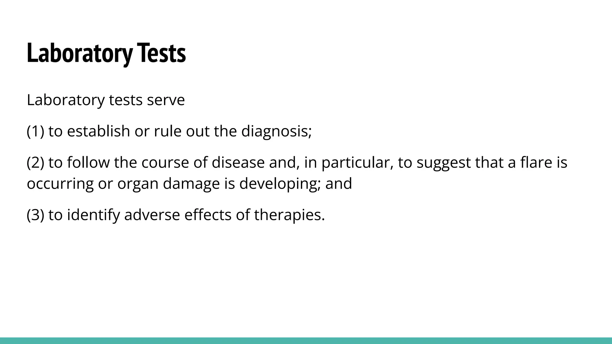

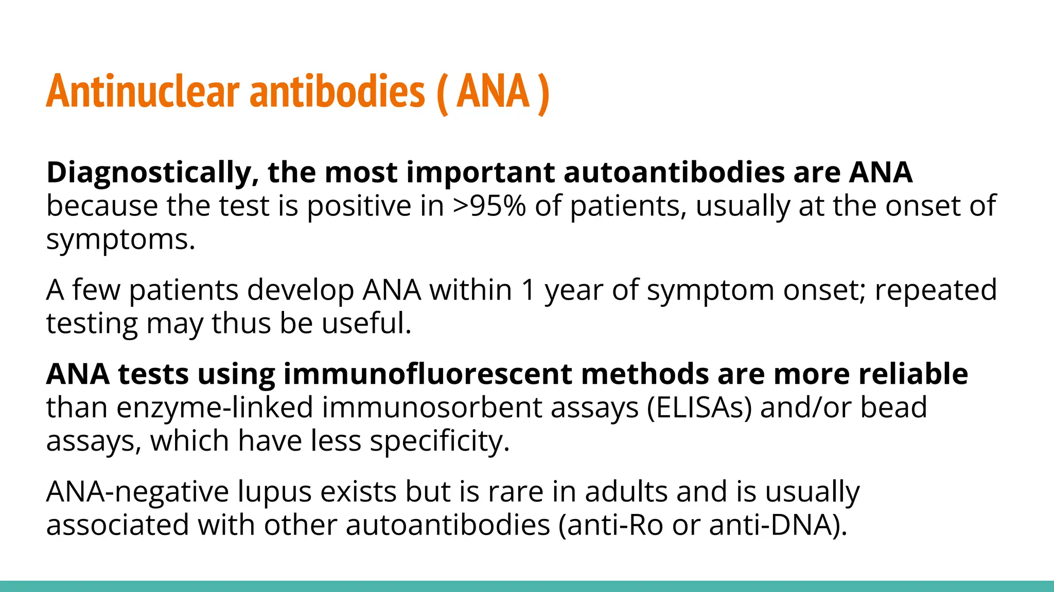
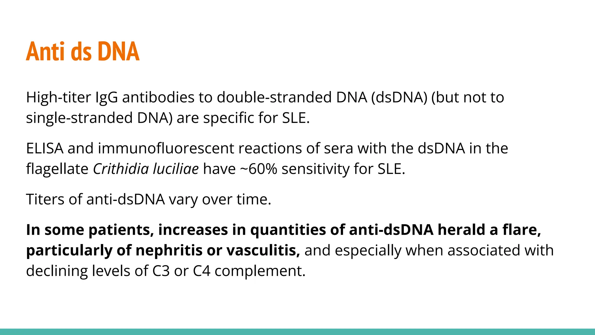


The document discusses laboratory tests for systemic lupus erythematosus (SLE), highlighting their roles in diagnosis, monitoring disease progression, and identifying treatment side effects. Key tests include antinuclear antibodies (ANA), anti-dsDNA, antiphospholipid antibodies, and anti-Ro/SS-A, each associated with specific clinical implications. It emphasizes the importance of regular monitoring as antibody levels can vary and may indicate disease flares, necessitating adjustments in therapy.


