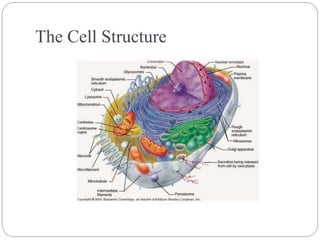
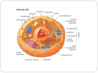
The cell is the basic unit of life. Animal cells lack cell walls and contain organelles like the nucleus, mitochondria, endoplasmic reticulum, Golgi bodies, lysosomes, and ribosomes surrounded by a plasma membrane. The nucleus contains DNA and controls cell activities. Mitochondria generate energy through cellular respiration. The endoplasmic reticulum and Golgi bodies process and transport proteins. Ribosomes synthesize proteins using mRNA. Together, these organelles allow animal cells to carry out specialized functions and maintain homeostasis.
![History
1595 – Jansen credited with 1st compound microscope
1655 – Hooke described ‘cells’ in cork.
1674 – Leeuwenhoek discovered protozoa. He saw
bacteria some 9 years later.
1833 – Brown descibed the cell nucleus in cells of the
orchid.
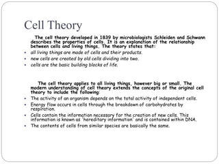
1838 – Schleiden and Schwann proposed cell theory.
1840 – Albrecht von Roelliker realized that sperm cells
and egg cells are also cells.
1856 – N. Pringsheim observed how a sperm cell
penetrated an egg cell.
1858 – Rudolf Virchow (physician, pathologist and
anthropologist) expounds his famous conclusion: omnis
cellula e cellula, that is cells develop only from existing
cells [cells come from preexisting cells]](https://image.slidesharecdn.com/structureofcell-componentsfunctions-201001093532/85/Structure-of-cell-components-and-functions-3-320.jpg)