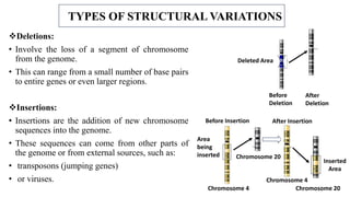
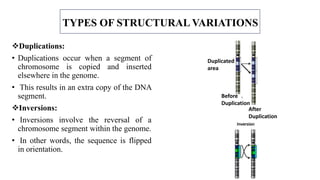
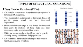
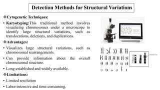
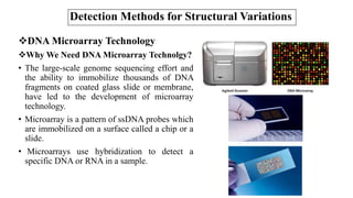
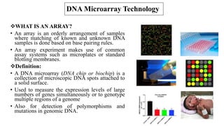
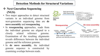
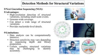
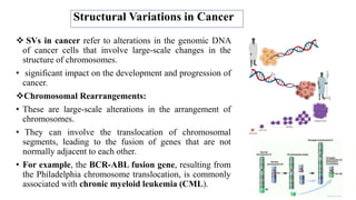
2. TABLE OF CONTENTS
Introduction
Types of Structural Variations
Deletions
Duplications
Insertions
Inversion
Translocations
Copy-number variants
Methods Of Detection SVs
Karyotyping
Microarray
SVs In Cancer
Applications
Future Direction Of SVs
Conclusion
3. Structural variations (SVs)
What Are SVs?
• Refer to alterations in the DNA sequence
larger than 50 KB.
• Opposed to single nucleotide changes
(mutations).
• It consists of many kinds of variation:
• Deletions
• Duplications
• Insertions
• Inversion
• Translocations
• Copy-number variants
cnv
4. TYPES OF STRUCTURAL VARIATIONS
Deletions:
• Involve the loss of a segment of chromosome
from the genome.
• This can range from a small number of base pairs
to entire genes or even larger regions.
Insertions:
• Insertions are the addition of new chromosome
sequences into the genome.
• These sequences can come from other parts of
the genome or from external sources, such as:
• transposons (jumping genes)
• or viruses.
Before
Deletion
After
Deletion
Deleted Area
Chromosome 4
Chromosome 4
Chromosome 20
Before Insertion After Insertion
Area
being
inserted
Chromosome 20
Inserted
Area
5. TYPES OF STRUCTURAL VARIATIONS
Duplications:
• Duplications occur when a segment of
chromosome is copied and inserted
elsewhere in the genome.
• This results in an extra copy of the DNA
segment.
Inversions:
• Inversions involve the reversal of a
chromosome segment within the genome.
• In other words, the sequence is flipped
in orientation.
Duplicated
area
Before
Duplication
After
Duplication
6. TYPES OF STRUCTURAL VARIATIONS
Translocations:
• Translocations occur when a segment of
chromosome is transferred from one location in
the genome to another.
• This can involve interchromosomal (between
different chromosomes) or intrachromosomal
(within the same chromosome) rearrangements.
• Tandem Repeats:
• Tandem repeats are sequences of DNA that are
repeated one after the other in the genome.
• Expansions or contractions of these repeat
regions can lead to structural variations, such as
in the case of trinucleotide repeat disorders.
After translocation
Before translocation
Chromosome 4
Chromosome 20
Derivative
Chromosome 20
Derivative
Chromosome 4
7. TYPES OF STRUCTURAL VARIATIONS
Copy Number Variations (CNVs):
• CNVs refer to variations in the number of copies of a
particular DNA segment.
• This can result in an increased or decreased dosage of
specific genes, which can have functional
implications.
• These variations can range in size from a few thousand
base pairs to millions of base pairs and may
encompass entire genes or multiple genes.
• CNVs are known to play a significant role in genetic
diversity among individuals and populations.
• CNVs led to a better understanding of their roles in
health, disease, and evolution.
8. Detection Methods for Structural Variations
Cytogenetic Techniques:
• Karyotyping:This traditional method involves
visualizing chromosomes under a microscope to
identify large structural variations, such as
translocations, deletions, and duplications.
Advantages:
• Visualizes large structural variations, such as
chromosomal rearrangements.
• Can provide information about the overall
chromosomal structure.
• Long-established and widely available.
Limitations:
• Limited resolution
• Labor-intensive and time-consuming.
9. Detection Methods for Structural Variations
DNA Microarray Technology
Why We Need DNA Microarray Technolgy?
• The large-scale genome sequencing effort and
the ability to immobilize thousands of DNA
fragments on coated glass slide or membrane,
have led to the development of microarray
technology.
• Microarray is a pattern of ssDNA probes which
are immobilized on a surface called a chip or a
slide.
• Microarrays use hybridization to detect a
specific DNA or RNA in a sample.
10. DNA Microarray Technology
WHAT IS AN ARRAY?
• An array is an orderly arrangement of samples
where matching of known and unknown DNA
samples is done based on base pairing rules.
• An array experiment makes use of common
assay systems such as microplates or standard
blotting membranes.
Definition:
• A DNA microarray (DNA chip or biochip) is a
collection of microscopic DNA spots attached to
a solid surface.
• Used to measure the expression levels of large
numbers of genes simultaneously or to genotype
multiple regions of a genome
• Also for detection of polymorphisms and
mutations in genomic DNA.
11. DNA Microarray Technology
History:
• Microarray technology evolved from Southern
blotting.
• The concept of microarrays was first proposed in the
late 1980s by Mark Schena and his colleagues.
• They spotted 4000 cDNA sequences on nitrocellulose
membrane and used radioactive labeling to analyze
differences in gene expression patterns among
different types of colon tumors in various stages of
malignancy.
Principle:
• The core principle behind microarrays is
hybridization between two DNA strands.
• Fluorescent labeled target sequences that bind to a
probe sequence generate a signal that depends on the
strength of the hybridization determined by the
number of paired bases.
12. DNA Microarray Technology
Steps of DNA Microarray Technology:
1. Sample Preparation
The first step in DNA microarray analysis is sample
preparation. The sample can be RNA, DNA, or protein.
2. Hybridization
The sample is labeled with fluorescent dyes, and then
hybridized to the probes on the microarray chip.
3. Scanning
The microarray chip is scanned using a fluorescence
scanner
4.Data Analysis
The data obtained from the microarray chip are analysed
using various software tools to identify differentially
expressed genes, detect DNA mutations, and identify
genetic variations.
13. DNA Microarray Technology
Types of DNA Microarray
• There are two main types of DNA
microarrays.
cDNA Microarray
• Cells are cultured
• RNA Extraction
• The cDNA fragments are prepared by
reverse-transcribing mRNA molecules into
cDNA using reverse transcriptase.
• The cDNA fragments are then labelled with
fluorescent dyes and hybridized to the probes
on the microarray chip.
• Results are analysed based on colors.
14. DNA Microarray Technology
Oligonucleotide Microarray
• Oligonucleotide microarrays are prepared
by synthesizing oligonucleotides directly on
the microarray chip.
• The oligonucleotides are designed to be
complementary to specific DNA or RNA
sequences that are to be analysed.
• The target molecules are labelled with
fluorescent dyes
• And hybridized to the probes on the
microarray chip.
15. DNA Microarray Technology
Advantages:
• High throughput for detecting copy
number variations (CNVs).
• Suitable for analyzing multiple samples
simultaneously.
• Can identify CNVs across the entire
genome.
Limitations:
• Limited resolution.
• May miss complex structural variations.
• This technique only identifies sequences
that the based array was supposed to
detect.
16.
17. Detection Methods for Structural Variations
Next Gneration Sequencing
(NGS):
• Two major approaches to detect structural
variants in an individual genome from
next-generation sequencing data are de
novo assembly and resequencing.
• In resequencing approaches, reads from
the individual genome are aligned to a
closely related reference genome.
Examination of the resulting alignments
reveals differences between the individual
genome and the reference genome.
• In de novo assembly, the individual
genome sequence is constructed by
examining overlaps between reads.
18. Detection Methods for Structural Variations
Next Gneration Sequencing (NGS):
Advantages:
• High-resolution detection of structural
variations, including small-scale events.
• Genome-wide coverage.
• Can detect a wide range of structural
variations.
• Provides nucleotide-level details.
Limitations:
• Data analysis can be computationally
intensive.
• Detection sensitivity may vary depending
on sequencing depth and library
preparation.
• Certain complex structural variations
may be challenging to identify
accurately.
19. Structural Variations in Cancer
SVs in cancer refer to alterations in the genomic DNA
of cancer cells that involve large-scale changes in the
structure of chromosomes.
• significant impact on the development and progression of
cancer.
Chromosomal Rearrangements:
• These are large-scale alterations in the arrangement of
chromosomes.
• They can involve the translocation of chromosomal
segments, leading to the fusion of genes that are not
normally adjacent to each other.
• For example, the BCR-ABL fusion gene, resulting from
the Philadelphia chromosome translocation, is commonly
associated with chronic myeloid leukemia (CML).
20. Applications Of SVs
SVs have a wide range of applications across
different fields,
Cancer Diagnosis: Detection of SVs in cancer
genomes is crucial for diagnosing and classifying
different types of cancer.
Functional Genomics: SVs can impact gene
expression and regulation.
Crop Improvement: Identifying SVs in plant
genomes is valuable for crop breeding programs.
Forensic Identification: SVs can be used for
human identification in forensic investigations.
Drug Response: SVs in the human genome can
affect an individual's response to certain medications
21. Conclusion
Key takeaways from our discussion include:
• Genomic Diversity: Structural variations represent a fundamental source of genomic
diversity, contributing to the uniqueness of individuals and populations.
• Disease Susceptibility: Structural variations have been associated with various genetic
disorders and diseases.
• Evolutionary Significance: Structural variations are essential drivers of evolution, enabling
species to adapt to changing environments and contributing to speciation.
• Technological Advances: The development of advanced genomic technologies, such as
high-throughput sequencing and bioinformatics tools, has revolutionized our ability to detect
and analyze structural variations with unprecedented precision.
• Clinical Implications: The identification of structural variations in the genome has paved
the way for personalized medicine, where tailored treatments and therapies can be
developed based on an individual's genetic profile.
• Ethical Considerations: we must address ethical concerns related to genetic privacy,
discrimination, and we navigate the era of genomics and precision medicine.
1)Genome structural variations (SVs) in the human genome are defined as DNA sequence polymorphisms of at least a few dozen or few hundred bases in length.
2)Structural variation (SV) groups different forms of mutations that involve longer stretches of DNA, including full chromosome fusion, fission or loss.
1)A deletion, as related to genomics, is a type of sv that involves the loss of one or more nucleotides from a segment of DNA. A deletion can involve the loss of any number of nucleotides, from a single nucleotide to an entire piece of a chromosome.Deletion are actually the cause for a large number of genetic diseases.
2) insertion involves the addition of one or more nucleotides into a segment of DNA. An insertion can involve the addition of any number of nucleotides, from a single nucleotide to an entire piece of a chromosome. Insertion means that a number of nucleotides have been erroneously added to the genome, most often during the process of DNA replication. This number can be as small as a single nucleotide or up to thousands or even millions of nucleotides. The effect of an insertion likewise varies. Some may cause no effect at all, whereas others, even single nucleotide insertions, can completely disrupt the function of a gene and lead to a pathogenic variant associated with a genetic disease.
1)Duplication, refers to a type of sv in which one or more copies of a DNA segment (which can be as small as a few bases or as large as a major chromosomal region) is produced. Duplications occur in all organisms. For example, they are especially prominent in plants, although they can also cause genetic diseases in humans. Duplications have been an important mechanism in the evolution of the genomes of humans and other organisms.
2)An inversion in a chromosome occurs when a segment breaks off and reattaches within the same chromosome, but in reverse orientation. DNA may or may not be lost in the process. An inversion occurs when part of your chromosome breaks off and then reattaches, but sometimes pieces along the edges are lost. Think of it as though you are reading the newspaper. Sometimes an internal page can get reversed. All the information is still there, but when you go to read the page, it doesn't make any sense. Therefore, having an inversion is an important complication.
1)Translocation occurs when a chromosome breaks and the (typically two) fragmented pieces re-attach to different chromosomes. The detection of chromosomal translocations can be important for the diagnosis of certain genetic diseases and disorders.
2) They are thus an important source of genetic variation. This variation is highly informative for population and conservation genetics.
Due to slippage in replication Polymerase get confused and add rong nucleotides again and again.
Copy number variation (abbreviated CNV) refers to a circumstance in which the number of copies of a specific segment of DNA varies among different individuals’ genomes. The individual variants may be short or include thousands of bases. These structural differences may have come about through duplications, deletions or other changes and can affect long stretches of DNA. Such regions may or may not contain a gene(s).
CNV. Changes in the number of copies of small sections of our genomes can have big consequences. One interesting example is a gene called amylase. This gene is important for digesting starchy foods like potatoes or grains. Scientists found copy number variations of the amylase gene in different groups of people. People from places where starchy foods were historically very important usually have more copies of the amylase gene than people from backgrounds where starchy foods were less common. Places where meat and fish were more important parts of the diet relative to starches. This suggests that ancient people who had more copies of the amylase gene were able to get more energy from starchy foods, and so were able to thrive in regions where starchy food sources were important.
1)Cytogenetics is the study of the structural and functional properties of the chromosome. It includes chromosomal structure, function, behavior – during mitosis and meiosis, and influence on the phenotype of an organism. It is used as a tool to understand genetic phenomena due to chromosomal .
2) Karyotype analysis is a culture-based technique whereby fresh viable tissue cells are grown and arrested in the metaphase stage of cell division. The nucleus is dissolved and the chromosomes are banded using various staining techniques and subsequently evaluated for numerical and gross abnormalities including aberrations in chromosome copy numbers (aneuploidies) as well as chromosomal translocations, segmental deletions, and inversions. Conventional karyotyping is most useful for detecting congenital genetic diseases and is often used in conjunction with medical autopsies of stillborns.
The principle behind microarrays is that complementary sequences will bind to each other. The unknown DNA molecules are cut into fragments by restriction endonucleases; fluorescent markers are attached to these DNA fragments. These are then allowed to react with probes of the DNA chip.
1) This step requires the control and target mRNA to be extracted - i.e., a non-cancerous cell line versus a cancerous cell line, respectively. The RNA is then converted into cDNA and labeled with a fluorescent dye. Typically, Cy3 (a red fluorescent dye) and Cy5 (a green fluorescent dye) are used to label the target and control cDNA
2) In this step the sample DNA is hybridized with complementary probe sequences on the array chip. DNA will strongly or weakly hybridize - or not at all .
3) Microarray scanners usually have red, green, and blue excitation wavelengths and a wide choice of up to 8 emission filters that enable imaging of an extensive variety of fluorophores (e.g. Cy3, Cy5, FITC, Texas Red).
What is cDNA in microarray?
The cDNA microarray is the most powerful tool for studying gene expression in many different organisms. It has been successfully applied to the simultaneous expression of many thousands of genes and to large-scale gene discovery, as well as polymorphism screening and mapping of genomic DNA clones
Oligonucleotide Arrays. Oligonucleotide microarrays are created either by in situ synthesis or deposition of presynthesized oligonucleotides ranging in size from 25- to 60-mers. Oligonucleotides can be synthesized directly in situ using photolithography techniques adapted from the microelectronics industry.