Tensor fascia lata[tfl] muscle pedicle grafting for avn hip dr mohamed ashraf...drashraf369
slide presentation of a very promising surgical technic for a very elusive condition called avascular necrosis of femoral head.good clinical and surgical demo by dr mohamed ashraf,HOD, govt TD medical college ,alleppey,kerala, india
Case Review #2: 66 year old female with severe Flatback SyndromeRobert Pashman
A 66 year old female presented with severe Flat back Syndrome, Kyphosis, and critical stenosis. Dr. Pashman treated the patient with a posterior spinal fusion T2 to the pelvis. The patient was able to stand up straight following surgery.
Case Review #7: 51 year old female with severe flatback after multiple surge...Robert Pashman
A 51 year old female status post multiple spine surgeries presented to Dr. Pashman with severe Flatback Syndrome and psueoarthrosis. Dr. Pashman treated the patient with a Posterior Spinal Fusion T4-Pelvis.
In thoracolumbar spine trauma, the biomechanical goals in minimally invasive fracture treatment include decompression of the spinal canal, reduction of spinal deformities, and maintenance of stable fixation of the spine to permit early mobilization.
This was a teaching lecture given by Prof. Mohamed Mohi Eldin, professor of neurosurgery, in the Multi- Institutional Neurosurgical Meeting, Kasr El Aini Hospital, Cairo University, April 2nd, 2009.
Urinary bladder collects urine from the kidney which is then passed through the urethra. Cancer is abnormal growth of cells leading to tumour in urinary bladder. Bladder Cancer is diagnosed with cystoscopy and biopsy . Treatment of Bladder cancer is done as per stage. It includes Radical Cystectomy, Plevic Lymphadenectomy, Ileal conduit, Neobladder as surgical options.
At the end of this lecture you will be able to:-
Describe the anatomy of the ligaments stabilising the wrist, DRUJ and the MCP joints
Assess confidently the stability of these joints and identify the anatomy of the lesions
Identify and provide a management plan for patients with ligament injuries and their post-op rehabilitation.
Avascular necrosis of Hip - treatment modalities and current concepts.pptxVivek Jadawala
Slide 1 - Treatment modalities of Avascular Necrosis of Hip
JOURNAL CLUB PRESENTATION
Dr. Vivek Jadawala
PGY-3, Dept. of Orthopaedics,
JNMC, DMIHER
Slide 2 - image
slide 3 - image
slide 4 - Osteonecrosis of Hip - Osteonecrosis is death of living elements of involved bone (cells including marrow) with progressive destruction and alteration of bone architecture as a result of compromised vascularity.
Usually aseptic but may be incited by loss of vascularity from infection.
Slide 5 - Epidemiology - Male > Female
Average age group – 35 to 50 years
Bilateral Hip joints – 80 % of the cases
Most common site – Antero-lateral aspect of femoral head
Slide 6 - Blood supply of femoral head
Slide 7 - Classification of AVN: Ficat and Arlet -STAGE 0 :
X-ray : normal
MRI: normal
clinical symptoms: nil
STAGE I :
X-ray : normal or minor osteopenia
MRI: edema
bone scan: increased uptake
clinical symptoms: pain typically in the groin
Slide 8 - Stage I
Slide 9 - Stage II -
X-ray: mixed osteopenia and/or sclerosis and/or subchondral cysts, without any subchondral lucency (crescent sign)
MRI: geographic defect
Bone scan: increased uptake
clinical symptoms: pain and stiffness
Slide 10 - Stage III - X-ray: Crescent sign and eventual cortical collapse
MRI: same as plain radiograph
clinical symptoms: pain and stiffness +/- radiation to knee and limp
Slide 11 - Stage IV - X-ray: end-stage with evidence of secondary degenerative change
MRI: same as plain radiograph
clinical symptoms: pain and limp
Slide 12 - Stage IV
Slide 13 - image
Slide 14 - Steinberg staging of AVN
Slide 15 - Steinberg staging - STAGE 0:
- normal or non-diagnostic radiographs, MRI and bone scan of at risk hip (often contralateral hip involved, or patient has risk factors and hip pain)
STAGE I:
normal radiograph, abnormal bone scan and/or MRI
STAGE II:
- cystic and sclerotic radiographic changes
STAGE I AND II
A, mild: <15% head involvement as seen on radiograph or MRI
B, moderate: 15% to 30%
C, severe: >30%
Slide 16 - STAGE III:
- subchondral lucency or crescent sign
A, mild: subchondral collapse (crescent) beneath <15% of articular surface
B, moderate: crescent beneath 15% to 30%
C, severe: crescent beneath >30%
STAGE IV:
flattening of femoral head, with depression graded into
A, mild: <15% of surface has collapsed and depression is <2 mm
B, moderate: 15% to 30% collapsed or 2-4 mm depression
C, severe: >30% collapsed or >4 mm depression
Slide 17 - STAGE V:
- joint space narrowing with or without acetabular involvement
STAGE VI:
- advanced degenerative changes
Slide 18 - Association Research Circulation Osseous classification
Slide 19 - image
Slide 20 - Kerboul angle - Original classification was proposed on radiographs where he divided the necrotic region into small, medium and large regions:
Small - less than or equal to 160°
Medium - 161 to 199°
Large - 200 or more degrees.
Slide 21 - Modified Kerboul angle - based on MRI has much higher values as the MRI overestimates the necrotic region
Foot and ankle trauma, common pitfalls, imaging modalities and radiographic occult fractures. The concept of the PITFL or "pitiful injury" an easily overlooked ligamentous injury of the talocrural joint
Presentations from Professor Adnan Saithna at the North West Upper Limb Group Meeting January 2018, focusing on current concepts in the diagnosis and management of long head of biceps tendon pathology, with an emphasis on the young, active patient with anterior shoulder pain
Similar to Stand alone x lif one year outcome (20)
Cancer cell metabolism: special Reference to Lactate PathwayAADYARAJPANDEY1
Normal Cell Metabolism:
Cellular respiration describes the series of steps that cells use to break down sugar and other chemicals to get the energy we need to function.
Energy is stored in the bonds of glucose and when glucose is broken down, much of that energy is released.
Cell utilize energy in the form of ATP.
The first step of respiration is called glycolysis. In a series of steps, glycolysis breaks glucose into two smaller molecules - a chemical called pyruvate. A small amount of ATP is formed during this process.
Most healthy cells continue the breakdown in a second process, called the Kreb's cycle. The Kreb's cycle allows cells to “burn” the pyruvates made in glycolysis to get more ATP.
The last step in the breakdown of glucose is called oxidative phosphorylation (Ox-Phos).
It takes place in specialized cell structures called mitochondria. This process produces a large amount of ATP. Importantly, cells need oxygen to complete oxidative phosphorylation.
If a cell completes only glycolysis, only 2 molecules of ATP are made per glucose. However, if the cell completes the entire respiration process (glycolysis - Kreb's - oxidative phosphorylation), about 36 molecules of ATP are created, giving it much more energy to use.
IN CANCER CELL:
Unlike healthy cells that "burn" the entire molecule of sugar to capture a large amount of energy as ATP, cancer cells are wasteful.
Cancer cells only partially break down sugar molecules. They overuse the first step of respiration, glycolysis. They frequently do not complete the second step, oxidative phosphorylation.
This results in only 2 molecules of ATP per each glucose molecule instead of the 36 or so ATPs healthy cells gain. As a result, cancer cells need to use a lot more sugar molecules to get enough energy to survive.
introduction to WARBERG PHENOMENA:
WARBURG EFFECT Usually, cancer cells are highly glycolytic (glucose addiction) and take up more glucose than do normal cells from outside.
Otto Heinrich Warburg (; 8 October 1883 – 1 August 1970) In 1931 was awarded the Nobel Prize in Physiology for his "discovery of the nature and mode of action of the respiratory enzyme.
WARNBURG EFFECT : cancer cells under aerobic (well-oxygenated) conditions to metabolize glucose to lactate (aerobic glycolysis) is known as the Warburg effect. Warburg made the observation that tumor slices consume glucose and secrete lactate at a higher rate than normal tissues.
Observation of Io’s Resurfacing via Plume Deposition Using Ground-based Adapt...Sérgio Sacani
Since volcanic activity was first discovered on Io from Voyager images in 1979, changes
on Io’s surface have been monitored from both spacecraft and ground-based telescopes.
Here, we present the highest spatial resolution images of Io ever obtained from a groundbased telescope. These images, acquired by the SHARK-VIS instrument on the Large
Binocular Telescope, show evidence of a major resurfacing event on Io’s trailing hemisphere. When compared to the most recent spacecraft images, the SHARK-VIS images
show that a plume deposit from a powerful eruption at Pillan Patera has covered part
of the long-lived Pele plume deposit. Although this type of resurfacing event may be common on Io, few have been detected due to the rarity of spacecraft visits and the previously low spatial resolution available from Earth-based telescopes. The SHARK-VIS instrument ushers in a new era of high resolution imaging of Io’s surface using adaptive
optics at visible wavelengths.
Richard's aventures in two entangled wonderlandsRichard Gill
Since the loophole-free Bell experiments of 2020 and the Nobel prizes in physics of 2022, critics of Bell's work have retreated to the fortress of super-determinism. Now, super-determinism is a derogatory word - it just means "determinism". Palmer, Hance and Hossenfelder argue that quantum mechanics and determinism are not incompatible, using a sophisticated mathematical construction based on a subtle thinning of allowed states and measurements in quantum mechanics, such that what is left appears to make Bell's argument fail, without altering the empirical predictions of quantum mechanics. I think however that it is a smoke screen, and the slogan "lost in math" comes to my mind. I will discuss some other recent disproofs of Bell's theorem using the language of causality based on causal graphs. Causal thinking is also central to law and justice. I will mention surprising connections to my work on serial killer nurse cases, in particular the Dutch case of Lucia de Berk and the current UK case of Lucy Letby.
(May 29th, 2024) Advancements in Intravital Microscopy- Insights for Preclini...Scintica Instrumentation
Intravital microscopy (IVM) is a powerful tool utilized to study cellular behavior over time and space in vivo. Much of our understanding of cell biology has been accomplished using various in vitro and ex vivo methods; however, these studies do not necessarily reflect the natural dynamics of biological processes. Unlike traditional cell culture or fixed tissue imaging, IVM allows for the ultra-fast high-resolution imaging of cellular processes over time and space and were studied in its natural environment. Real-time visualization of biological processes in the context of an intact organism helps maintain physiological relevance and provide insights into the progression of disease, response to treatments or developmental processes.
In this webinar we give an overview of advanced applications of the IVM system in preclinical research. IVIM technology is a provider of all-in-one intravital microscopy systems and solutions optimized for in vivo imaging of live animal models at sub-micron resolution. The system’s unique features and user-friendly software enables researchers to probe fast dynamic biological processes such as immune cell tracking, cell-cell interaction as well as vascularization and tumor metastasis with exceptional detail. This webinar will also give an overview of IVM being utilized in drug development, offering a view into the intricate interaction between drugs/nanoparticles and tissues in vivo and allows for the evaluation of therapeutic intervention in a variety of tissues and organs. This interdisciplinary collaboration continues to drive the advancements of novel therapeutic strategies.
This pdf is about the Schizophrenia.
For more details visit on YouTube; @SELF-EXPLANATORY;
https://www.youtube.com/channel/UCAiarMZDNhe1A3Rnpr_WkzA/videos
Thanks...!
Mammalian Pineal Body Structure and Also Functions
Stand alone x lif one year outcome
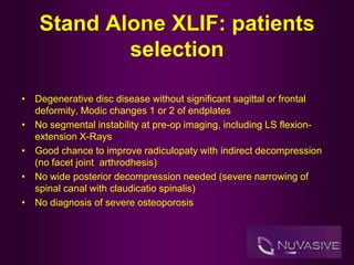
1. Stand Alone XLIF: patients
selection
• Degenerative disc disease without significant sagittal or frontal
deformity, Modic changes 1 or 2 of endplates
• No segmental instability at pre-op imaging, including LS flexion-
extension X-Rays
• Good chance to improve radiculopaty with indirect decompression
(no facet joint arthrodhesis)
• No wide posterior decompression needed (severe narrowing of
spinal canal with claudicatio spinalis)
• No diagnosis of severe osteoporosis
2. Stand alone XLIF: why?
• Short operative time: better for elderly patients with co-morbidities
• Minimal blood loss
• Shorter hospitalization
• Good cost/effectiveness
Stand alone XLIF: handicaps
• Persistent radiculopaty : indirect decompression alone insufficient
• Risk of subsidence of the cage (in particular 18 mm cages)
• Amount of bone growth: biomechanical stability of cage alone
less than circumferential constructs
• Risk of two stages surgery
3. Stand alone XLIF: FU criteria
• Pre-operative clinical assessment completed with ODI
questionnaire, VAS B/VAS L ; pre-operative imaging with LS MRI +
LS lateral and a-p flexion-extension Radiographs
• Clinical evaluation + LS lateral and a-p X-Ray at one month
• ODI/VAS evaluation + LS lateral Flexion-extension X-Ray at three
months
• ODI/VAS evaluation + LS lateral flexion-extension X-Rays at six
months
• In poor grade outcomes, LS MRI was performed
4. Case collection
• October 2011-August 2013: 48 patients treated with XLIF approach: 17
male and 31 females, mean age 62 (range 39-81)
• Of the 48 patients, 37 were treated with stand alone XLIF, 10 with
circumferential approach and one with XLIF + Lateral Plating
• In 3 cases stand alone XLIF + posterior decompression without
instrumentation
• 31 out of 37 patients completed the FU and were enrolled for the study
• 21 patients single level procedure, 10 patients double level procedure
livelli trattati
L1-L2
L2-L3
L3-L4
L4-L5
L1-L2/L2-L3
L2-L3/L3-L4
L3-L4/L4-L5
7. Stand alone XLIF: results
after 3-months FU
0
1
2
3
4
5
6
7
8
9
1 2 3 4 5 6 7 8 9
VB pre
VB post
8. Stand alone XLIF: results
after 3-months FU
0
1
2
3
4
5
6
7
1 2 3 4 5
VL pre
VL post
9. Stand alone XLIF: results
after 6-months FU
0
10
20
30
40
50
60
70
1 3 5 7 9 11 13 15 17 19 21
ODI pre
ODI post 3
ODI post 6
10. Stand alone XLIF: results
after 6-months FU
0
1
2
3
4
5
6
7
8
9
10
1 3 5 7 9 11 13 15 17 19 21
VB pre
VB post 3
VB post 6
11. Stand alone XLIF: results
after 6-months FU
0
1
2
3
4
5
6
7
8
9
1 2 3 4 5 6 7 8 9
VL pre
VL post 3
VL post 6
12. Results analysis
• Most cases show good outcome with progressive improvement at six month
FU
• Unmodified or worsened ODI and VAS scores are classified as poor
outcome
• Limited improving of scores at three-six months FU that doesn’t lead to
category shifting is considered as no satisfactory (poor outcome)
• After 3 months FU, 4 out of 9 patients did not significantly improve; of these,
three had limited improvement but didn’t change ODI category, one had bad
outcome with ODA/VAS scores worsening.
• After six month, 1 out of 22 patients didn’t improve significantly
• Data matching at three and six months shows progressive outcome
improving
13. Stand alone XLIF: pitfalls
• Radiological study at three and six months with lateral Flexion-Extension X-
Rays didn’t show significant bone formation
• 8 out of 23 patients (34,8%) showed radiological evidence of subsidence of
the cage at six months FU
• 3 out of 9 patients (33%) showed radiological evidence of subsidence of the
cage at three months FU
• 7 out of 11 cages were 18 mm wide.
• Subsidence was identified in one case of poor outcome at three month FU
(22mm CoRoent XL)
• No subsidence in the case of poor outcome after 6-months FU
• In 10 cases subsidence was clinically silent
14. Poor outcome analysis
• Back pain in three cases, back pain + radiculopathy in 2 cases
• Subsidence in one case (BP + RP)
• Single level (L3-L4) interbody fusion in two levels degenerative disc
disease (L3-L4/L4-L5): procedure aborted in L4-L5.
• Persistent foraminal stenosis in one case
• No clear causes of persistent symptoms in two cases
15. Stand alone XLIF: implant
failure
• GR, female, 66 Years-old, previous L4-L5 PL arthrodesis in L5-S1
grade two spondylolishtesis, osteoporosis
• Symptoms: invalidating low back pain, lower limbs radicular pain
with cladicatio spinalis, walking severely restricted
• Clinical examination on admission: segmental paresis in extension
of left foot, Lasegue + 40° in left lower limb
• Pre-op LS MRI: L5-S1 grade II spondylolisthesis with spontaneous
fusion, L4-L5 pedicle screws with left L5 screw malposition, adjacent
level discopaty with Modic 1 changes of the endplate, right convex
scoliosis with L3-L4 apex.
• Surgical planning: L3-L4 stand alone XLIF to achieve mild coronal
deformity correction and treat adjacent level discopathy, L4-L5
laminectomy to decompress left L5 nerve root
16. Peri-operative complications
• Right side surgical approach with MAXcess, standard fashion
discectomy, 18x55x8 mm parallel trial followed by 18x50x10 mm
parallel trial then 22x50x10 mm lordotic trial
• No evidence of subsidence during the discectomy and trial
introduction, but significant bleeding from the disk space started
after last steps
• Cage dislocated in the cranial third of L4 vertebral body (22x50x10
lordoticl); bleeding stopped just after cage insertion, the implant was
tightly positioned in the L4 body
• We decided to leave the implant there and go on with posterior
decompression