What is a Brain CT Imaging Perfusion Study?Carestream
Computed tomography perfusion (aka CTP) imaging shows which areas of the brain are supplied or perfused adequately with blood and provides detailed information on delivery of blood or blood flow to the brain. Here are 10 things you need to know about the procedure.
A computed tomography (CT) scan uses X-rays to produce detailed images of the inside of the body. During the scan, the patient lies on a table that slides into a large circular machine. The machine rapidly rotates around the patient and takes images from different angles, which are combined by a computer to produce cross-sectional images of tissues and organs. CT scans can help diagnose medical conditions by revealing abnormalities that may not be visible on regular X-rays.
IGRT is a radiation therapy process that uses imaging to ensure accurate patient positioning and alignment. Frequent imaging during treatment, such as with CBCT, allows corrections to be made for set-up errors and organ motion. This improves the precision of radiation delivery to the target volume while reducing doses to healthy tissues, leading to better treatment outcomes and fewer side effects.
DEXA is an effective technique to measure bone mineral density. It uses two low-dose X-ray beams of different energies to create a 2D image and measure the density of bones. The soft tissue measurement is subtracted to determine the bone mineral content. These measurements are compared to normal ranges based on age and gender using T-scores and Z-scores to assess fracture risk. DEXA requires minimal radiation exposure and is a quick, non-invasive procedure to accurately measure bone density.
SPECT (Single Photon Emission Computed Tomography) is a nuclear imaging test that uses radioactive substances and a special camera to create 3D pictures of organs inside the body. It can detect issues like brain disorders, heart problems, and bone disorders. During a SPECT scan, a person is injected with a radioactive substance like iodine-123 or technetium-99m that emits gamma rays. A scanner then detects these gamma rays and uses the images to create 3D pictures of the area being examined. The radioactive substances used are generally safe and will clear from the body after the scan is complete.
Pet presentation, positron emission tomography emicica
PET is a nuclear medicine scan that uses radioactive tracers to visualize metabolic processes in the body. It works by administering a radioactive tracer that accumulates in tissues and organs, emitting gamma rays that are detected by a ring of scintillation detectors. This allows reconstruction of 2D images showing tracer concentration. Common tracers include carbon-11, nitrogen-13, oxygen-15 and fluorine-18, which are produced by a cyclotron. PET scans are used to detect and monitor cancer, heart disease, and brain disorders. The scans provide functional information that can be fused with anatomical CT or MRI images. While exposing patients to radiation, PET offers high sensitivity for disease detection at early stages.
Basic principle of ct and ct generationsTarunGoyal66
This document provides information about the history and development of computed tomography (CT) scanning technology. It discusses the key innovations and generations of CT scanners, including:
- The first generation translate-rotate scanner with a single detector and pencil beam.
- Second generation scanners that used a fan beam and multiple detectors to reduce scan time.
- Third generation rotate-rotate scanners that eliminated translation by using a rotating x-ray tube and detector array.
- Fourth generation rotate-fixed scanners with a stationary detector ring and rotating x-ray tube.
It also covers the basic components and functioning of modern CT scanners, image reconstruction principles, and factors that influence image quality.
Single Photon Emission Computed Tomography (SPECT) is a nuclear imaging technique that produces 3D images using gamma rays emitted from radiotracers injected into the body. SPECT cameras rotate around the patient to capture 2D images that are reconstructed into cross-sectional slices. SPECT provides physiological information through functional imaging of organ systems like the heart or brain. While it has lower resolution than PET, SPECT machines are less expensive and more widely available. SPECT finds medical use in areas like myocardial perfusion imaging and evaluating thyroid or renal function.
Medical imaging is the process of creating visual representations of the interior of the body for clinical analysis and medical intervention. It involves techniques such as computed tomography (CT), magnetic resonance imaging (MRI), positron emission tomography (PET), and ultrasound. X-ray machines produce beams of radiation that pass through the body and are used to capture images of areas like bones and soft tissues. CT scans take multiple X-ray images from different angles to construct cross-sectional views of the inside of the body. MRI uses strong magnetic fields and radio waves to generate images of organs and tissues. PET scans use radioactive tracers to show how tissues and organs are functioning.
Computed Tomography (CT) is a medical imaging technique that uses X-ray technology to produce detailed cross-sectional images of the body.
It is a valuable tool for diagnosing and monitoring a wide range of medical conditions
This document provides an overview of PET/MRI technology, including its current and future status. It discusses:
1. The history and evolution of PET and MRI from the 1960s onwards, leading to the development of simultaneous PET/MRI systems in the late 1990s.
2. Examples of whole-body PET/MRI images from 2011 demonstrating the technique's ability to provide molecular and anatomical data.
3. The paradigm shift brought by PET/MRI's ability to provide integrated information on structure, function and tissue environment for applications in oncology, neurology and other areas.
4. Future directions for PET/MRI including 'whole body mapping' to characterize metastatic disease, improved data analysis techniques, and
Dual energy CT uses two x-ray spectra to distinguish materials based on their differential attenuation properties. This allows reconstruction of various image sets and material-specific images without contrast. Key applications include bone removal, virtual non-calcium imaging, uric acid stone differentiation, gout detection, perfusion imaging, and differentiating enhancing lesions from calcification. Dual energy CT provides material-specific information useful for diagnosis and treatment planning in various clinical contexts.
Breast tomosynthesis is an advanced form of mammography that uses low-dose x-rays and 3D reconstruction to create three-dimensional images of the breast. Multiple x-ray images are taken over a limited sweep angle and digitally reconstructed into slices to view tissue at different depths within the breast. This allows for detection of smaller tumors and greater accuracy in diagnosis compared to traditional mammography. While exposure to radiation is a risk, the benefits include improved cancer detection rates and fewer unnecessary biopsies.
MRI of the breast has certain contraindications including the presence of metallic implants, those unable to lie prone, or with large body habitus. Normal breast tissue may enhance asymmetrically, so scheduling during days 7-20 of the menstrual cycle can provide less enhancement. Dedicated breast coils are used with patients lying prone, and protocols involve unilateral or bilateral imaging with pre-and post-contrast sequences to analyze enhancement kinetics. Morphological features like irregular shapes and enhancement kinetics help identify lesions, with Type I curves associated with benign lesions and Type III with malignancy. MRI is useful for screening, determining tumor extent, assessing recurrence or residual disease, and providing information not available from other imaging methods.
Image reconstruction in CT is mostly a mathematical process however, this presentation tries to explain the complicated process of image reconstruction in a visual way, mainly focusing om Filtered back projection, Iterative Reconstruction and AI based image reconstruction.
This document discusses the basics of CT scanning, including its history and key components. It describes how CT scanning works, from the x-ray tube emitting radiation that is detected after passing through the body, to the computer using this data to reconstruct cross-sectional images. It outlines the main parts of a CT system, including the gantry, detector, and control console. It also explains different scanning methods and how image quality is determined.
This document discusses magnetic resonance angiography (MRA) and its advantages and disadvantages compared to catheter angiography. It describes different MRA techniques including contrast enhanced MRA, time of flight angiography, phase contrast angiography, and non-contrast techniques. It also discusses artifacts that can appear on MRA such as metal artifacts and blooming artifacts. Key features and images of each technique are provided.
The document summarizes the history and development of computed tomography (CT) scanning technology. It describes the key events and innovations such as the development of the first CT scanner by Godfrey Hounsfield in 1972 (1), the introduction of whole body scanning in 1975 (2), and Hounsfield and Cormack being awarded the Nobel Prize in 1979 (3). Subsequent generations of CT scanners incorporated improvements like faster scanning speeds, multiple detectors, and eliminating moving parts to enable ultra-fast scanning.
The document discusses various aspects of image display in CT scanning. It describes different types of display monitors used, such as CRT and LCD. It also discusses window width and window level settings, which are used to adjust the contrast and brightness of CT images by mapping CT number ranges to grayscale values. Region of interest tools allow measuring values within a selected area of an image. Distance measurements, annotations, reference images, and magnification can also be used to analyze CT scans.
Focused ultrasound surgery uses high-intensity focused ultrasound guided by MRI to heat and destroy targeted tissue, such as uterine fibroids, without the need for invasive surgery. MRI is used to identify the target tissue and guide the ultrasound beams to heat small spots within the tissue to ablate cells. The procedure is repeated around 50 times to treat the required volume, taking around 3 hours. It provides symptom relief as the body removes the treated tissue over months without impacting surrounding areas.
This document discusses the capabilities of dual energy CT, including direct angiography and bone removal with plaque highlighting. Dual energy CT can directly visualize arteries and branching vessels by removing bone based on differentiating iodine and bone through spectral analysis. This allows dual energy CT to be used for angiographic applications as a minimally invasive alternative to digital subtraction angiography. Examples given include carotid and aortic angiography to assess conditions like aneurysms. Dual energy CT also enables characterization of plaques and assessment of endoleaks after endovascular procedures.
Computed Tomography Dose Index, Includes various CTDI parameters and the way of calculating effective dose from various Computed Tomography procedures along with their conversion factor.
Magnetic resonance imaging (MRI) uses strong magnetic fields and radio waves to produce detailed images of the inside of the body. Dr. Raymond Damadian discovered in the 1970s that MRI could distinguish between healthy and cancerous tissue, and he filed the first patent for using MRI for medical diagnosis. An MRI scanner aligns hydrogen atoms in the body with a strong magnetic field and uses radio waves to flip their spins, and sensors detect the radio signal emitted as the spins return to normal, allowing an image of tissues and structures to be produced. MRI is used to diagnose conditions like tumors, strokes, and musculoskeletal disorders.
Post processing of computed tomography images allows radiologists to view images in different planes and highlight key anatomical structures. Techniques like multiplanar reconstruction generate coronal and sagittal views from axial scans, while maximum intensity projection highlights contrast-filled vessels. Together, these techniques provide additional diagnostic information beyond the original axial images.
PET/CT is a medical imaging technique that combines a positron emission tomography (PET) scanner and an x-ray computed tomography (CT) scanner into a single gantry system. This allows it to obtain both functional metabolic information from PET and anatomic information from CT in a single imaging session. The PET data provides physiological functional imaging while the CT data provides accurate structural information. By combining the PET and CT images, diagnostic accuracy and localization of lesions is improved for conditions like cancer, infections, and inflammation. The PET/CT scan involves intravenous injection of FDG, a CT scan, a PET scan, and generation of thousands of fused PET/CT images which are reconstructed, reformatted and analyzed.
This document provides positioning and exposure guidelines for performing an axiolateral hip view using the Clements-Nakayama modification. Key points include:
1) The patient is supine with the leg in a neutral or slightly externally rotated position and the image receptor is angled 15 degrees posterior to be perpendicular to the femoral neck.
2) The hip and proximal femur are imaged using a 24x30cm cassette with a stationary grid at 100cm SID/FFD at 80kVp and 40mAs.
3) Proper positioning and angulation of the image receptor and central ray are important to demonstrate the femoral neck and avoid grid cutoff.
Neke opće smjernice za izradu prezentacija, i kako ih uspješno prezentirati. Tips & tricks kako sve skupa uspješno obaviti.
Preuzeto uz dozvolu autora, dr. sc. Vedrana Šege, PMF-MO, Sveučilišta u Zagrebu.
2. Вовед
• Компјутерската томографија (КТ) претставува современ начин на
визуелно навлегување во длабочината на човековите органи ,
прикажување на нивната функција во тродимензионалната слика со
употреба на рентгенски зраци. Со системот на компјутерска
томографија се добива прецизна медицинска дијагностика на
човековото тело по системи.
• Во 21-от век во медицината особено важно е да се предвидат
идните здравствени проблеми кај секој пациент на начин кој е
безболен и кој трае само неколку секунди. Токму тоа го овозможува
компјутерската томографија со помош на томограф кој дава детална
слика и овозможува да се види што се случува внатре во организмот
на пациентите давајќи тродимензионална слика на сите органи (срце,
бели дробови, органите во стомачната празнина итн ).
3. Компјутерска томографија (КТ – скен)
Компјутерската томографија (КТ) користи посебна опрема со рендген зраци за добивање на слики од
телото од различни агли. Податоците се анализираат и се обработуваат со помош на компјутер. Така се
добиваат дигитални слики кои прикажуваат пресеци на телото низ органите и ткивата. На тој начин се
овозможува тродимензионален детален приказ на надолжни и на напречни пресеци на сите ткива и органи. КТ
е најпрецизна дијагностичка метода.
КТ бара посебни просторни услови и посебно обучен кадар (радиолошки технолози и лекари – радиолози) и
се изведува во дијагностички или клинички медицински центри. Со помош на КТ скенот многу полесно се
дијагностицираат разни заболувања како: карциномите, кардиоваскуларните болести, инфективните болести,
повредите и болестите на мускулно-скелетниот систем.
КТ дава морфолошки изглед на туморот, овозможува да се измери неговата големина, да се утврди
прецизната локализација на туморот како и неговата проширеност во околните ткива и структури.
КТ се користи, покрај за дијагноза, и за планирање и за соодветно администрирање на радиотерапијата,
за што се користи т.н. КТ симулатор.
КТ се користи за прецизно водење на биопсијата и на другите минимално инвазивни процедури, како и за
планирање на хируршкиот третман и за утврдување на операбилноста на туморот.
Оваа дијагностичка процедура е безболна и не дава несакани ефекти.
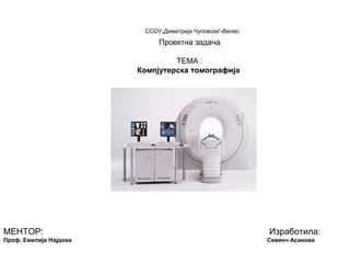
4. • Како изгледа уредот за компјутреска томографија?
КТ е голема машина со облик на коцка, која има дупката во средина. Пациентот лежи на
кревет кој може да се движи горе или долу и кој се движи во центарот на дупката при
прегледот. Во машината е вградена рентгенска цевка која ротира околу телото на
пациентот и дава слики од различни агли на телото. Иако радиолошкиот технолог може да
му зборува и да го гледа пациентот во текот на прегледот, пациентот лежи сам во
просторијата во која е КТ апаратот.Многу мали, контролирани количини на рентген
зрачење поминува низ телото и различни ткива апсорбираат различни количини од
зрачењето. Кај обичните рентген снимања, сликата од рентген се добива кога специјален
филм е изложен на рентген зраци.
Во КТ скенерот рентген цевката ротира а на спротивната страна се наоѓаат детекторите.
Секогаш кога рентген цевката ќе направи свртување од 360 степени, рентген зраците
поминуваат низ одреден дел од телото и се добива тенок пресек на телото (пресекот
обично изнесува неколку милиметри и во текот на прегледот се прават голем број на вакви
пресеци, надолжни или напречни на различни нивоа од делот од телотот кој се
испитува).За време на секоја поединечна ротација детекторот снима околу 1000 слики кои
потоа се обработуваат од компјутерски до добивање на дво и тро-димензионални слики. За
управување на целиот систем на КТ се користат компјутери и посебни софтверски
програми.
5. • Како се изведува прегледот на КТ уредот, дали е КТ скенот безбеден ?
Радиолошкиот технолог го позиционира пациентот на креветот на КТ- скенот. Пациентот може да лежи на грб
или на стомак, во зависност од тоа кој дел од телото треба да се скенира. Откога правилно ќе се позиционира
пациентот, за што се кориси ситем на ласери, пациентот се внесува во отворот на на КТ уредот. Контрастниот
материјал, во зависност од испитувањето може да се испие (орално контрасно средство), а во некои случаи е
потребно да се даде и интравенско контрасното средство, во вид на инекција. Пред давање на контрастен
материјал потребно е да праша пациентот дали е алергичен на некои лекови, посебно на лекови кои содржат
јод. Исто така треба да се земе и детална анамнеза на пациентот во смисло на постоење на некои хронични
заболувања како на пример: шеќерна болест, астма, кардиоваскуларни заболувања или бубрежни и тироидни
болести. Овие состојби се поврзани со повисок ризик на реакција на пациентот кон контрастниот материјал како
и во поспора елиминација на контрастот по прегледот.КТ прегледот обично трае од неколку минути до половина
час. Кога прегледот ќе заврши, пациентот треба да почека за да се оцени дали се потребни уште некои
дополнителни снимања. КТ е безболна, неинвазивна метода. Таа е брза и едноставна метода и не е потребна
некоја посебна поготовка на пациентот. КТ доведува до изложеност на х-зраци, но корисноста од оваа
дијагностичка метода, како и нејзината голема точност во поставувањето на дијагнозата, во голема мера го
надминува ризикот од зрачењето. Зрачењето од КТ е контролирано и точно дозирано и одговара на
зрачењето на околината (телевизија и сл.) на кое сме изложени во период од три години. Сепак, жените треба
секогаш да го информираат лекарот доколку постои можност да се бремени. Доилките треба да го прекинат
доењето најмалку 24 часа по инјектирањето на инекција со контаст.
6. • Квантитативна компјутерска томографија (ККТ) неинвазивна
дијагностичка метода
Квантитативната компјутерска томографија е брза, неинвазивна метода за одредување на
густината на коскеното минерализирано ткиво, која користи компјутерска томографска (КТ)
технологија. Оваа метода е многу прецизна и се користи за рано откривање на
остеопорозата.
Квантитативната компјутерска томографија детектира намалена густина на коскено ткиво
и, исто така, со неа може да се следи ефектот од терапијата на остеопорозата.
За време на прегледот се испитуваат `рбетниот столб и карлицата за утврдување на
намалена густина на коскеното ткиво, односно остеопороза. ККТ е единствениот преглед
со кој е можен тродимензионален коскен преглед.
Со ККТ се одредува посебно густината на метаболно-активниот дел од коската-
трабекуларниот дел и посебно цврстиот, надворешен дел на коските.
Бидејќи „мекиот“ дел од коските е порано зафатен од остеопороза во однос на цврстиот
дел, ККТ овозможува поставување на дијагноза на остеопороза во најраните фази.
7. • Подготовка и изведба на преглед
• За изведување на прегледот не се потребни посебни
подготовки на пациентот. Потребно е пациентот да не носи
метални објекти при изведување на прегледот.
Пациентот лежи нормално облечен на маса, над која се
наоѓа апаратурата на ККТ. ККТ работи на принцип на рентген
зраци, но зрачењето е минимално. Коскената густина се
одредува и на рбетниот столб и на карлицата, бидејќи
коскената минерализација не е иста во сите коски во телото.
Така додека во некои коски, коскената густина е нормална, во
други делови може да има остеопороза и со тоа зголемена
склоност кон скршеници. ККТ прегледот трае околу 15 минути.
8. • Заклучок
• Компјутерската томографија (КТ) користи посебна опрема со рендген
зраци за добивање на слики од телото од различни агли. Податоците се
анализираат и се обработуваат со помош на компјутер. Така се добиваат
дигитални слики кои прикажуваат пресеци на телото низ органите и ткивата. На
тој начин се овозможува тродимензионален детален приказ на надолжни и на
напречни пресеци на сите ткива и органи. КТ е најпрецизна дијагностичка
метода. КТ скенот е посебно корисен бидејќи прикажува повеќе различни типови
на ткива - бели дробови, коскено ткиво, меко ткиво (мускули и сврзно ткиво) и
крвни садови и тоа со голема јасност. Со помош на КТ скенот многу полесно се
дијагностицираат разни заболувања како: карциномите, кардиоваскуларните
болести, инфективните болести, повредите и болестите на мускулно-скелетниот
систем. КТ се користи, покрај за дијагноза, и за планирање и за соодветно
администрирање на радиотерапијата, за што се користи т.н. КТ симулатор. КТ
се користи за прецизно водење на биопсијата и на другите минимално
инвазивни процедури, како и за планирање на хируршкиот третман и за
утврдување на операбилноста на туморот. Оваа дијагностичка процедура е
безболна и не дава несакани ефекти.
• За секоја активирана слика, рентген цевката ротира околу пациентот и рентген
зраците се движат во означена област при што се прават илјадници рентген
мерења.
• Компјутерот потоа ги процесира податоците, давајќи соодветни пресеци на
екранот .