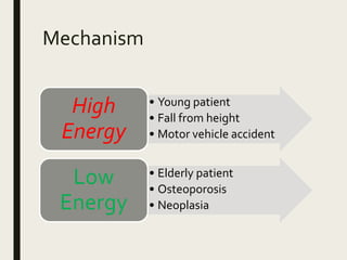
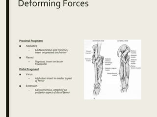
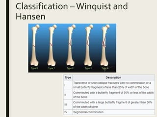
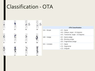
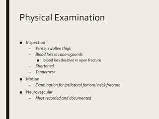
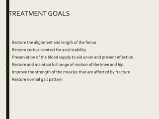
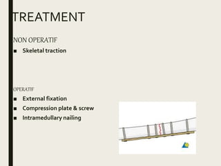
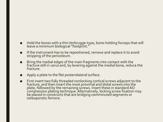
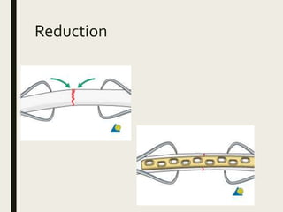
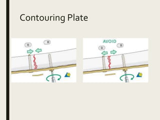
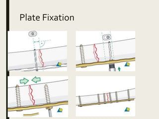
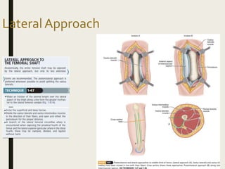
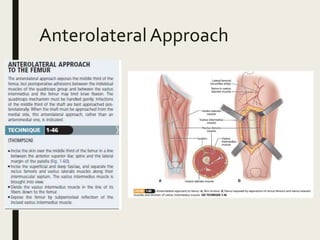
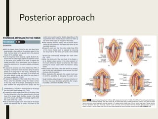
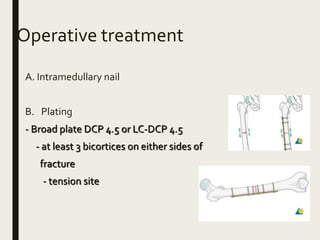
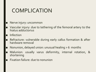
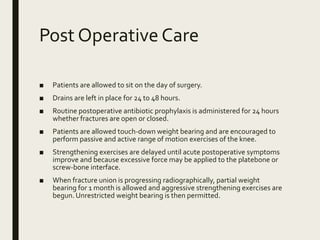
SHAFT FEMUR
FRACTURE
Herryanto Agustriadi S
Introduction
■ High energy injuries frequently associated with life-
threatening conditions
■ Incidence 37.1 per 100.000 person per year
Mechanism
• Young patient
• Fall from height
• Motor vehicle accident
High
Energy
• Elderly patient
• Osteoporosis
• Neoplasia
Low
Energy
Associated Condition
Ipsilateral Femoral Neck
Fracture
■ 2-6 incidence
■ Missed 19-31% of time
■ Often basicervical,
vertical and non
displaced
Bilateral Femur Fracture
■ Significant risk of
pulmonary complication
■ Increased rate of mortality
as compared to unilateral
fractures
Anatomy
■ Largest and strongest bone
■ Femur has an anterior bow
■ Has 3 muscle compartment
– Anterior
■ Sartorius
■ Quadriceps
– Posterior
■ Biceps femoris
■ Semitendineous
■ Semimembranosus
– Medial
■ Gracillis
■ Adductor Longus
■ Adductor brevis
■ Adductor Magnus
Deforming Forces
Proximal Fragment
■ Abducted
– Gluteus medius and minimus,
insert on greated trochanter
■ Flexed
– Iliopsoas, insert on lesser
trochanter
Distal Fragment
■ Varus
– Adductors insert in medial aspect
of femur
■ Extension
– Gastrocnemius, attached on
posterior aspect of distal femur
Classification –Winquist and
Hansen
Classification - OTA
Physical Examination
■ Inspection
– Tense, swollen thigh
– Blood loss is 1000-1500mls
■ Blood loss doubled in open fracture
– Shortened
– Tenderness
■ Motion
– Examination for ipsilateral femoral neck fracture
■ Neurovascular
– Must recorded and documented
Imaging
■ Radiograph
– AP and Lateral views of entire femur
– AP and Lateral views of ipsilateral hip
– AP and lateral views of ipsilateral knee
BIOMECHANICS
TREATMENT GOALS
• Restore the alignment and length of the femur
• Restore cortical contact for axial stability
• Preservation of the blood supply to aid union and prevent infection
• Restore and maintain full range of motion of the knee and hip
• Improve the strength of the muscles that are affected by fracture
• Restore normal gait pattern
TREATMENT
NON OPERATIF
■ Skeletal traction
OPERATIF
■ External fixation
■ Compression plate & screw
■ Intramedullary nailing
Surgical approach
■ Lateral
■ Posterolateral
■ Anterolateral
■ Posterior
Position
Place the patient supine on a
radiolucent table if fluoroscopy is
planned for the procedure.
Place a well-padded bump under
the ipsilateral hip.
Prepare the entire leg, including the
iliac crest and groin.
Alternative: Lateral Position
Skin incision
An incision is made along an
imaginary line between the lateral
femoral epicondyle and the greater
trochanter, along the length of the
femur required by the specific
fracture pattern
Separation of vastus
lateralis from fascia lata
using blunt dissection
Incision of the fascia vastus
lateralis
The vastus lateralis is now retracted
anteromedially.
The muscle fascia investing the vastus
lateralis is incised about 1 cm anterior
to the intermuscular septum
Elevate the vastus lateralis muscle
anteriorly off of the intermuscular
septum, which can be readily
accomplished with an elevator
Perforating vessels run perpendicular to the shaft of the femur
approximately every 3 cm. Identify and ligate the perforating vessels
prior to transection. Small or medium vascular clips are useful at this
stage of the exposure. Cauterization alone often is inadequate because
retraction can cause disruption of the clot.
■ Use only rakes as retractors, and avoid retractors placed medially
because this may injure the soft tissue attachments of fracture
fragments and theoretically increase the risk for difficulties with
healing.
■ Elevate only a minimal amount of periosteum to view the fracture
reduction. Care should be taken to expose the lateral femur in an
extraperiosteal fashion and only enough to permit implant
placement.
■ Overlap the fracture ends, and displace them into varus to allow a
direct view of the medullary canal
■ Débride the fracture ends of frayed soft tissue that is impaled on
sharp bone edges, and irrigate
■ Hold the bones with a thinVerbrugge-type, bone-holding forceps that will
leave a minimum biological “footprint.”
■ If the instrument has to be repositioned, remove and replace it to avoid
stripping of the periosteum.
■ Bring the medial edges of the main fragments into contact with the
fracture still in varus and, by levering against the medial bone, reduce the
fracture.
■ Apply a plate to the flat posterolateral surface.
■ First insert two fully threaded nonlocking cortical screws adjacent to the
fracture, and then insert the most proximal and distal screws into the
plate, followed by the remaining screws. Insert these in standard AO
compression plating technique.Alternatively, locking screw fixation may
be placed in constructs that are bridging comminuted segments or
osteoporotic femora.
Reduction
Contouring Plate
Plate Fixation
LateralApproach
PosterolateralApproach
AnterolateralApproach
Posterior approach
Operative treatment
A. Intramedullary nail
B. Plating
- Broad plate DCP 4.5 or LC-DCP 4.5
- at least 3 bicortices on either sides of
fracture
- tension site
COMPLICATION
■ Nerve injury: uncommon
■ Vascular injury: due to tethering of the femoral artery to the
hiatus adductorius
■ Infection
■ Refracture: vulnerable during early callus formation & after
hardware removal
■ Nonunion, delayed union: unusual healing > 6 months
■ Malunion: usually varus deformity, internal rotation, &
shortening
■ Fixation failure: due to nonunion
Post Operative Care
■ Patients are allowed to sit on the day of surgery.
■ Drains are left in place for 24 to 48 hours.
■ Routine postoperative antibiotic prophylaxis is administered for 24 hours
whether fractures are open or closed.
■ Patients are allowed touch-down weight bearing and are encouraged to
perform passive and active range of motion exercises of the knee.
■ Strengthening exercises are delayed until acute postoperative symptoms
improve and because excessive force may be applied to the platebone or
screw-bone interface.
■ When fracture union is progressing radiographically, partial weight
bearing for 1 month is allowed and aggressive strengthening exercises are
begun. Unrestricted weight bearing is then permitted.
ThankYou
References
■ Canale ST, Beaty JH, & CampbellWC. 2013. Campbell’s
Operative Orthopaedics, Philadelphia, PA: Elsevier/Mosby

Shaft femur

  • 1.
  • 2.
    Introduction ■ High energyinjuries frequently associated with life- threatening conditions ■ Incidence 37.1 per 100.000 person per year
  • 3.
    Mechanism • Young patient •Fall from height • Motor vehicle accident High Energy • Elderly patient • Osteoporosis • Neoplasia Low Energy
  • 4.
    Associated Condition Ipsilateral FemoralNeck Fracture ■ 2-6 incidence ■ Missed 19-31% of time ■ Often basicervical, vertical and non displaced Bilateral Femur Fracture ■ Significant risk of pulmonary complication ■ Increased rate of mortality as compared to unilateral fractures
  • 5.
    Anatomy ■ Largest andstrongest bone ■ Femur has an anterior bow ■ Has 3 muscle compartment – Anterior ■ Sartorius ■ Quadriceps – Posterior ■ Biceps femoris ■ Semitendineous ■ Semimembranosus – Medial ■ Gracillis ■ Adductor Longus ■ Adductor brevis ■ Adductor Magnus
  • 6.
    Deforming Forces Proximal Fragment ■Abducted – Gluteus medius and minimus, insert on greated trochanter ■ Flexed – Iliopsoas, insert on lesser trochanter Distal Fragment ■ Varus – Adductors insert in medial aspect of femur ■ Extension – Gastrocnemius, attached on posterior aspect of distal femur
  • 7.
  • 8.
  • 9.
    Physical Examination ■ Inspection –Tense, swollen thigh – Blood loss is 1000-1500mls ■ Blood loss doubled in open fracture – Shortened – Tenderness ■ Motion – Examination for ipsilateral femoral neck fracture ■ Neurovascular – Must recorded and documented
  • 10.
    Imaging ■ Radiograph – APand Lateral views of entire femur – AP and Lateral views of ipsilateral hip – AP and lateral views of ipsilateral knee
  • 11.
  • 12.
    TREATMENT GOALS • Restorethe alignment and length of the femur • Restore cortical contact for axial stability • Preservation of the blood supply to aid union and prevent infection • Restore and maintain full range of motion of the knee and hip • Improve the strength of the muscles that are affected by fracture • Restore normal gait pattern
  • 13.
    TREATMENT NON OPERATIF ■ Skeletaltraction OPERATIF ■ External fixation ■ Compression plate & screw ■ Intramedullary nailing
  • 14.
    Surgical approach ■ Lateral ■Posterolateral ■ Anterolateral ■ Posterior
  • 15.
    Position Place the patientsupine on a radiolucent table if fluoroscopy is planned for the procedure. Place a well-padded bump under the ipsilateral hip. Prepare the entire leg, including the iliac crest and groin. Alternative: Lateral Position
  • 16.
    Skin incision An incisionis made along an imaginary line between the lateral femoral epicondyle and the greater trochanter, along the length of the femur required by the specific fracture pattern
  • 17.
    Separation of vastus lateralisfrom fascia lata using blunt dissection
  • 18.
    Incision of thefascia vastus lateralis The vastus lateralis is now retracted anteromedially. The muscle fascia investing the vastus lateralis is incised about 1 cm anterior to the intermuscular septum
  • 19.
    Elevate the vastuslateralis muscle anteriorly off of the intermuscular septum, which can be readily accomplished with an elevator
  • 20.
    Perforating vessels runperpendicular to the shaft of the femur approximately every 3 cm. Identify and ligate the perforating vessels prior to transection. Small or medium vascular clips are useful at this stage of the exposure. Cauterization alone often is inadequate because retraction can cause disruption of the clot.
  • 21.
    ■ Use onlyrakes as retractors, and avoid retractors placed medially because this may injure the soft tissue attachments of fracture fragments and theoretically increase the risk for difficulties with healing. ■ Elevate only a minimal amount of periosteum to view the fracture reduction. Care should be taken to expose the lateral femur in an extraperiosteal fashion and only enough to permit implant placement. ■ Overlap the fracture ends, and displace them into varus to allow a direct view of the medullary canal ■ Débride the fracture ends of frayed soft tissue that is impaled on sharp bone edges, and irrigate
  • 22.
    ■ Hold thebones with a thinVerbrugge-type, bone-holding forceps that will leave a minimum biological “footprint.” ■ If the instrument has to be repositioned, remove and replace it to avoid stripping of the periosteum. ■ Bring the medial edges of the main fragments into contact with the fracture still in varus and, by levering against the medial bone, reduce the fracture. ■ Apply a plate to the flat posterolateral surface. ■ First insert two fully threaded nonlocking cortical screws adjacent to the fracture, and then insert the most proximal and distal screws into the plate, followed by the remaining screws. Insert these in standard AO compression plating technique.Alternatively, locking screw fixation may be placed in constructs that are bridging comminuted segments or osteoporotic femora.
  • 23.
  • 24.
  • 25.
  • 26.
  • 27.
  • 28.
  • 29.
  • 30.
    Operative treatment A. Intramedullarynail B. Plating - Broad plate DCP 4.5 or LC-DCP 4.5 - at least 3 bicortices on either sides of fracture - tension site
  • 31.
    COMPLICATION ■ Nerve injury:uncommon ■ Vascular injury: due to tethering of the femoral artery to the hiatus adductorius ■ Infection ■ Refracture: vulnerable during early callus formation & after hardware removal ■ Nonunion, delayed union: unusual healing > 6 months ■ Malunion: usually varus deformity, internal rotation, & shortening ■ Fixation failure: due to nonunion
  • 32.
    Post Operative Care ■Patients are allowed to sit on the day of surgery. ■ Drains are left in place for 24 to 48 hours. ■ Routine postoperative antibiotic prophylaxis is administered for 24 hours whether fractures are open or closed. ■ Patients are allowed touch-down weight bearing and are encouraged to perform passive and active range of motion exercises of the knee. ■ Strengthening exercises are delayed until acute postoperative symptoms improve and because excessive force may be applied to the platebone or screw-bone interface. ■ When fracture union is progressing radiographically, partial weight bearing for 1 month is allowed and aggressive strengthening exercises are begun. Unrestricted weight bearing is then permitted.
  • 33.
  • 34.
    References ■ Canale ST,Beaty JH, & CampbellWC. 2013. Campbell’s Operative Orthopaedics, Philadelphia, PA: Elsevier/Mosby

Editor's Notes

  • #25 Overbending the plate With short oblique, or transverse fractures, the plate should be slightly overbent (more convex), so that it fits imperfectly onto the bone. Any micromotion results in excessive strain on the healing tissues at the fracture site and also increases the risk of fatigue failure of the plates if the union is delayed.