SlideShare a Scribd company logo
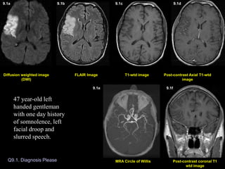
Q9.1. Diagnosis Please
Post-contrast Axial T1-wtd
image
T1-wtd image
FLAIR Image
Post-contrast coronal T1
wtd image
MRA Circle of Willis
Diffusion weighted image
(DWI)
Diffusion weighted image (DWI)
9.1a 9.1b 9.1c 9.1d
9.1e 9.1f
47 year-old left
handed gentleman
with one day history
of somnolence, left
facial droop and
slurred speech.
Q9.2. Diagnosis Please
Post-contrast Axial T1-wtd
image
T1-wtd image
FLAIR Image
(DWI) image
51 year-old patient multiple myeloma presented with acute onset of right side weakness leading to MRI of the brain 3 days later.
51 year-old patient with multiple myeloma presented
with acute onset of right sided weakness leading to MRI
of the brain 3 days later.
9.2a 9.2b 9.2c 9.2d
Q9.3. Diagnosis Please
Post-contrast Axial T1-wtd
image
T1-wtd image
FLAIR Image
Post-contrast coronal T1
wtd image
MR Angiography Neck
Diffusion weighted image
(DWI)
72 year-old left handed white man
with history of chest pain, 5 days
prior to MRI, which subsided.
Patient went to a gas station and
could not read the credit card and
signs in the gas station and was
unable to read the magazines in
the doctor’s office. Neurological
examination revealed alexia
without agraphia.
9.3a 9.3b 9.3c 9.3d
9.3e 9.3f
9.4a. Non-contrast CT Brain 9.4b. Non-contrast CT Brain
Q9.4. Diagnosis Please
9.4c. Non-contrast CT Brain
June 29, 2004 July 30, 2004
Patient is status post recent right temporal craniectomy for extensive
right middle fossa skull-based meningioma, with follow-up post-
operative CT images.
9.4a 9.4b 9.4c
Q9.5. Diagnosis Please
58 year-old male with history of renal cell carcinoma
presented with 3 weeks history of dressing apraxia consisting
of difficulty in performing routine tasks such as getting
dressed, tying his shoes, difficulty with recall and inferior
quadrantopsia with no focal motor deficits. Symptoms slowly
improved. Differential diagnosis: Stroke versus metastasis.
Post-contrast Axial T1-wtd
image
T1-wtd image
FLAIR Image
Post-contrast sagittal T1
wtd image
Diffusion weighted image
(DWI)
9.5a 9.5b 9.5c 9.5d
9.5e
Q9.6. Diagnosis Please
July 31, 2003
T1-wtd image
DW Image FLAIR Image Post-contrast Axial T1-wtd
image
9.6a 9.6b 9.6c 9.6d
December 31, 2003
DW Image FLAIR Image T1-wtd image Post-contrast Axial T1-wtd
image
9.6e 9.6f 9.6g 9.6h
73 year-old male with stage IV non-small cell carcinoma presented with 2 weeks history of
sudden onset of speech difficulty with difficulty in word finding, symptoms gradually
improved. Clinical diagnosis: Stroke versus metastasis.
Diffusion weighted image (DWI)
9.1a 9.1b 9.1c 9.1d 9.1e 9.1f
Diagnosis: Acute one day old infarction involving the right middle
cerebral artery (MCA) territory.
Acute infarction is seen as an area of increased signal intensity on
DWI (arrow in A), FLAIR image (arrow in B), with no evidence of
hemorrhage on T1-wtd image (C) and no enhancement on post
contrast images (D). Intravascular enhancement also an indication
of acute stroke is shown on coronal T1 weighted image (arrow in F).
MR angiography of circle of Willis demonstrates small caliber of
right Sylvian branches of MCA (arrows in E) when compared to the
normal side.
Post-contrast Axial T1-wtd
image
T1-wtd image
FLAIR Image
DWI
9.2a 9.2b 9.2c 9.2d
Diagnosis: Small acute 3 day old infarction involving the left
insular cortex, the territory of left MCA, best noted on diffusion
weighted image (arrow in A) and with enhancement (arrow in D).
9.3a 9.3b 9.3c 9.3d 9.3e 9.3f
Diagnosis: Acute infarction (5 day old) involving the left posterior
cerebral artery (PCA) territory.
The area of infarction is seen as an area of increased signal intensity
on DWI (arrow in A) and FLAIR image (arrow in B) involving the
left posterior temporal-occipital lobe. Mild enhancement seen on
post contrast images (arrow in D and F). Figure E represents
screening 2D time of flight MR angiogram of the neck vessels
revealing no obvious occlusion of major vessels in the neck.
Diagnosis: Acute infarction (5 day old) involving the left posterior cerebral artery (PCA) territory.
9.4a. Non-contrast CT Brain 9.4b. Non-contrast CT Brain 9.4c. Non-contrast CT Brain
June 29, 2004 July 30, 2004
Non-contrast CT brain on 8 days post-operatively demonstrated a
focal area of low attenuation within the right frontal lobe cortex
(arrow in A) and adjacent white matter representing acute infarct.
Scan done a month later (B & C) demonstrated a massive
acute/subacute infarct involving the right MCA territory (white
arrows in B & C) and ACA (anterior cerebral artery) territory
(yellow arrow in C) with mass effect and subfalcine herniation to
the left (fig. B).
Q9.5. Diagnosis Please
Post-contrast Axial T1-wtd
image
T1-wtd image
FLAIR Image
Post-contrast sagittal T1
wtd image
Diffusion weighted image
(DWI)
9.5a 9.5b 9.5c 9.5d
9.5e
Diagnosis: Subacute infarct 3 weeks old involving the
right middle cerebral (MCA) artery territory.
Abnormal T2 wtd hyperintensity involving the right posterior temporal-
parietal-occipital lobe with gyral thickening best noted on FLAIR image
(arrows in B). Subacute blood outlining the cortex is best seen on pre-
contrast T1-wtd image (arrows in C). There is no definite contrast
enhancement obscured by bright signal intensity of blood. The right MCA
territory infarct is also shown on sagittal post contrast T1-wtd image
(arrows in E). Diffusion weighted image reveals bright signal intensity
(arrows in A) involving the cortex from restricted diffusion, an important
sequence in the diagnosis of acute stroke.
Q9.6. Diagnosis Please
July 31, 2003
December 31, 2003
Diagnosis: Non-hemorrhagic subacute enhancing infarct (2 weeks old) involving
the left basal ganglia region. Subacute infarct is seen as an area of increased signal
intensity on FLAIR image (arrow in B) and bright signal intensity on DWI (arrow in
A). Enhancement of the infarct is shown on post contrast image (arrow in D).
A repeat MRI scan done 5 months later
showed resolution of infarct and no
evidence of bright signal intensity on
diffusion weighted image E.
5 months old infarct.
9.6a 9.6b 9.6c 9.6d
9.6e 9.6f 9.6g 9.6h
1a
2a
5b
6d
4b
6e
Imaging Findings of Stroke:
• MR imaging of the brain is far more sensitive than CT imaging to recognize acute
infarction.
• Diffusion wtd. pulse sequence (DW imaging) is the most sensitive MR sequence to
demonstrate stroke. This sequence is sensitive to restricted diffusion within the cell from
stroke-induced cytotoxic edema and the region of acute stroke is seen as an area of bright
signal on DWI (Figs. 1a, 2a). Cytotoxic edema can occur immediately after the initial insult
thus DWI images can reveal, the area of acute infarct immediately after the insult.
• Intravascular contrast enhancement, another sign of early stroke (Figure 1f).
• Sulcal effacement, gyral edema (Fig. 5b), loss of gray-white matter interface can occur
within 12 hours of stroke.
• Parenchymal contrast enhancement (Fig. 6d), mass effect (Fig. 4b) and hemorrhage can
occur within 1-7 days of insult.
Subacute infarct: (1 week to 8 weeks)
•Focal area of encephalomalacia
•Porencephalic dilatation of adjacent ventricle.
• Residual old blood products may be present.
Old Infarct:
• Contrast enhancement slowly decreases in time but can persist for 8 weeks, with
decreasing mass effect and abnormal signal intensity:
Acute Stroke (up to 7 days)

More Related Content

Similar to set9_20041008.ppt

imagingofspinaltrauMA
imagingofspinaltrauMAimagingofspinaltrauMA
imagingofspinaltrauMA
Sandra710258
 
Casos Clínicos
Casos ClínicosCasos Clínicos
Casos Clínicos
Inês Ferreira
 
Stroke
StrokeStroke
Stroke
Adila Iqbal
 
Presentation2, radiological imaging of phakomatosis.
Presentation2, radiological imaging of phakomatosis.Presentation2, radiological imaging of phakomatosis.
Presentation2, radiological imaging of phakomatosis.
Abdellah Nazeer
 
Presentation1, radiological imaging of corpus callosum lesios.
Presentation1, radiological imaging of corpus callosum lesios.Presentation1, radiological imaging of corpus callosum lesios.
Presentation1, radiological imaging of corpus callosum lesios.
Abdellah Nazeer
 
Ariunaa spine trauma
Ariunaa spine traumaAriunaa spine trauma
Ariunaa spine trauma
Battulga Munkhtsetseg
 
Stroke and stroke mimics
Stroke and stroke mimicsStroke and stroke mimics
Stroke and stroke mimics
Nathaliazuos
 
Diagnostic Imaging of Cerebral Trauma
Diagnostic Imaging of Cerebral TraumaDiagnostic Imaging of Cerebral Trauma
Diagnostic Imaging of Cerebral Trauma
Mohamed M.A. Zaitoun
 
First Clinical Results of Coronary CT Subtraction
First Clinical Results of Coronary CT SubtractionFirst Clinical Results of Coronary CT Subtraction
First Clinical Results of Coronary CT Subtraction
Canon Medical Systems Europe
 
3. mri in acute stroke 2017 vietnam v2
3. mri in acute stroke 2017 vietnam v23. mri in acute stroke 2017 vietnam v2
3. mri in acute stroke 2017 vietnam v2
Đường Nguyễn
 
Clinical correlation and interpretation of Brain MRI by dr.Sagor
Clinical correlation and interpretation of Brain MRI by dr.SagorClinical correlation and interpretation of Brain MRI by dr.Sagor
Clinical correlation and interpretation of Brain MRI by dr.Sagor
Chittagong Medical College Hospital
 
Susceptibility Weighted Imaging (SWI)
Susceptibility Weighted Imaging (SWI)Susceptibility Weighted Imaging (SWI)
Susceptibility Weighted Imaging (SWI)
Nija Panchal
 
Role of magnetic resonance imaging in coronary artery disease MRCA part 7 Dr ...
Role of magnetic resonance imaging in coronary artery disease MRCA part 7 Dr ...Role of magnetic resonance imaging in coronary artery disease MRCA part 7 Dr ...
Role of magnetic resonance imaging in coronary artery disease MRCA part 7 Dr ...
AHMED ESAWY
 
4D-flow MRI of double aortic arch in a 14-year-old patient Authors: Nadya Al-...
4D-flow MRI of double aortic arch in a 14-year-old patient Authors: Nadya Al-...4D-flow MRI of double aortic arch in a 14-year-old patient Authors: Nadya Al-...
4D-flow MRI of double aortic arch in a 14-year-old patient Authors: Nadya Al-...
Cardiovascular Diagnosis and Therapy (CDT)
 
MRI in obstetric practice part 2
MRI in obstetric practice   part 2MRI in obstetric practice   part 2
MRI in obstetric practice part 2
Shivamurthy Hm
 
Errores En Interpretacion De Tc Cráneo
Errores En Interpretacion De Tc CráneoErrores En Interpretacion De Tc Cráneo
Errores En Interpretacion De Tc Cráneorahterrazas
 
Traumatic Brain Injury Pearls and Pitfalls (2014)
Traumatic Brain Injury Pearls and Pitfalls (2014)Traumatic Brain Injury Pearls and Pitfalls (2014)
Traumatic Brain Injury Pearls and Pitfalls (2014)
Rathachai Kaewlai
 
Thrombotic Occlusion Of The Common Carotid Artery
Thrombotic Occlusion Of The Common Carotid ArteryThrombotic Occlusion Of The Common Carotid Artery
Thrombotic Occlusion Of The Common Carotid ArteryMedicineAndHealthNeurolog
 
23205056
2320505623205056
23205056
radgirl
 
Marchiafava–Bignami disease (MBD)
Marchiafava–Bignami disease (MBD) Marchiafava–Bignami disease (MBD)
Marchiafava–Bignami disease (MBD) Dr Surendra Khosya
 

Similar to set9_20041008.ppt (20)

imagingofspinaltrauMA
imagingofspinaltrauMAimagingofspinaltrauMA
imagingofspinaltrauMA
 
Casos Clínicos
Casos ClínicosCasos Clínicos
Casos Clínicos
 
Stroke
StrokeStroke
Stroke
 
Presentation2, radiological imaging of phakomatosis.
Presentation2, radiological imaging of phakomatosis.Presentation2, radiological imaging of phakomatosis.
Presentation2, radiological imaging of phakomatosis.
 
Presentation1, radiological imaging of corpus callosum lesios.
Presentation1, radiological imaging of corpus callosum lesios.Presentation1, radiological imaging of corpus callosum lesios.
Presentation1, radiological imaging of corpus callosum lesios.
 
Ariunaa spine trauma
Ariunaa spine traumaAriunaa spine trauma
Ariunaa spine trauma
 
Stroke and stroke mimics
Stroke and stroke mimicsStroke and stroke mimics
Stroke and stroke mimics
 
Diagnostic Imaging of Cerebral Trauma
Diagnostic Imaging of Cerebral TraumaDiagnostic Imaging of Cerebral Trauma
Diagnostic Imaging of Cerebral Trauma
 
First Clinical Results of Coronary CT Subtraction
First Clinical Results of Coronary CT SubtractionFirst Clinical Results of Coronary CT Subtraction
First Clinical Results of Coronary CT Subtraction
 
3. mri in acute stroke 2017 vietnam v2
3. mri in acute stroke 2017 vietnam v23. mri in acute stroke 2017 vietnam v2
3. mri in acute stroke 2017 vietnam v2
 
Clinical correlation and interpretation of Brain MRI by dr.Sagor
Clinical correlation and interpretation of Brain MRI by dr.SagorClinical correlation and interpretation of Brain MRI by dr.Sagor
Clinical correlation and interpretation of Brain MRI by dr.Sagor
 
Susceptibility Weighted Imaging (SWI)
Susceptibility Weighted Imaging (SWI)Susceptibility Weighted Imaging (SWI)
Susceptibility Weighted Imaging (SWI)
 
Role of magnetic resonance imaging in coronary artery disease MRCA part 7 Dr ...
Role of magnetic resonance imaging in coronary artery disease MRCA part 7 Dr ...Role of magnetic resonance imaging in coronary artery disease MRCA part 7 Dr ...
Role of magnetic resonance imaging in coronary artery disease MRCA part 7 Dr ...
 
4D-flow MRI of double aortic arch in a 14-year-old patient Authors: Nadya Al-...
4D-flow MRI of double aortic arch in a 14-year-old patient Authors: Nadya Al-...4D-flow MRI of double aortic arch in a 14-year-old patient Authors: Nadya Al-...
4D-flow MRI of double aortic arch in a 14-year-old patient Authors: Nadya Al-...
 
MRI in obstetric practice part 2
MRI in obstetric practice   part 2MRI in obstetric practice   part 2
MRI in obstetric practice part 2
 
Errores En Interpretacion De Tc Cráneo
Errores En Interpretacion De Tc CráneoErrores En Interpretacion De Tc Cráneo
Errores En Interpretacion De Tc Cráneo
 
Traumatic Brain Injury Pearls and Pitfalls (2014)
Traumatic Brain Injury Pearls and Pitfalls (2014)Traumatic Brain Injury Pearls and Pitfalls (2014)
Traumatic Brain Injury Pearls and Pitfalls (2014)
 
Thrombotic Occlusion Of The Common Carotid Artery
Thrombotic Occlusion Of The Common Carotid ArteryThrombotic Occlusion Of The Common Carotid Artery
Thrombotic Occlusion Of The Common Carotid Artery
 
23205056
2320505623205056
23205056
 
Marchiafava–Bignami disease (MBD)
Marchiafava–Bignami disease (MBD) Marchiafava–Bignami disease (MBD)
Marchiafava–Bignami disease (MBD)
 

More from hussainAltaher

11-Sexually transmitted disease (2).pptx
11-Sexually transmitted disease (2).pptx11-Sexually transmitted disease (2).pptx
11-Sexually transmitted disease (2).pptx
hussainAltaher
 
Gyneco_Dr_Ezdehar_L2_physiological_Changes_in_pregnancy_part_2.pptx
Gyneco_Dr_Ezdehar_L2_physiological_Changes_in_pregnancy_part_2.pptxGyneco_Dr_Ezdehar_L2_physiological_Changes_in_pregnancy_part_2.pptx
Gyneco_Dr_Ezdehar_L2_physiological_Changes_in_pregnancy_part_2.pptx
hussainAltaher
 
Gyneco.Dr.Ezdehar.L1-physiological Changes in pregnancy.pptx
Gyneco.Dr.Ezdehar.L1-physiological Changes in pregnancy.pptxGyneco.Dr.Ezdehar.L1-physiological Changes in pregnancy.pptx
Gyneco.Dr.Ezdehar.L1-physiological Changes in pregnancy.pptx
hussainAltaher
 
Gyneco. Dr. Ezdehar.L3-DIC.ppertyuiopptx
Gyneco. Dr. Ezdehar.L3-DIC.ppertyuiopptxGyneco. Dr. Ezdehar.L3-DIC.ppertyuiopptx
Gyneco. Dr. Ezdehar.L3-DIC.ppertyuiopptx
hussainAltaher
 
Gyneco. D. Ezdehar L5 Fetal Assessment in Labor.pptx
Gyneco. D. Ezdehar L5 Fetal Assessment in Labor.pptxGyneco. D. Ezdehar L5 Fetal Assessment in Labor.pptx
Gyneco. D. Ezdehar L5 Fetal Assessment in Labor.pptx
hussainAltaher
 
Gynea.D.Izdihar.L3-Liver disease.pTTTTptx
Gynea.D.Izdihar.L3-Liver disease.pTTTTptxGynea.D.Izdihar.L3-Liver disease.pTTTTptx
Gynea.D.Izdihar.L3-Liver disease.pTTTTptx
hussainAltaher
 
طلاب كروب D Osce slides in gynaecology-1 (Muhadharaty).pptx
طلاب كروب D Osce slides in gynaecology-1 (Muhadharaty).pptxطلاب كروب D Osce slides in gynaecology-1 (Muhadharaty).pptx
طلاب كروب D Osce slides in gynaecology-1 (Muhadharaty).pptx
hussainAltaher
 
D. Ihsan Rotator Cuff Disorders-4 (Muhadharaty).pptx
D. Ihsan Rotator Cuff Disorders-4 (Muhadharaty).pptxD. Ihsan Rotator Cuff Disorders-4 (Muhadharaty).pptx
D. Ihsan Rotator Cuff Disorders-4 (Muhadharaty).pptx
hussainAltaher
 
د. عمار الياسري shoulder and pectoral girdle-5 (Muhadharaty).pptx
د. عمار الياسري shoulder and pectoral girdle-5 (Muhadharaty).pptxد. عمار الياسري shoulder and pectoral girdle-5 (Muhadharaty).pptx
د. عمار الياسري shoulder and pectoral girdle-5 (Muhadharaty).pptx
hussainAltaher
 
د. عمار الياسري shoulder and pectoral girdle-5 (Muhadharaty).pptx
د. عمار الياسري shoulder and pectoral girdle-5 (Muhadharaty).pptxد. عمار الياسري shoulder and pectoral girdle-5 (Muhadharaty).pptx
د. عمار الياسري shoulder and pectoral girdle-5 (Muhadharaty).pptx
hussainAltaher
 
PPH-6 (Muhadharaty).pptx , tyuiop[oiuytr
PPH-6 (Muhadharaty).pptx , tyuiop[oiuytrPPH-6 (Muhadharaty).pptx , tyuiop[oiuytr
PPH-6 (Muhadharaty).pptx , tyuiop[oiuytr
hussainAltaher
 
4- LUNG ABSCESS &EMPYEMA (2)...............pptx
4- LUNG ABSCESS &EMPYEMA (2)...............pptx4- LUNG ABSCESS &EMPYEMA (2)...............pptx
4- LUNG ABSCESS &EMPYEMA (2)...............pptx
hussainAltaher
 
endometrial cancer #.ppt.......................
endometrial cancer #.ppt.......................endometrial cancer #.ppt.......................
endometrial cancer #.ppt.......................
hussainAltaher
 
Assisted RT22.ppt.......................
Assisted RT22.ppt.......................Assisted RT22.ppt.......................
Assisted RT22.ppt.......................
hussainAltaher
 
4- LUNG ABSCESS &EMPYEMA (2).........pptx
4- LUNG ABSCESS &EMPYEMA (2).........pptx4- LUNG ABSCESS &EMPYEMA (2).........pptx
4- LUNG ABSCESS &EMPYEMA (2).........pptx
hussainAltaher
 
Chest Trauma (2).pptx....................
Chest Trauma (2).pptx....................Chest Trauma (2).pptx....................
Chest Trauma (2).pptx....................
hussainAltaher
 
congnital anomaly (2).pptx..............
congnital anomaly (2).pptx..............congnital anomaly (2).pptx..............
congnital anomaly (2).pptx..............
hussainAltaher
 
BURN.pptx.............general ............
BURN.pptx.............general ............BURN.pptx.............general ............
BURN.pptx.............general ............
hussainAltaher
 
5- BRONCHIACTASIS..pptx thoracic surgery
5- BRONCHIACTASIS..pptx thoracic surgery5- BRONCHIACTASIS..pptx thoracic surgery
5- BRONCHIACTASIS..pptx thoracic surgery
hussainAltaher
 
Soft tissues tumors. general surgerypptx
Soft tissues tumors. general surgerypptxSoft tissues tumors. general surgerypptx
Soft tissues tumors. general surgerypptx
hussainAltaher
 

More from hussainAltaher (20)

11-Sexually transmitted disease (2).pptx
11-Sexually transmitted disease (2).pptx11-Sexually transmitted disease (2).pptx
11-Sexually transmitted disease (2).pptx
 
Gyneco_Dr_Ezdehar_L2_physiological_Changes_in_pregnancy_part_2.pptx
Gyneco_Dr_Ezdehar_L2_physiological_Changes_in_pregnancy_part_2.pptxGyneco_Dr_Ezdehar_L2_physiological_Changes_in_pregnancy_part_2.pptx
Gyneco_Dr_Ezdehar_L2_physiological_Changes_in_pregnancy_part_2.pptx
 
Gyneco.Dr.Ezdehar.L1-physiological Changes in pregnancy.pptx
Gyneco.Dr.Ezdehar.L1-physiological Changes in pregnancy.pptxGyneco.Dr.Ezdehar.L1-physiological Changes in pregnancy.pptx
Gyneco.Dr.Ezdehar.L1-physiological Changes in pregnancy.pptx
 
Gyneco. Dr. Ezdehar.L3-DIC.ppertyuiopptx
Gyneco. Dr. Ezdehar.L3-DIC.ppertyuiopptxGyneco. Dr. Ezdehar.L3-DIC.ppertyuiopptx
Gyneco. Dr. Ezdehar.L3-DIC.ppertyuiopptx
 
Gyneco. D. Ezdehar L5 Fetal Assessment in Labor.pptx
Gyneco. D. Ezdehar L5 Fetal Assessment in Labor.pptxGyneco. D. Ezdehar L5 Fetal Assessment in Labor.pptx
Gyneco. D. Ezdehar L5 Fetal Assessment in Labor.pptx
 
Gynea.D.Izdihar.L3-Liver disease.pTTTTptx
Gynea.D.Izdihar.L3-Liver disease.pTTTTptxGynea.D.Izdihar.L3-Liver disease.pTTTTptx
Gynea.D.Izdihar.L3-Liver disease.pTTTTptx
 
طلاب كروب D Osce slides in gynaecology-1 (Muhadharaty).pptx
طلاب كروب D Osce slides in gynaecology-1 (Muhadharaty).pptxطلاب كروب D Osce slides in gynaecology-1 (Muhadharaty).pptx
طلاب كروب D Osce slides in gynaecology-1 (Muhadharaty).pptx
 
D. Ihsan Rotator Cuff Disorders-4 (Muhadharaty).pptx
D. Ihsan Rotator Cuff Disorders-4 (Muhadharaty).pptxD. Ihsan Rotator Cuff Disorders-4 (Muhadharaty).pptx
D. Ihsan Rotator Cuff Disorders-4 (Muhadharaty).pptx
 
د. عمار الياسري shoulder and pectoral girdle-5 (Muhadharaty).pptx
د. عمار الياسري shoulder and pectoral girdle-5 (Muhadharaty).pptxد. عمار الياسري shoulder and pectoral girdle-5 (Muhadharaty).pptx
د. عمار الياسري shoulder and pectoral girdle-5 (Muhadharaty).pptx
 
د. عمار الياسري shoulder and pectoral girdle-5 (Muhadharaty).pptx
د. عمار الياسري shoulder and pectoral girdle-5 (Muhadharaty).pptxد. عمار الياسري shoulder and pectoral girdle-5 (Muhadharaty).pptx
د. عمار الياسري shoulder and pectoral girdle-5 (Muhadharaty).pptx
 
PPH-6 (Muhadharaty).pptx , tyuiop[oiuytr
PPH-6 (Muhadharaty).pptx , tyuiop[oiuytrPPH-6 (Muhadharaty).pptx , tyuiop[oiuytr
PPH-6 (Muhadharaty).pptx , tyuiop[oiuytr
 
4- LUNG ABSCESS &EMPYEMA (2)...............pptx
4- LUNG ABSCESS &EMPYEMA (2)...............pptx4- LUNG ABSCESS &EMPYEMA (2)...............pptx
4- LUNG ABSCESS &EMPYEMA (2)...............pptx
 
endometrial cancer #.ppt.......................
endometrial cancer #.ppt.......................endometrial cancer #.ppt.......................
endometrial cancer #.ppt.......................
 
Assisted RT22.ppt.......................
Assisted RT22.ppt.......................Assisted RT22.ppt.......................
Assisted RT22.ppt.......................
 
4- LUNG ABSCESS &EMPYEMA (2).........pptx
4- LUNG ABSCESS &EMPYEMA (2).........pptx4- LUNG ABSCESS &EMPYEMA (2).........pptx
4- LUNG ABSCESS &EMPYEMA (2).........pptx
 
Chest Trauma (2).pptx....................
Chest Trauma (2).pptx....................Chest Trauma (2).pptx....................
Chest Trauma (2).pptx....................
 
congnital anomaly (2).pptx..............
congnital anomaly (2).pptx..............congnital anomaly (2).pptx..............
congnital anomaly (2).pptx..............
 
BURN.pptx.............general ............
BURN.pptx.............general ............BURN.pptx.............general ............
BURN.pptx.............general ............
 
5- BRONCHIACTASIS..pptx thoracic surgery
5- BRONCHIACTASIS..pptx thoracic surgery5- BRONCHIACTASIS..pptx thoracic surgery
5- BRONCHIACTASIS..pptx thoracic surgery
 
Soft tissues tumors. general surgerypptx
Soft tissues tumors. general surgerypptxSoft tissues tumors. general surgerypptx
Soft tissues tumors. general surgerypptx
 

Recently uploaded

Azure Interview Questions and Answers PDF By ScholarHat
Azure Interview Questions and Answers PDF By ScholarHatAzure Interview Questions and Answers PDF By ScholarHat
Azure Interview Questions and Answers PDF By ScholarHat
Scholarhat
 
Model Attribute Check Company Auto Property
Model Attribute  Check Company Auto PropertyModel Attribute  Check Company Auto Property
Model Attribute Check Company Auto Property
Celine George
 
2024.06.01 Introducing a competency framework for languag learning materials ...
2024.06.01 Introducing a competency framework for languag learning materials ...2024.06.01 Introducing a competency framework for languag learning materials ...
2024.06.01 Introducing a competency framework for languag learning materials ...
Sandy Millin
 
The Diamond Necklace by Guy De Maupassant.pptx
The Diamond Necklace by Guy De Maupassant.pptxThe Diamond Necklace by Guy De Maupassant.pptx
The Diamond Necklace by Guy De Maupassant.pptx
DhatriParmar
 
Thesis Statement for students diagnonsed withADHD.ppt
Thesis Statement for students diagnonsed withADHD.pptThesis Statement for students diagnonsed withADHD.ppt
Thesis Statement for students diagnonsed withADHD.ppt
EverAndrsGuerraGuerr
 
The Accursed House by Émile Gaboriau.pptx
The Accursed House by Émile Gaboriau.pptxThe Accursed House by Émile Gaboriau.pptx
The Accursed House by Émile Gaboriau.pptx
DhatriParmar
 
Advantages and Disadvantages of CMS from an SEO Perspective
Advantages and Disadvantages of CMS from an SEO PerspectiveAdvantages and Disadvantages of CMS from an SEO Perspective
Advantages and Disadvantages of CMS from an SEO Perspective
Krisztián Száraz
 
1.4 modern child centered education - mahatma gandhi-2.pptx
1.4 modern child centered education - mahatma gandhi-2.pptx1.4 modern child centered education - mahatma gandhi-2.pptx
1.4 modern child centered education - mahatma gandhi-2.pptx
JosvitaDsouza2
 
Embracing GenAI - A Strategic Imperative
Embracing GenAI - A Strategic ImperativeEmbracing GenAI - A Strategic Imperative
Embracing GenAI - A Strategic Imperative
Peter Windle
 
A Survey of Techniques for Maximizing LLM Performance.pptx
A Survey of Techniques for Maximizing LLM Performance.pptxA Survey of Techniques for Maximizing LLM Performance.pptx
A Survey of Techniques for Maximizing LLM Performance.pptx
thanhdowork
 
How libraries can support authors with open access requirements for UKRI fund...
How libraries can support authors with open access requirements for UKRI fund...How libraries can support authors with open access requirements for UKRI fund...
How libraries can support authors with open access requirements for UKRI fund...
Jisc
 
Marketing internship report file for MBA
Marketing internship report file for MBAMarketing internship report file for MBA
Marketing internship report file for MBA
gb193092
 
Digital Artifact 2 - Investigating Pavilion Designs
Digital Artifact 2 - Investigating Pavilion DesignsDigital Artifact 2 - Investigating Pavilion Designs
Digital Artifact 2 - Investigating Pavilion Designs
chanes7
 
Overview on Edible Vaccine: Pros & Cons with Mechanism
Overview on Edible Vaccine: Pros & Cons with MechanismOverview on Edible Vaccine: Pros & Cons with Mechanism
Overview on Edible Vaccine: Pros & Cons with Mechanism
DeeptiGupta154
 
Language Across the Curriculm LAC B.Ed.
Language Across the  Curriculm LAC B.Ed.Language Across the  Curriculm LAC B.Ed.
Language Across the Curriculm LAC B.Ed.
Atul Kumar Singh
 
Francesca Gottschalk - How can education support child empowerment.pptx
Francesca Gottschalk - How can education support child empowerment.pptxFrancesca Gottschalk - How can education support child empowerment.pptx
Francesca Gottschalk - How can education support child empowerment.pptx
EduSkills OECD
 
Operation Blue Star - Saka Neela Tara
Operation Blue Star   -  Saka Neela TaraOperation Blue Star   -  Saka Neela Tara
Operation Blue Star - Saka Neela Tara
Balvir Singh
 
Mule 4.6 & Java 17 Upgrade | MuleSoft Mysore Meetup #46
Mule 4.6 & Java 17 Upgrade | MuleSoft Mysore Meetup #46Mule 4.6 & Java 17 Upgrade | MuleSoft Mysore Meetup #46
Mule 4.6 & Java 17 Upgrade | MuleSoft Mysore Meetup #46
MysoreMuleSoftMeetup
 
How to Make a Field invisible in Odoo 17
How to Make a Field invisible in Odoo 17How to Make a Field invisible in Odoo 17
How to Make a Field invisible in Odoo 17
Celine George
 
Exploiting Artificial Intelligence for Empowering Researchers and Faculty, In...
Exploiting Artificial Intelligence for Empowering Researchers and Faculty, In...Exploiting Artificial Intelligence for Empowering Researchers and Faculty, In...
Exploiting Artificial Intelligence for Empowering Researchers and Faculty, In...
Dr. Vinod Kumar Kanvaria
 

Recently uploaded (20)

Azure Interview Questions and Answers PDF By ScholarHat
Azure Interview Questions and Answers PDF By ScholarHatAzure Interview Questions and Answers PDF By ScholarHat
Azure Interview Questions and Answers PDF By ScholarHat
 
Model Attribute Check Company Auto Property
Model Attribute  Check Company Auto PropertyModel Attribute  Check Company Auto Property
Model Attribute Check Company Auto Property
 
2024.06.01 Introducing a competency framework for languag learning materials ...
2024.06.01 Introducing a competency framework for languag learning materials ...2024.06.01 Introducing a competency framework for languag learning materials ...
2024.06.01 Introducing a competency framework for languag learning materials ...
 
The Diamond Necklace by Guy De Maupassant.pptx
The Diamond Necklace by Guy De Maupassant.pptxThe Diamond Necklace by Guy De Maupassant.pptx
The Diamond Necklace by Guy De Maupassant.pptx
 
Thesis Statement for students diagnonsed withADHD.ppt
Thesis Statement for students diagnonsed withADHD.pptThesis Statement for students diagnonsed withADHD.ppt
Thesis Statement for students diagnonsed withADHD.ppt
 
The Accursed House by Émile Gaboriau.pptx
The Accursed House by Émile Gaboriau.pptxThe Accursed House by Émile Gaboriau.pptx
The Accursed House by Émile Gaboriau.pptx
 
Advantages and Disadvantages of CMS from an SEO Perspective
Advantages and Disadvantages of CMS from an SEO PerspectiveAdvantages and Disadvantages of CMS from an SEO Perspective
Advantages and Disadvantages of CMS from an SEO Perspective
 
1.4 modern child centered education - mahatma gandhi-2.pptx
1.4 modern child centered education - mahatma gandhi-2.pptx1.4 modern child centered education - mahatma gandhi-2.pptx
1.4 modern child centered education - mahatma gandhi-2.pptx
 
Embracing GenAI - A Strategic Imperative
Embracing GenAI - A Strategic ImperativeEmbracing GenAI - A Strategic Imperative
Embracing GenAI - A Strategic Imperative
 
A Survey of Techniques for Maximizing LLM Performance.pptx
A Survey of Techniques for Maximizing LLM Performance.pptxA Survey of Techniques for Maximizing LLM Performance.pptx
A Survey of Techniques for Maximizing LLM Performance.pptx
 
How libraries can support authors with open access requirements for UKRI fund...
How libraries can support authors with open access requirements for UKRI fund...How libraries can support authors with open access requirements for UKRI fund...
How libraries can support authors with open access requirements for UKRI fund...
 
Marketing internship report file for MBA
Marketing internship report file for MBAMarketing internship report file for MBA
Marketing internship report file for MBA
 
Digital Artifact 2 - Investigating Pavilion Designs
Digital Artifact 2 - Investigating Pavilion DesignsDigital Artifact 2 - Investigating Pavilion Designs
Digital Artifact 2 - Investigating Pavilion Designs
 
Overview on Edible Vaccine: Pros & Cons with Mechanism
Overview on Edible Vaccine: Pros & Cons with MechanismOverview on Edible Vaccine: Pros & Cons with Mechanism
Overview on Edible Vaccine: Pros & Cons with Mechanism
 
Language Across the Curriculm LAC B.Ed.
Language Across the  Curriculm LAC B.Ed.Language Across the  Curriculm LAC B.Ed.
Language Across the Curriculm LAC B.Ed.
 
Francesca Gottschalk - How can education support child empowerment.pptx
Francesca Gottschalk - How can education support child empowerment.pptxFrancesca Gottschalk - How can education support child empowerment.pptx
Francesca Gottschalk - How can education support child empowerment.pptx
 
Operation Blue Star - Saka Neela Tara
Operation Blue Star   -  Saka Neela TaraOperation Blue Star   -  Saka Neela Tara
Operation Blue Star - Saka Neela Tara
 
Mule 4.6 & Java 17 Upgrade | MuleSoft Mysore Meetup #46
Mule 4.6 & Java 17 Upgrade | MuleSoft Mysore Meetup #46Mule 4.6 & Java 17 Upgrade | MuleSoft Mysore Meetup #46
Mule 4.6 & Java 17 Upgrade | MuleSoft Mysore Meetup #46
 
How to Make a Field invisible in Odoo 17
How to Make a Field invisible in Odoo 17How to Make a Field invisible in Odoo 17
How to Make a Field invisible in Odoo 17
 
Exploiting Artificial Intelligence for Empowering Researchers and Faculty, In...
Exploiting Artificial Intelligence for Empowering Researchers and Faculty, In...Exploiting Artificial Intelligence for Empowering Researchers and Faculty, In...
Exploiting Artificial Intelligence for Empowering Researchers and Faculty, In...
 

set9_20041008.ppt

  • 1. Q9.1. Diagnosis Please Post-contrast Axial T1-wtd image T1-wtd image FLAIR Image Post-contrast coronal T1 wtd image MRA Circle of Willis Diffusion weighted image (DWI) Diffusion weighted image (DWI) 9.1a 9.1b 9.1c 9.1d 9.1e 9.1f 47 year-old left handed gentleman with one day history of somnolence, left facial droop and slurred speech.
  • 2. Q9.2. Diagnosis Please Post-contrast Axial T1-wtd image T1-wtd image FLAIR Image (DWI) image 51 year-old patient multiple myeloma presented with acute onset of right side weakness leading to MRI of the brain 3 days later. 51 year-old patient multiple myeloma presented with acute onset of right side weakness leading to MRI of the brain 3 days later. 51 year-old patient with multiple myeloma presented with acute onset of right sided weakness leading to MRI of the brain 3 days later. 9.2a 9.2b 9.2c 9.2d
  • 3. Q9.3. Diagnosis Please Post-contrast Axial T1-wtd image T1-wtd image FLAIR Image Post-contrast coronal T1 wtd image MR Angiography Neck Diffusion weighted image (DWI) 72 year-old left handed white man with history of chest pain, 5 days prior to MRI, which subsided. Patient went to a gas station and could not read the credit card and signs in the gas station and was unable to read the magazines in the doctor’s office. Neurological examination revealed alexia without agraphia. 9.3a 9.3b 9.3c 9.3d 9.3e 9.3f
  • 4. 9.4a. Non-contrast CT Brain 9.4b. Non-contrast CT Brain Q9.4. Diagnosis Please 9.4c. Non-contrast CT Brain June 29, 2004 July 30, 2004 Patient is status post recent right temporal craniectomy for extensive right middle fossa skull-based meningioma, with follow-up post- operative CT images. 9.4a 9.4b 9.4c
  • 5. Q9.5. Diagnosis Please 58 year-old male with history of renal cell carcinoma presented with 3 weeks history of dressing apraxia consisting of difficulty in performing routine tasks such as getting dressed, tying his shoes, difficulty with recall and inferior quadrantopsia with no focal motor deficits. Symptoms slowly improved. Differential diagnosis: Stroke versus metastasis. Post-contrast Axial T1-wtd image T1-wtd image FLAIR Image Post-contrast sagittal T1 wtd image Diffusion weighted image (DWI) 9.5a 9.5b 9.5c 9.5d 9.5e
  • 6. Q9.6. Diagnosis Please July 31, 2003 T1-wtd image DW Image FLAIR Image Post-contrast Axial T1-wtd image 9.6a 9.6b 9.6c 9.6d December 31, 2003 DW Image FLAIR Image T1-wtd image Post-contrast Axial T1-wtd image 9.6e 9.6f 9.6g 9.6h 73 year-old male with stage IV non-small cell carcinoma presented with 2 weeks history of sudden onset of speech difficulty with difficulty in word finding, symptoms gradually improved. Clinical diagnosis: Stroke versus metastasis.
  • 7. Diffusion weighted image (DWI) 9.1a 9.1b 9.1c 9.1d 9.1e 9.1f Diagnosis: Acute one day old infarction involving the right middle cerebral artery (MCA) territory. Acute infarction is seen as an area of increased signal intensity on DWI (arrow in A), FLAIR image (arrow in B), with no evidence of hemorrhage on T1-wtd image (C) and no enhancement on post contrast images (D). Intravascular enhancement also an indication of acute stroke is shown on coronal T1 weighted image (arrow in F). MR angiography of circle of Willis demonstrates small caliber of right Sylvian branches of MCA (arrows in E) when compared to the normal side.
  • 8. Post-contrast Axial T1-wtd image T1-wtd image FLAIR Image DWI 9.2a 9.2b 9.2c 9.2d Diagnosis: Small acute 3 day old infarction involving the left insular cortex, the territory of left MCA, best noted on diffusion weighted image (arrow in A) and with enhancement (arrow in D).
  • 9. 9.3a 9.3b 9.3c 9.3d 9.3e 9.3f Diagnosis: Acute infarction (5 day old) involving the left posterior cerebral artery (PCA) territory. The area of infarction is seen as an area of increased signal intensity on DWI (arrow in A) and FLAIR image (arrow in B) involving the left posterior temporal-occipital lobe. Mild enhancement seen on post contrast images (arrow in D and F). Figure E represents screening 2D time of flight MR angiogram of the neck vessels revealing no obvious occlusion of major vessels in the neck. Diagnosis: Acute infarction (5 day old) involving the left posterior cerebral artery (PCA) territory.
  • 10. 9.4a. Non-contrast CT Brain 9.4b. Non-contrast CT Brain 9.4c. Non-contrast CT Brain June 29, 2004 July 30, 2004 Non-contrast CT brain on 8 days post-operatively demonstrated a focal area of low attenuation within the right frontal lobe cortex (arrow in A) and adjacent white matter representing acute infarct. Scan done a month later (B & C) demonstrated a massive acute/subacute infarct involving the right MCA territory (white arrows in B & C) and ACA (anterior cerebral artery) territory (yellow arrow in C) with mass effect and subfalcine herniation to the left (fig. B).
  • 11. Q9.5. Diagnosis Please Post-contrast Axial T1-wtd image T1-wtd image FLAIR Image Post-contrast sagittal T1 wtd image Diffusion weighted image (DWI) 9.5a 9.5b 9.5c 9.5d 9.5e Diagnosis: Subacute infarct 3 weeks old involving the right middle cerebral (MCA) artery territory. Abnormal T2 wtd hyperintensity involving the right posterior temporal- parietal-occipital lobe with gyral thickening best noted on FLAIR image (arrows in B). Subacute blood outlining the cortex is best seen on pre- contrast T1-wtd image (arrows in C). There is no definite contrast enhancement obscured by bright signal intensity of blood. The right MCA territory infarct is also shown on sagittal post contrast T1-wtd image (arrows in E). Diffusion weighted image reveals bright signal intensity (arrows in A) involving the cortex from restricted diffusion, an important sequence in the diagnosis of acute stroke.
  • 12. Q9.6. Diagnosis Please July 31, 2003 December 31, 2003 Diagnosis: Non-hemorrhagic subacute enhancing infarct (2 weeks old) involving the left basal ganglia region. Subacute infarct is seen as an area of increased signal intensity on FLAIR image (arrow in B) and bright signal intensity on DWI (arrow in A). Enhancement of the infarct is shown on post contrast image (arrow in D). A repeat MRI scan done 5 months later showed resolution of infarct and no evidence of bright signal intensity on diffusion weighted image E. 5 months old infarct. 9.6a 9.6b 9.6c 9.6d 9.6e 9.6f 9.6g 9.6h
  • 13. 1a 2a 5b 6d 4b 6e Imaging Findings of Stroke: • MR imaging of the brain is far more sensitive than CT imaging to recognize acute infarction. • Diffusion wtd. pulse sequence (DW imaging) is the most sensitive MR sequence to demonstrate stroke. This sequence is sensitive to restricted diffusion within the cell from stroke-induced cytotoxic edema and the region of acute stroke is seen as an area of bright signal on DWI (Figs. 1a, 2a). Cytotoxic edema can occur immediately after the initial insult thus DWI images can reveal, the area of acute infarct immediately after the insult. • Intravascular contrast enhancement, another sign of early stroke (Figure 1f). • Sulcal effacement, gyral edema (Fig. 5b), loss of gray-white matter interface can occur within 12 hours of stroke. • Parenchymal contrast enhancement (Fig. 6d), mass effect (Fig. 4b) and hemorrhage can occur within 1-7 days of insult. Subacute infarct: (1 week to 8 weeks) •Focal area of encephalomalacia •Porencephalic dilatation of adjacent ventricle. • Residual old blood products may be present. Old Infarct: • Contrast enhancement slowly decreases in time but can persist for 8 weeks, with decreasing mass effect and abnormal signal intensity: Acute Stroke (up to 7 days)