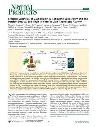
![molecule,9
and blocked cell cycle progression in mitosis due to
mitotic spindle damage.8−10
Recently it was found that GVA
interacts with both α/β- and γ-tubulins, whereas its 7-O-benzyl
synthetic analogue, gatastatin, selectively affects γ-tubulin,
thereby inhibiting γ-tubulin-dependent microtubule nuclea-
tion.11
The isoflavones ferrugone and preferrugone (Figure 1),
which are structurally similar to GVA, were isolated from the
root bark, stem bark, and seeds of African plants of the genus
Millettia; however their biological effects were not studied.12−14
Considering these limited data, the aim of the present work was
to devise a concise synthetic route toward GVA and its
derivatives in order to expand structure−activity relationship
studies and to investigate their antimitotic effect using an in
vivo phenotypic sea urchin embryo assay.15
A common route toward the isoflavone scaffold involves
Friedel−Crafts acylation of respective alkoxyphenols with
electron-rich phenylacetic acids followed by acid-mediated
cyclization of the resulting ortho-phenol intermediate to yield
isoflavones.16,17
A noted drawback of this protocol is the rather
limited access to diverse methoxyphenylacetic acids. The
recently reported alternative approaches include (i) intra-
molecular cyclization of diarylketoaldehydes with o-benzoy-
loxy18
or o-bromo substituents19
and (ii) the Suzuki coupling
reaction.20,21
However, starting building blocks for these
synthetic protocols are not readily accessible as well. In order
to address this issue, a facile synthesis of diverse alkoxy-
substituted isoflavones starting with readily available natural
precursors isolated from plant essential oils was developed and
validated. Specifically, we focused on preparing several
structural analogues of GVA varying both the nature and
position of the alkoxyphenyl substituents in both the A- and B-
rings of the molecule. The goal was both to improve antimitotic
activity of the molecule and to study the isoflavone substitution
pattern leading to the optimal antiproliferative activity.
■ RESULTS AND DISCUSSION
The bromochalcone-mediated route was considered as the
most attractive for the synthesis of alkoxyisoflavones.
Metabolites 1 (Scheme 1) can be readily isolated from different
crop plants, namely, dill [Anetum graveolens L. (Apiaceae)],
parsley [Petroselinum sativum Hoffm. (Apiaceae)],22,23
tarragon
[Aretmisia dracunculus L. (Asteraceae)],24
and lemongrass
[Cymbopogon flexuosus W. Watson (Poaceae)].25
The technol-
ogy for planting and harvesting of these species is well
developed, and the targeted allyl derivatives 1 are scaled-up
readily. Rings A and B could be constructed from the same
aldehydes 2, accessible from allylbenzenes 1, which are the
major components (30−80%) of plant essential oils (Scheme
1).22,24
Methoxybenzaldehydes 2 were selectively o-brominated
to yield intermediates 3. These products were treated with
MeMgI followed by oxidation of the resulting o-bromobenzyl
alcohols 4 to afford bromoacetophenones 5. The subsequent
conversion of 5 to chalcones 6 followed by their epoxidation
and subsequent epoxide rearrangement of 7 resulted in
ketoaldehydes 8 in high yields (60−90%) for each step.
Cyclization of ketoaldehydes 8 in the presence of CuI19
afforded the targeted isoflavones 9 in moderate yields. The
attempted synthesis of isoflavone 9fc via cyclization of 8fc was
Figure 1. Structures of alkoxyisoflavones, combretastatins, and podophyllotoxin.
Scheme 1a
a
Reagents and conditions:23
(a) (i) KOH, 100 °C, 40 min; (ii) O3, CHCl3−MeOH−pyridine (80:20:3 v/v), 15 °C, 1−2 h.
Journal of Natural Products Article
DOI: 10.1021/acs.jnatprod.6b00173
J. Nat. Prod. XXXX, XXX, XXX−XXX
B](data:image/gif;base64,R0lGODlhAQABAIAAAAAAAP///yH5BAEAAAAALAAAAAABAAEAAAIBRAA7)
Recommended
More Related Content
What's hot
What's hot (20)
Viewers also liked
Viewers also liked (20)
Similar to semenov2016
Similar to semenov2016 (20)
More from Alex Kiselyov
More from Alex Kiselyov (13)
semenov2016
- 1. Efficient Synthesis of Glaziovianin A Isoflavone Series from Dill and Parsley Extracts and Their in Vitro/in Vivo Antimitotic Activity Victor V. Semenov,*,† Dmitry V. Tsyganov,† Marina N. Semenova,‡,§ Roman N. Chuprov-Netochin,⊥ Mikhail M. Raihstat,† Leonid D. Konyushkin,† Polina B. Volynchuk,⊥ Elena I. Marusich,⊥ Vera V. Nazarenko,⊥ Sergey V. Leonov,⊥,∥ and Alex S. Kiselyov⊥ † N. D. Zelinsky Institute of Organic Chemistry, RAS, Leninsky Prospect, 47, 119991, Moscow, Russian Federation ‡ Institute of Developmental Biology, RAS, Vavilov Street, 26, 119334, Moscow, Russian Federation § Chemical Block Ltd., 3 Kyriacou Matsi, 3723, Limassol, Cyprus ⊥ Life Sciences Center, Moscow Institute of Physics and Technology, Institutsky Per., 9, Dolgoprudny, Moscow Region 141700, Russian Federation ∥ Institute of Cell Biophysics, RAS, Institutskaya Street, 3, Pushchino, Moscow Region 142290, Russian Federation *S Supporting Information ABSTRACT: A concise six-step protocol for the synthesis of isoflavone glaziovianin A (GVA) and its alkoxyphenyl derivatives 9 starting with readily available plant metabolites from dill and parsley seeds was developed. The reaction sequence involved an efficient conversion of the key intermediate epoxides 7 into the respective β-ketoaldehydes 8 followed by their Cu(I)-mediated cyclization into the target series 9. The biological activity of GVA and its derivatives was evaluated using a panel of seven human cancer cell lines and an in vivo sea urchin embryo assay. Both screening platforms confirmed the antimitotic effect of the parent GVA (9cg) and its alkoxy derivatives. Structure−activity relationship studies suggested that compounds 9cd and 9cf substituted with trimethoxy- and dillapiol-derived B-rings, respectively, were less active than the parent 9cg. Of the evaluated human cancer cell lines, the A375 melanoma cell line was the most sensitive to the tested molecules. Notably, the target compounds were not cytotoxic against human peripheral blood mononuclear cells up to 10 μM concentration. Phenotypic readouts from the sea urchin assay unequivocally suggest a direct microtubule-destabilizing effect of isoflavones 9cg, 9cd, and 9cf. Natural compounds and their analogues account for almost 80% of all anticancer drugs approved between 1981 and 2010.1 Several antimitotic agents isolated from plants, including colchicine, combretastatins, and podophyllotoxin, as well as their semisynthetic and synthetic analogues, contain a trimethoxyphenyl moiety.2−6 Flavonoids are abundant in nature and exhibit both topological and pharmacophore resemblance to pharmacologically active combretastatin and podophyllotox- in derivatives.7 It is reasonable to assume that properly substituted flavonoids and their structural analogues may show comparable antimitotic activity. Of these, isoflavones deserve particular attention. There is limited literature data on alkoxyisoflavones with antiproliferative activity. A potent cytotoxic compound, glaziovianin A (GVA), and less potent analogues I−III (Figure 1) were isolated from the leaves of the Brazilian tree Ateleia glazioviana Baill. (Leguminosae).8 The leaves of this plant were reported to induce abortions, cardiac failure, and central nervous system injuries in live stock.8 Parent GVA and related isoflavones I−III inhibited the growth of HL-60 human promyelocytic leukemia cells with IC50 values of 0.29, 16, 23, and 8.5 μM, respectively.8 Furthermore, GVA displayed high cytotoxicity against a panel of 39 human cancer cell lines (average IC50 = 0.66 μM),8 disrupted microtubule structure and dynamics by binding to the colchicine site of the tubulin Received: February 28, 2016 Article pubs.acs.org/jnp © XXXX American Chemical Society and American Society of Pharmacognosy A DOI: 10.1021/acs.jnatprod.6b00173 J. Nat. Prod. XXXX, XXX, XXX−XXX
- 2. molecule,9 and blocked cell cycle progression in mitosis due to mitotic spindle damage.8−10 Recently it was found that GVA interacts with both α/β- and γ-tubulins, whereas its 7-O-benzyl synthetic analogue, gatastatin, selectively affects γ-tubulin, thereby inhibiting γ-tubulin-dependent microtubule nuclea- tion.11 The isoflavones ferrugone and preferrugone (Figure 1), which are structurally similar to GVA, were isolated from the root bark, stem bark, and seeds of African plants of the genus Millettia; however their biological effects were not studied.12−14 Considering these limited data, the aim of the present work was to devise a concise synthetic route toward GVA and its derivatives in order to expand structure−activity relationship studies and to investigate their antimitotic effect using an in vivo phenotypic sea urchin embryo assay.15 A common route toward the isoflavone scaffold involves Friedel−Crafts acylation of respective alkoxyphenols with electron-rich phenylacetic acids followed by acid-mediated cyclization of the resulting ortho-phenol intermediate to yield isoflavones.16,17 A noted drawback of this protocol is the rather limited access to diverse methoxyphenylacetic acids. The recently reported alternative approaches include (i) intra- molecular cyclization of diarylketoaldehydes with o-benzoy- loxy18 or o-bromo substituents19 and (ii) the Suzuki coupling reaction.20,21 However, starting building blocks for these synthetic protocols are not readily accessible as well. In order to address this issue, a facile synthesis of diverse alkoxy- substituted isoflavones starting with readily available natural precursors isolated from plant essential oils was developed and validated. Specifically, we focused on preparing several structural analogues of GVA varying both the nature and position of the alkoxyphenyl substituents in both the A- and B- rings of the molecule. The goal was both to improve antimitotic activity of the molecule and to study the isoflavone substitution pattern leading to the optimal antiproliferative activity. ■ RESULTS AND DISCUSSION The bromochalcone-mediated route was considered as the most attractive for the synthesis of alkoxyisoflavones. Metabolites 1 (Scheme 1) can be readily isolated from different crop plants, namely, dill [Anetum graveolens L. (Apiaceae)], parsley [Petroselinum sativum Hoffm. (Apiaceae)],22,23 tarragon [Aretmisia dracunculus L. (Asteraceae)],24 and lemongrass [Cymbopogon flexuosus W. Watson (Poaceae)].25 The technol- ogy for planting and harvesting of these species is well developed, and the targeted allyl derivatives 1 are scaled-up readily. Rings A and B could be constructed from the same aldehydes 2, accessible from allylbenzenes 1, which are the major components (30−80%) of plant essential oils (Scheme 1).22,24 Methoxybenzaldehydes 2 were selectively o-brominated to yield intermediates 3. These products were treated with MeMgI followed by oxidation of the resulting o-bromobenzyl alcohols 4 to afford bromoacetophenones 5. The subsequent conversion of 5 to chalcones 6 followed by their epoxidation and subsequent epoxide rearrangement of 7 resulted in ketoaldehydes 8 in high yields (60−90%) for each step. Cyclization of ketoaldehydes 8 in the presence of CuI19 afforded the targeted isoflavones 9 in moderate yields. The attempted synthesis of isoflavone 9fc via cyclization of 8fc was Figure 1. Structures of alkoxyisoflavones, combretastatins, and podophyllotoxin. Scheme 1a a Reagents and conditions:23 (a) (i) KOH, 100 °C, 40 min; (ii) O3, CHCl3−MeOH−pyridine (80:20:3 v/v), 15 °C, 1−2 h. Journal of Natural Products Article DOI: 10.1021/acs.jnatprod.6b00173 J. Nat. Prod. XXXX, XXX, XXX−XXX B
- 3. not successful, presumably due to the instability of the 5-OMe group of the A-ring under experimental conditions (Supporting Information, pp S3, S4). Specifically, NMR spectra of the crude reaction mixture revealed the presence of compounds 9fc and 9ec (13:10 ratio, respectively). Compound 9ec was successfully isolated from the reaction mixture via chromatography. It should be noted that this six-step sequence is considerably shorter than the reported route,20,21 which comprises nine steps including the synthesis of the intermediate 2,3,4-trimethox- ybenzaldehyde.26 In addition, as opposed to our synthetic route, which relies on readily available natural precursors 1, protocols published earlier employed expensive 2-hydroxy-4,5- dimethoxyacetophenone and a considerable quantity of Pd- catalyst (342 mg to obtain 714 mg of the targeted GVA). Literature data suggest that the 7-alkoxy substituents of the A-ring and the 2′,5′-dimethoxy-3′,4′-methylenedioxy B-ring are the essential features for antimitotic microtubule-modulating properties of GVA and its analogues.10,21 Specifically, GVA derivatives with O-allyl, O-propargyl, and O-benzyl groups at C- 7 showed the highest cytotoxicity against HeLa S3 human cervical cancer cells and caused cell cycle arrest in the G2/M phase.10,21 The B-ring modification effects were investigated to a lesser extent. It was shown that both removal of 2′- and 5′- methoxy groups and the replacement of the 3′,4′-methyl- enedioxy moiety with two individual methoxy groups resulted in a significant decrease of cytotoxicity.21 In the next series of experiments the antimitotic activity of GVA and its analogues was evaluated, using the in vivo sea urchin embryo assay.15 Combretastatin A4 disodium phosphate (CA4P) served as reference compound. The results are summarized in Table 1. The sea urchin embryo tests confirmed that GVA directly affects tubulin/microtubule dynamics and structure.8−11,21 The parent compound 9cg was the most active of the tested series. It induced sea urchin embryo spinning indicative of a microtubule destabilizing mode of action.15 GVA analogues 9cd, 9ce, 9cf, and 9ia caused cleavage arrest and initiated the formation of tuberculate eggs, thus providing the indirect evidence of their microtubule-destabilizing properties.15 The B- ring substition pattern of GVA derivatives exhibited strong effects on activity. As suggested by the assay results, the antimitotic activity decreased in the following order: the parent GVA (9cg) > myristicin (9ce) ≥ 3,4,5-trimethoxyphenyl (9cd) = 4-methoxyphenyl (9ca) > dillapiol (9cf) > 3-methoxyphenyl (9cb) > 3,4-dimethoxyphenyl (9cc) > 2,3,4,5-tetramethox- yphenyl (9ch) derivatives. Any definitive relationship between the number of B-ring alkoxy groups and respective antimitotic activity was not observed. A methylenedioxy moiety was essential for the activity of compounds substituted with four B- ring alkoxy groups. Thus, a compound containing an apiol moiety (GVA, 9cg) exhibited stronger activity than the respective dillapiol-derived analogue 9cf. In addition, the myristicin derivative with a methylenedioxy moiety, 9ce, was more active than isoflavonoid 9cd, with a 3,4,5-trimethoxy B- ring. A compound with a 4-methoxy group was more potent than the respective 3-methoxy-analogue (compare 9ca and 9cb). Dimethoxy substitution of the A-ring was also important. The replacement of 7-OMe with a hydroxy group in 9ca resulted in the less active isoflavone 9ia. Introduction of the third MeO group as in 9dc led to low activity and inactivity in compound 9ja. As opposed to GVA, iso-GVA (9gc) featuring swapped A- and B-rings had no effect on the sea urchin embryos up to 4 μM concentration. Isoflavone 9cc with dimethoxy-substituted B-ring displayed low activity. The inhibitory effects of isoflavones 9ca, 9cb, 9cc, 9cd, 9ce, 9ch, 9cf, 9cg, and 9gc on cell growth were evaluated using seven human cancer cell lines, namely, A549 lung carcinoma, A375 melanoma, DU145 and PC3 prostate cancer, HCT116 Scheme 2a a Reagents and conditions: (a) NBS, DMF, rt, 8 h; (b) MeMgI, ether, rt, 0.5 h; (c) pyridinium chlorochromate, CH2Cl2, rt, 18 h. Scheme 3a a Reagents and conditions: (a) NaOH, EtOH, rt, 6 h; (b) H2O2, EtOH−NaOH, rt, 54 h; (c) BF3−Et2O, CH2Cl2, rt, 3 h; (d) CuI−K2CO3, 2-picolinic acid, DMF, 135−140 °C, 8 h. Journal of Natural Products Article DOI: 10.1021/acs.jnatprod.6b00173 J. Nat. Prod. XXXX, XXX, XXX−XXX C
- 4. colon cancer, MDA-MB-231 breast adenocarcinoma, and SK- OV-3 ovarian carcinoma. The MTS cell viability assay was employed with CA4P as a positive control. For comparison, the cytotoxicity of compounds against nonmalignant human peripheral blood mononuclear cells (PBMCs) was assessed (Table 1). The maximal tested concentration of isoflavones was 10 μM due to their restricted solubility. GVA (9cg) displayed the highest inhibitory activity, with IC50 values ranging from 0.27 (A375 cells) to 2.2 μM (MDA-MB-231 cells). The data obtained for the six cancer cell lines by the MTS assay correlated well with the published data acquired using the SRB method (Table 1).8 Compounds 9cd and 9cf, containing 3,4,5- trimethoxy and dillapiol-derived B-rings, respectively, were less active. Isoflavones 9ca, 9cb, 9cc, 9ch, and 9gc did not affect cancer cell growth up to 10 μM concentration. These results correlated well with published data. GVA (9cg) was significantly more active than 6,7,3′,4′-tetramethoxyisoflavone 9cc, exhibiting cytotoxic effects against HL-60 human leukemia cells and HeLa S3 human cervical cancer cells with GI50 values of 0.29 vs 23 μM and 0.59 vs 22 μM, respectively.8,21 Of the evaluated human cancer cell lines, A375 melanoma cells were most sensitive to the tested compounds. On the contrary, compounds 9 showed little to no effect on A549 lung carcinoma cells. GVA (9cg) was the only compound that inhibited growth of A549 cells (Table 1). Importantly, none of the synthesized isoflavones 9 demonstrated cytotoxicity in Table 1. Effects of GVA and its Analogues on Sea Urchin Embryos and Human Cancer Cellsa a A549: lung carcinoma; A375: melanoma; DU145 and PC3: prostate cancer; HCT116: colon cancer; MDA-MB-231: breast adenocarcinoma; SK- OV-3: ovarian carcinoma; PBMCs: human peripheral blood mononuclear cells. b The sea urchin embryo assay was conducted as described previously.15 Fertilized eggs and hatched blastulae were exposed to 2-fold decreasing concentrations of compounds. Duplicate measurements showed no differences in effective threshold concentration (EC) values. c IC50 values were determined by sigmoidal curve interpolation of cell survival data after 48 h of treatment. d TE: tuberculate eggs. e Data in bparentheses are from ref 8, obtained for natural GVA using the SRB assay. f ND: not determined. IC50 values are expressed as means ± SD based on dose−response curves of three independent experiments that involved each compound concentration tested in quadruplicates. Journal of Natural Products Article DOI: 10.1021/acs.jnatprod.6b00173 J. Nat. Prod. XXXX, XXX, XXX−XXX D
- 5. nonmalignant cells, namely, human peripheral blood mono- nuclear cells, up to 10 μM concentration. It should be noted that the antiproliferative effects of isoflavones 9 observed in both the sea urchin embryo model and human cancer cell lines correlated well. Specifically, GVA (9cg) was found to be the most potent agent in both systems. Swapping rings A and B as well as the replacement of the methylenedioxy moiety with two individual methoxy groups in GVA (9cg) resulted in inactive compounds 9gc and 9ch. Both trimethoxy- and dillapiol-derived compounds 9cd and 9cf were less active than the parent GVA (9cg). Compounds 9cb and 9cc were inactive in human cancer cells and showed only marginal activity in the sea urchin embryo assay. However, the sea urchin embryos were more sensitive to isoflavones 9ca and 9ce than the cancer cells. In summary, GVA (9cg) and its analogues were synthesized via a scaleable six-step reaction sequence. The synthetic route started with readily available allylpolyalkoxybenzenes 1, which were isolated from dill and parsley seed essential oils. The parent GVA (9cg) and its analogues 9cd and 9cf were found to be promising antimitotic microtubule destabilizing agents with low toxicity against human nonmalignant cells. ■ EXPERIMENTAL SECTION General Experimental Procedures. Melting points were measured on a Boetius melting point apparatus and were uncorrected. 1 H NMR spectra were recorded on a Bruker DRX-500 (500.13 MHz) instrument. Chemical shifts are reported in parts per million (ppm) and referenced to the appropriate NMR solvent peak(s). 2D NMR experiments {1 H−1 H} NOESY, {1 H−13 C} HMBC-qs, and {1 H−13 C} HSQC were used where necessary in assigning NMR spectra. Spin− spin coupling constants (J) are reported in hertz (Hz). 13 C NMR spectra were recorded on a Bruker DRX-500 (75.47 MHz) instrument. Chemical shifts are reported in ppm and referenced to the appropriate solvent peak(s) and were assigned C, CH, CH2, and CH3 as determined using HSQC and HMBC experiments. NMR spectra (Supporting Information) were produced using original software designed at the N. D. Zelinsky Institute of Organic Chemistry RAS (Moscow, Russian Federation) (http://nmr.ioc.ac.ru:8080/SDF2PDF. kl1). Low-resolution mass spectra (m/z) were recorded on a Finnigan MAT/INCOS 50 mass spectrometer at 70 eV using direct probe injection. Elemental analysis was performed on the automated PerkinElmer 2400 CHN microanalyzer. Flash chromatography was carried out on silica gel (Acros 0.035−0.070 mm, 60 Å). TLC was performed on Merck 60 F254 plates. Nonanhydrous solvents and reagents were purchased at the highest commercial quality and used as received. The starting materials, 4- methoxybenzaldehyde (2a), 3-methoxybenzaldehyde (2b), 3,4-dime- thoxybenzaldehyde (2c), 3,4,5-trimethoxybenzaldehyde (2d), and 2- bromo-4,5-dimethoxybenzaldehyde (3c), were purchased from Acros Organics (Belgium). Allylmethoxybenzenes 1d−h with 98−99% purity were obtained by high-efficiency distillation of CO2 extracts of parsley and dill seeds using a pilot plant device at the N. D. Zelinsky Institute of Organic Chemistry RAS (Moscow, Russian Federation).22 General Procedure for the Synthesis of 2-Bromobenzalde- hydes 3. Freshly recrystallized and free from bromine N- bromosuccinimide (0.02 mol) was added to a solution of the corresponding benzaldehyde 2 (0.02 mol) in DMF (30 mL) cooled with ice water. The reaction mixture was stirred at room temperature for 8 h and diluted with ice water (150 mL). The precipitate was filtered, washed with water (3 × 10 mL), and dried to afford 2- bromobenzaldehyde 3. 2-Bromo-3,4,5-trimethoxybenzaldehyde (3d): 4.73 g, 86% yield; colorless crystals; mp 68−70 °C (50% EtOH) (lit.27 70.5−71.5 °C); 1 H NMR (DMSO-d6) δ 10.17 (1H, s, CHO), 7.28 (1H, s, H-6), 3.90 (3H, s, OCH3), 3.88 (3H, s, OCH3), 3.83 (3H, s, OCH3). 4-Bromo-6,7-dimethoxy-2H-1,3-benzodioxole-5-carbaldehyde (3f): 4.85 g, 84% yield; colorless crystals; mp 103 °C (50% EtOH); 1 H NMR (DMSO-d6) δ 10.07 (1H, s, CHO), 6.22 (2H, s, OCH2O), 3.96 (3H, s, OCH3-6), 3.84 (3H, s, OCH3-5); EIMS m/z 290 [M + 1] (100), 289 [M] + (25), 288 (98), 287 (14), 275 (22), 273 (22), 272 (10), 260 (13), 258 (13), 257 (11), 243 (7), 241 (5), 93 (6); anal. C 41.55; H 3.14; Br 27.64%, calcd for C10H9BrO5, C 41.67; H 3.17; Br 27.55%. 6-Bromo-4,7-dimethoxy-2H-1,3-benzodioxole-5-carbaldehyde (3g): 4.16 g, 72% yield; colorless crystals; mp 112−113 °C (EtOH) (lit.28 94−95 °C); 1 H NMR (DMSO-d6) δ 10.10 (1H, s, CH(O)), 6.23 (2H, s, OCH2O), 3.92 (3H, s, OCH3-6), 3.87 (3H, s, OCH3-3); EIMS m/z 290 [M + 1] (4), 288 (4), 133 (14), 131 (14), 107 (13), 95 (19), 93 (32), 92 (31), 77 (100), 76 (25); anal. C 41.55; H 3.14; Br 27.64%, calcd for C10H9BrO5, C 41.63; H 3.17; Br 27.57%. General Procedure for the Synthesis of 2-Bromo Alcohols 4.29,30 A solution of 2-bromobenzaldehyde 3 (0.1 mmol) in THF (140 mL) was added dropwise to MeMgBr (0.15 mol) in ether (50 mL). The reaction mixture was stirred for 2 h, evaporated to ca. 150 mL, and gently poured into aqueous NH4CI with ice (300 g). The organic layer was separated, and the aqueous layer was extracted with Et2O (200 mL). The combined organic extracts were washed with H2O and brine, dried over anhydrous Na2SO4, and concentrated to afford crude 2-bromo alcohols 4 used for the next step without further purification. 1-(2-Bromo-4,5-dimethoxyphenyl)ethan-1-ol (4c): 16.98 g, 65% yield; colorless crystals; mp 59−60 °C, n-hexane (lit.29 60−62 °C). 1-(2-Bromo-3,4,5-trimethoxyphenyl)ethan-1-ol (4d): 2.1 g, 72% yield; colorless oil; 1 H NMR spectra as in lit.30 ; 1 H NMR (CDCl3) δ 6.99 (1H, s, H-6), 5.22−5.25 (1H, dd,J = 6.3 Hz, CH), 3.87 (3H, s, OCH3), 3.88 (3H, s, OCH3), 3.89 (3H, s, OCH3), 2.09 (1H, s, OH), 1.46 (3H, d, J = 6.3 Hz, CH3). 1-(2-Bromo-5,6-dimethoxy-3,4-methylendioxyphenyl)ethan-1-ol (4f): 2.32 g, 76% yield; colorless oil; 1 H NMR (CDCl3) δ 5.99 (2H, s, OCH2O), 5.17 (1H, m, CH), 3.78 (1H, d, J = 6.2 Hz, OH), 3.83 (3H, s, OCH3), 3.82 (3H, s, OCH3), 1.53 (3H, d, J = 6.7 Hz, CH3); anal. C 43.33; H 4.27; Br 26.16%, calcd for C11H13BrO5, C 43.40; H 4.32; Br 26.09%. 1-(6-Bromo-4,7-dimethoxy-2H-1,3-benzodioxol-5-yl)ethan-1-ol (4g): 1.95 g, yield 64%; colorless crystals; mp 98−100 °C (EtOAc−n- hexane, 1:3); 1 H NMR (DMSO-d6) δ 6.09 (1H, s, OCH2O) and 6.07 (1H, s, OCH2O), 5.20 (1H, quint, J = 6.4, J = 6.6 Hz, CH), 4.89 (1H, d, J = 6.0 Hz, OH), 3.83 (3H, s, OCH3), 3.82 (3H, s, OCH3), 1.40 (3H, d, J = 6.6 Hz, CH3); EIMS m/z 306 [M + 1] (31), 305 [M]+ (4), 304 (31), 292 (11), 291 (98), 290 (11), 289 (100), 276 (9), 274 (9), 261 (6), 259 (6), 210 (6), 182 (16); anal. C 43.30; H 4.29; Br 26.19%, calcd for C11H13BrO5, C 43.40; H 4.32; Br 26.09%. General Procedure for the Synthesis of Bromoacetophe- nones 5. The crude bromo alcohol 4 (0.01 mol) dissolved in CH2Cl2 (20 mL) was added rapidly to a suspension of pyridinium chlorochromate (3.23 g, 0.015 mol) in CH2Cl2 (10 mL). The resulting mixture was stirred for 18 h, diluted with 15 mL of CH2Cl2, and filtered through Celite. The residual gum was treated with boiling CH2Cl2 (3 × 25 mL). The combined organic extracts were washed with 15% aqueous KOH, aqueous NH4I, and brine. The extracts were dried over anhydrous Na2SO4, concentrated, and crystallized or separated by column chromatography (silica gel 0.035−0.070 mm, 60 Å) to yield the corresponding bromoacetophenones 5. 1-(2-Bromo-4,5-dimethoxyphenyl)ethan-1-one (5c): 16.8 g, 65% yield; colorless crystals; mp 75−77 °C (EtOAc−n-hexane, 1:1) (lit.29 73−75 °C); 1 H NMR (DMSO-d6) δ 7.28 (1H, s, H-6), 7.21 (1H, s, H- 3), 3.83 (3H, s, OCH3), 3.81 (3H, s, OCH3), 2.57 (3H, s, CH3); EIMS m/z 260 [M + 1] (36), 258 (36), 245 (97), 243 (100), 108 (16), 93 (15), 77 (12). 1-(2-Bromo-3,4,5-trimethoxyphenyl)ethan-1-one (5d): 2.20 g, 76% yield; colorless crystals; mp 33−34 °C (Et2O−n-hexane) (lit.30 34.5−35 °C); 1 H NMR (CDCl3) δ 6.82 (1H, s, H-6), 33.92 (3H, s, OCH3), 3.90 (3H, s, OCH3), 3.88 (3H, s, OCH3), 2.65 (3H, s, CH3); EIMS m/z 290 [M + 1] (27), 288 (27), 275 (43), 273 (43), 230 (10), 123 (10), 93 (20), 77 (21), 43 (100). Journal of Natural Products Article DOI: 10.1021/acs.jnatprod.6b00173 J. Nat. Prod. XXXX, XXX, XXX−XXX E
- 6. 11-(4-Bromo-6,7-dimethoxy-2H-1,3-benzodioxol-5-yl)ethan-1- one (5f): 2.27 g, 75% yield; colorless oil; 1 H NMR (CDCl3) δ 6.02 (2H, s, OCH2O), 4.02 (3H, s, OCH3-6), 3.77 (3H, s, OCH3-5), 2.50 (3H, s, CH3); EIMS m/z 304 [M + 1] (29), 302 (29), 289 (53), 287 (53), 92 (18), 77 (15), 43 (100); anal. C 43.59; H 3.66; Br 26.36%, calcd for C11H11BrO5, C 3.68; H 3.70; Br 26.27%. 1-(6-Bromo-4,7-dimethoxy-2H-1,3-benzodioxol-5-yl)ethan-1-one (5g): 2.58 g, 85% yield; colorless crystals; mp 55−57 °C (EtOAc−n- hexane, 1:1); 1 H NMR (DMSO-d6) δ 6.14 (2H, s, OCH2O), 3.86 (6H, s, OCH3-3,6), 2.40 (3H, s, CH3); EIMS m/z 304 [M + 1] (51), 302 (51), 289 (100), 288 (11), 287 (100), 274 (13), 272 (13), 43 (19); anal. C 43.59; H 3.66; Br 26.36%, calcd for C11H11BrO5, C 43.65; H 3.68; Br 26.29%. General Procedure for the Synthesis of Chalcones 6. NaOH (1.2 g, 30 mmol) was added to a vigorously stirred solution containing 2-bromoacetophenones 5 (10 mmol) and benzaldehyde 2 (10 mmol) in EtOH (30 mL) at 20 °C. The reaction mixture was stirred at room temperature for 6 h, kept overnight, acidified with 10% HCl to pH 3− 4, and strirred for 1 h. The residue was filtered, washed with H2O (3 × 20 mL), and crystallized from EtOAc to afford chalcones 6. (2E)-1-(2-Bromo-4,5-dimethoxyphenyl)-3-(4-methoxyphenyl)- prop-2-en-1-one (6ca): 3.13 g, 83% yield; light yellow solid; mp 171− 173 °C; 1 H NMR (DMSO-d6) δ 7.73 (2H, d, J = 8.8 Hz, H-2″,6″), 7.41 (1H, d, J = 16.0 Hz, H-2), 7.24 (1H, s, H-6′), 7.14 (1H, d, J = 16.0 Hz, H-3), 7.11 (1H, s, H-3′), 7.00 (2H, d, J = 8.8 Hz, H-3″,5″), 3.85 (3H, s, OCH3), 3.81 (3H, s, OCH3), 3.80 (3H, s, OCH3); EIMS m/z 379 [M + 2] (15), 378 [M + 1] (80), 377 [M]+ (30), 376 375 (14), 363 (12), 361 (12), 347 (13), 345 (13), 297 (34), 269 (11), 254 (14), 245 (19), 243 (19), 238 (14), 161 (100), 159 (10), 157 (10), 133 (45), 118 (23), 90 (27), 89 (30), 77 (29); anal. C 57.31; H 4.54; Br 21.18%, calcd for C18H17BrO4, C 57.24; H 4.51; Br 21.25%. (2E)-1-(2-Bromo-4,5-dimethoxyphenyl)-3-(3-methoxyphenyl)- prop-2-en-1-one (6cb): 3.17 g, 84% yield; light yellow solid; mp 139− 141 °C; 1 H NMR (DMSO-d6) δ 7.43 (1H, d, J = 16.0 Hz, H-2), 7.36− 7.34 (3H, m, H-2″,5″,6″), 7.31 (1H, d, J = 16.0 Hz, H-3), 7.26 (1H, s, H-6′), 7.14 (1H, s, H-3′), 7.04−7.01 (1H, m, H-4″), 3.85, 3.81, 3.80 (9H, 3s, 3OCH3-4′,5′,3″); EIMS m/z 379 [M + 2] (13), 378 [M + 1] (73), 377 [M]+ (34), 376 (73), 375 (20), 347 (36), 345 (36), 297 (44), 269 (71), 255 (19), 254 (47), 245 (48), 243 (50), 239 (25), 238 (57), 211 (21), 161 (100), 159 (26), 157 (26), 152 (25), 139 (22), 133 (46), 118 (68), 103 (23), 102 (33), 90 (54), 89 (44), 77 (53); anal. C 57.31; H 4.54; Br 21.18%, calcd for C18H17BrO4, C 57.22; H 4.50; Br 21.27%. (2E)-1-(2-Bromo-4,5-dimethoxyphenyl)-3-(3,4-dimethoxyphenyl)- prop-2-en-1-one (6cc): 3.51g, 86% yield; light yellow solid; mp 146− 148 °C; 1 H NMR (DMSO-d6) δ 7.39 (1H, s, H-2″), 7.36 (1H, d, J = 16.0 Hz, H-2), 7.30 (1H, d, J = 8.4 Hz, H-6″), 7.24 (1H, s, H-6′), 7.17 (1H, d, J = 16.0 Hz, H-3), 7.09 (1H, s, H-3′), 7.00 (1H, d, J = 8.4 Hz, H-5″), 3.85 (3H, s, OCH3), 3.81 (6H, s, OCH3), 3.80 (3H, s, OCH3); EIMS m/z 409 [M + 2] (21), 408 [M + 1] (100), 407 [M]+ (37), 406 (98), 393 (15), 391 (15), 377 (14), 375 (14), 327 (26), 299 (15), 284 (15), 269 (14), 268 (29), 245 (15), 243 (15), 241 (15), 225 (10), 191 (79), 165 (15), 163 (23), 159 (12), 157 (12), 152 (10), 150 (20), 139 (22), 119 (21), 118 (16), 105 (15), 102 (13), 93 (15), 91 (26), 89 (23), 77 (41); anal. C 56.04; H 4.70; Br 19.62%, calcd for C19H19BrO5, C 55.98; H 4.67; Br 19.67%. ( 2E )-1 -( 2-Br omo - 4,5 -d i metho xyp heny l) -3 -(3 , 4,5 - trimethoxyphenyl)prop-2-en-1-one (6cd): 3.63 g, 83% yield; yellow solid; mp 148−150 °C; 1 H NMR (DMSO-d6) δ 7.35 (1H, d, J = 16.0 Hz, H-2), 7.25 (1H, s, H-6′), 7.24 (1H, d, J = 16.0 Hz, H-3), 7.11 (2H, s, H-2″,6″), 7.09 (1H, s, H-3′), 3.85 (3H, s, OCH3), 3.82 (6H, s, OCH3), 3.80 (3H, s, OCH3), 3.70 (3H, s, OCH3); EIMS m/z 439 [M + 2] (21), 438 [M + 1] (97), 437 [M]+ (28), 436 (100), 423 (14), 421 (14), 407 (30), 405 (30), 358 (18), 357 (62), 314 (10), 299 (22), 298 (40), 283 (11), 271 (15), 245 (10), 243 (10), 221 (11), 157 (5), 149 (5); anal. C 54.93; H 4.84; Br 18.27%, calcd for C20H21BrO6, C 54.84; H 4.80; Br 18.33%. (2E)-1-(2-Bromo-4,5-dimethoxyphenyl)-3-(7-methoxy-2H-1,3- benzodioxol-5-yl)prop-2-en-1-one (6ce): 2.62 g, 62% yield; yellow solid; mp 179−181 °C; 1 H NMR (DMSO-d6) δ 7.35 (1H, d, J = 16.0 Hz, H-2), 7.24 (1H, s, H-6′), 7.18 (1H, d, J = 16.0 Hz, H-3), 7.16 (1H, s, H-3′), 7.10 (2H, s, H-4″,6″), 6.07 (2H, s, OCH2O), 3.85 (6H, s, OCH3), 3.80 (3H, s, OCH3); EIMS m/z 423 [M + 2] (20), 422 [M + 1] (100), 421 [M]+ (31), 420 (97), 419 (10), 391 (16), 389 (16), 342 (9), 341 (29), 313 (8), 311 (12), 298 (7), 284 (5), 283 (28), 245 (11), 243 (11), 205 (9); anal. C 54.17; H 4.07; Br 18.97%, calcd for C19H17BrO6, C 54.25; H 4.09; Br 19.07%. (2E)-1-(2-Bromo-4,5-dimethoxyphenyl)-3-(6,7-dimethoxy-2H-1,3- benzodioxol-5-yl)prop-2-en-1-one (6cf): 4.01 g, 89% yield; yellow solid; mp 124−126 °C; 1 H NMR (DMSO-d6) δ 7.58 (1H, d, J = 16.1 Hz, H-2), 7.25 (1H, s, H-6′), 7.23 (1H, s, H-3′), 7.17 (1H, d, J = 16.1 Hz, H-3), 7.13 (1H, s, H-4″), 6.08 (2H, s, OCH2O), 3.94 (3H, s, OCH3-6″), 3.85 (3H, s, OCH3), 3.80 (3H, s, OCH3), 3.70 (3H, s, OCH3-7″); EIMS m/z 452 [M + 1] (13), 450 (13), 422 (21), 421 (100), 420 (21), 419 (97), 405 (5), 404 (5), 245 (6), 243 (6), 209 (9); anal. C 53.23; H 4.24; Br 17.71%, calcd for C20H19BrO7, C 53.27; H 4.27; Br 17.66%. (2E)-1-(2-Bromo-4,5-dimethoxyphenyl)-3-(4,7-dimethoxy-2H-1,3- benzodioxol-5-yl)prop-2-en-1-one (6cg): 3.88 g, 86% yield; yellow solid; mp 158−161 °C; 1 H NMR (DMSO-d6) δ 7.58 (1H, d, J = 16.0 Hz, H-2), 7.26 (1H, d, J = 16.0 Hz, H-3), 7.25 (1H, s, H-6′), 7.13 (1H, s, H-3′), 7.11 (1H, s, H-6″), 6.10 (2H, s, OCH2O), 3.87 (3H, s, OCH3), 3.85 (3H, s, OCH3), 3.84 (3H, s, OCH3), 3.80 (3H, s, OCH3); EIMS m/z 452 [M + 1] (10), 450 (10), 422 (13), 421 (65), 420 (14), 419 (75), 306 (23), 292 (25), 245 (60), 243 (60), 235 (26), 220 (39), 206 (16), 205 (18), 192 (39), 191 (28), 181 (68), 177 (34), 175 (20), 165 (34), 164 (35), 157 (41), 149 (40), 147 (54), 135 (33), 121 (40), 119 (29), 108 (43), 107 (36), 106 (27), 105 (25), 93 (70), 92 (30), 91 (53), 90 (22), 79 (100), 78 (58), 77 (76), 75 (43); anal. C 53.23, H 4.24, Br 17.71%; calcd for C20H19BrO7; C 53.29; H 4.28; Br 17.67%. (2E)-1-(2-Bromo-4,5-dimethoxyphenyl)-3-(2,3,4,5- tetramethoxyphenyl)prop-2-en-1-one (6ch): 3.88 g, 83% yield; yellow solid; mp 94−96 °C; 1 H NMR (DMSO-d6) δ 7.58 (1H, d, J = 16.1 Hz, H-2), 7.29 (1H, d, J = 16.1 Hz, H-3), 7.26 (1H, s, H-6′), 7.22 (1H, s, H-3′), 7.12 (1H, s, H-6″), 3.86 (3H, s, OCH3), 3.84 (3H, s, OCH3), 3.82 (3H, s, OCH3), 3.81 (3H, s, OCH3), 3.80 (3H, s, OCH3), 3.71 (3H, s, OCH3); EIMS m/z 468 [M + 1] (11), 466 (11), 438 (23), 437 (100), 436 (31), 435 (98), 245 (26), 243 (31), 215 (10), 193 (10), 172 (14), 165 (13), 164 (17), 157 (12), 149 (6), 93 (11); anal. C 53.97; H 4.96; Br 17.10%, calcd for C21H23BrO7, C 53.84; H 4.90; Br 17.19%. (2E)-1-(2-Bromo-3,4,5-trimethoxyphenyl)-3-(3,4- dimethoxyphenyl)prop-2-en-1-one (6dc): 2.71 g, 62% yield; light yellow solid; mp 94−96 °C; 1 H NMR (DMSO-d6) δ 7.39 (1H, d, J = 1.9 Hz, H-2″), 7.35 (1H, d, J = 16.0 Hz, H-2), 7.30 (1H, dd, J = 8.4 Hz, J = 1.9 Hz, H-6″), 7.11 (1H, d, J = 16.0 Hz, H-3), 7.00 (1H, d, J = 8.4 Hz, H-5″), 6.95 (1H, s, H-6′), 3.84 (9H, s, OCH3), 3.80 (6H, s, OCH3); EIMS m/z 439 [M + 2] (13), 438 [M + 1] (69), 437 [M]+ (20), 436 (69), 423 (14), 421 (14), 407 (9), 405 (9), 358 (7), 357 (24), 315 (11), 299 (20), 298 (32), 191 (100), 163 (20), 157 (23), 149 (8), 133 (11), 132 (10), 119 (12), 118 (10), 91 (13), 77 (19); anal. C 54.93; H 4.84; Br 18.27%, calcd for C20H21BrO6, C 54.87; H 4.83; Br 18.32%. (2E)-1-(4-Bromo-6,7-dimethoxy-2H-1,3-benzodioxol-5-yl)-3-(3,4- dimethoxyphenyl)prop-2-en-1-one (6fc): 3.88 g, 86% yield; light yellow solid; mp 146−148 °C; 1 H NMR (DMSO-d6) δ 7.37 (1H, d, J = 2.0 Hz, H-2″), 7.28 (1H, dd, J = 8.4 Hz, J = 2.0 Hz, H-6″), 7.24 (1H, d, J = 16.1 Hz, H-2), 6.99 (1H, d, J = 16.1 Hz, H-3), 6.98 (1H, d, J = 8.4 Hz, H-5″), 6.17 (2H, s, OCH2O), 3.98 (3H, s, OCH3-6′), 3.81 (3H, s, OCH3), 3.80 (3H, s, OCH3),3.63 (3H, s, OCH3-7′); EIMS m/ z 453 [M + 2] (10), 452 [M + 1] (47), 451 [M]+ (10), 450 (47), 435 (7), 371 (11), 328 (13), 313 (24), 312 (58), 285 (18), 191 (100), 164 (41), 163 (32), 151 (46), 148 (19), 147 (18), 133 (27), 132 (19), 119 (25), 118 (20), 92 (29), 91 (32), 77 (50); anal. C 53.23; H 4.24; Br 17.71%, calcd for C20H19BrO7, C 53.28; H 4.27; Br 17.65%. (2E)-1-(6-Bromo-7-methoxy-4-methyl-2H-1,3-benzodioxol-5-yl)- 3-(3,4-dimethoxyphenyl)prop-2-en-1-one (6gc): 3.52 g, 78% yield; yellow solid; mp 125−127 °C; 1 H NMR (DMSO-d6) δ 7.37 (1H, d, J = 2.0 Hz, H-2″), 7.28 (1H, dd, J = 8.4 Hz, J = 2.0 Hz, H-6″), 7.23 (1H, Journal of Natural Products Article DOI: 10.1021/acs.jnatprod.6b00173 J. Nat. Prod. XXXX, XXX, XXX−XXX F
- 7. d, J = 16.1 Hz, H-2), 6.98 (1H, d, J = 8.4 Hz, H-5″), 6.97 (1H, d, J = 16.1 Hz, H-3), 6.17 (2H, s, OCH2O), 3.90 (3H, s, OCH3), 3.81 (3H, 3s, OCH3), 3.80 (3H, s, OCH3), 3.77 (3H, s, OCH3); EIMS m/z 453 [M + 2] (22), 452 [M + 1] (96), 451 [M]+ (31), 450 (96), 437 (10), 435 (10), 421 (10), 419 (10), 371 (45), 341 (17), 328 (22), 313 (58), 312 (79), 287 (16), 285 (16), 191 (100), 164 (27), 163 (21), 151 (15), 148 (10), 147 (10), 133 (11), 132 (8), 119 (8), 118 (7), 77 (7); anal. C 53.23; H 4.24; Br 17.71%, calcd for C20H19BrO7, C 53.30; H 4.27; Br 17.63%. General Procedure for the Synthesis of Epoxides 7.31 H2O2 (30%, 0.6 mL) was added to a vigorously stirred suspension of chalcone 6 (4 mmol) in EtOH (15 mL) and NaOH (1N, 1.9 mL) at room temperature. The reaction mixture was stirred at 30 °C for 3 h and left for 24 h at room temperature; then the second portion of NaOH (1N, 1.9 mL) and H2O2 (30%, 0.6 mL) was added, and the mixture was stirred for 6 h at room temperature. The third portion of NaOH (1N, 1.9 mL) and H2O2 (30%, 0.6 mL) was added and stirred for 24 h at room temperature. The residue was filtered, washed with EtOH and H2O, and dried at reduced pressure to afford epoxychalcones 7. (2-Bromo-4,5-dimethoxyphenyl)[3-(4-methoxyphenyl)oxiran-2- yl]methanone (7ca): 1.23 g, 78% yield; light yellow solid; mp 121− 122 °C; 1 H NMR (DMSO-d6) δ 7.36 (2H, d, J = 8.7 Hz, H-2″,6″), 7.27 (1H, s, H-6′), 7.25 (1H, s, H-3′), 6.96 (2H, d, J = 8.7 Hz, H- 3″,5″), 4.45 (1H, d, J = 1.8 Hz, H-2), 4.08 (1H, d, J = 1.6 Hz, H-3), 3.85 (3H, s, OCH3), 3.79 (3H, s, OCH3), 3.77 (3H, s, OCH3); EIMS m/z 395 [M + 2] (4), 394 [M + 1] (18), 393 [M]+ (4), 392 (18), 337 (31), 335 (31), 246 (10), 245 (97), 244 (10), 243 (100), 159 (12), 157 (17), 135 (28), 121 (88), 119 (16), 93 (24), 91 (38), 77 (61); anal. C 54.98; H 4.36; Br 20.32%, calcd for C18H17BrO5; C 54.92; H 4.32; Br 20.38%. (2-Bromo-4,5-dimethoxyphenyl)[3-(3-methoxyphenyl)oxiran-2- yl]methanone (7cb): 1.35 g, 86% yield; light yellow solid; mp 133− 136 °C; 1 H NMR (DMSO-d6) δ 7.32 (1H, t, J = 7.9 Hz, H-5″), 7.28 (1H, s, H-6′), 7.26 (1H, s, H-3′), 7.01 (1H, d, J = 7.9 Hz, H-6″), 6.97 (1H, br s, H-2″), 6.94 (1H, dd, J = 8.2 Hz, J = 2.4 Hz, H-4″), 4.46 (1H, d, J = 1.6 Hz, H-2), 4.13 (1H, d, J = 1.6 Hz, H-3), 3.85 (3H, s, OCH3), 3.79 (3H, s, OCH3), 3.76 (3H, s, OCH3); EIMS m/z 394 [M + 1] (20), 392 (20), 313 (6), 256 (21), 246 (10), 245 (96), 244 (10), 243 (100), 149 (19), 135 (10), 121 (10), 119 (5), 93 (6), 91 (10), 77 (14); anal. C 54.98; H 4.36; Br 20.32%, calcd for C18H17BrO5, C 54.90; H 4.33; Br 20.40%. (2-Bromo-4,5-dimethoxyphenyl)[3-(3,4-dimethoxyphenyl)oxiran- 2-yl]methanone (7cc): 1.10 g, 65% yield; light yellow solid; mp 108− 110 °C; 1 H NMR (DMSO-d6) δ 7.27 (1H, s, H-6′), 7.26 (1H, s, H- 3′), 7.00 (1H, d, J = 8.2 Hz, H-6″), 6.96 (1H, d, J = 8.2 Hz, H-5″), 6.96 (1H, br s, H-2″), 4.48 (1H, d, J = 1.5 Hz, H-2), 4.07 (1H, d, J = 1.5 Hz, H-3), 3.85 (3H, s, OCH3), 3.79 (3H, s, OCH3), 3.76 (3H, s, OCH3); EIMS m/z 425 [M + 2] (4), 424 [M + 1] (20), 423 [M]+ (4), 422 (20), 367 (19), 365 (19), 286 (13), 245 (97), 244 (12), 243 (100), 241 (11), 179 (13), 165 (24), 151 (78), 149 (39), 121 (13), 119 (15), 107 (24), 93 (22), 92 (20), 79 (27), 77 (37); anal. C 53.92; H 4.52; Br 18.88%, calcd for C19H19BrO6, C 53.87; H 4.50; Br 18.94%. (2-Bromo-4,5-dimethoxyphenyl)[3-(3,4,5-trimethoxyphenyl)- oxiran-2-yl]methanone (7cd): 1.41 g, 78% yield; yellow solid; mp 144−146 °C; 1 H NMR (DMSO-d6) δ 7.27 (1H, s, H-6′), 7.26 (1H, s, H-3′), 6.74 (2H, s, H-2″,6″), 4.48 (1H, d, J = 1.6 Hz, H-2), 4.09 (1H, d, J = 1.6 Hz, H-3), 3.85 (3H, s, OCH3), 3.80 (3H, s, OCH3), 3.78 (6H, 3s, OCH3), 3.66 (3H, s, OCH3); EIMS m/z 455 [M + 2] (2), 454 [M + 1] (12), 453 [M] + (2), 452 (12), 316 (15), 315 (13), 285 (14), 245 (95), 244 (11), 243 (100), 215 (10), 209 (34), 181 (66), 165 (18), 157 (11), 149 (25), 135 (10), 108 (24), 93 (13), 79 (14), 77 (11); anal. C 53.00; H 4.67; Br 17.63%, calcd for C20H21BrO7, C 52.87; H 4.63; Br 17.73%. (2-Bromo-4,5-dimethoxyphenyl)[3-(7-methoxy-1,3-benzodioxol- 5-yl)oxiran-2-yl]methanone (7ce): 1.21 g, 69% yield; yellow solid; mp 148−150 °C; 1 H NMR (DMSO-d6) δ 7.27 (1H, s, H-6′), 7.26 (1H, s, H-3′), 6.78 (1H, d, J = 1.3 Hz, H-4″), 6.65 (1H, d, J = 1.3 Hz, H-6″), 6.01 (2H, br s, OCH2O), 4.47 (1H, d, J = 1.9 Hz, H-2), 4.07 (1H, d, J = 1.9 Hz, H-3), 3.85 (3H, s, OCH3), 3.83 (3H, s, OCH3), 3.80 (3H, s, OCH3); EIMS m/z 439 [M + 2] (1), 438 [M + 1] (8), 437 [M]+ (1), 436 (8), 300 (24), 299 (16), 285 (6), 245 (96), 244 (10), 243 (100), 215 (10), 193 (19), 179 (21), 165 (42), 157 (11), 149 (25), 135 (11), 93 (12), 79 (12), 77 (15); anal. C 52.19, H 3.92, Br 18.27%, calcd for C19H17BrO7, C 52.27; H 3.96; Br 18.21%. (2-Bromo-4,5-dimethoxyphenyl)[3-(6,7-dimethoxy-1,3-benzo- dioxol-5-yl)oxiran-2-yl]methanone (7cf): 1.66 g, 89% yield; yellow solid; mp 179−181 °C; 1 H NMR (DMSO-d6) δ 7.32 (1H, s, H-6′), 7.27 (1H, s, H-3′), 6.42 (1H, s, H-4″), 6.02 (1H, s, OCH2O), 6.01 (1H, s, OCH2O), 4.44 (1H, d, J = 1.9 Hz, H-2), 4.15 (1H, d, J = 1.9 Hz, H-3), 3.94 (3H, s, OCH3-6″), 3.85 (3H, s, OCH3), 3.80 (3H, s, OCH3), 3.71 (3H, s, OCH3-7″); EIMS m/z 469 [M + 2] (8), 468 [M + 1] (36), 467 [M]+ (8), 466 (36), 421 (21), 419 (21), 411 (18), 409 (18), 245 (97), 244 (11), 243 (100), 223 (28), 195 (68), 180 (19), 165 (14), 157 (11), 149 (25), 121 (11), 93 (16), 79 (16), 77 (15); anal. C 51.41; H 4.10; Br 17.10%; calcd for C20H19BrO8, C 51.44; H 4.12; Br 17.02%. (2-Bromo-4,5-dimethoxyphenyl)[3-(4,7-dimethoxy-1,3-benzo- dioxol-5-yl)oxiran-2-yl]methanone (7cg): 1.44 g, 77% yield; yellow solid; mp 152−155 °C; 1 H NMR (DMSO-d6) δ 7.30 (1H, s, H-6′), 7.27 (1H, s, H-3′), 6.44 (1H, s, H-6″), 6.05 (1H, s, OCH2O), 6.04 (1H, s, OCH2O), 4.45 (1H, d, J = 1.9 Hz, H-2), 4.16 (1H, d, J = 1.9 Hz, H-3), 3.85 (3H, s, OCH3), 3.84 (3H, s, OCH3), 3.81 (3H, s, OCH3), 3.76 (3H, s, OCH3); EIMS m/z 469 [M + 2] (1), 468 [M + 1] (11), 467 [M]+ (1), 466 (11), 421 (3), 419 (3), 330 (12), 245 (97), 244 (10), 243 (100), 223 (45), 195 (72), 180 (11), 165 (14), 157 (8), 149 (20), 135 (15), 121 (10), 93 (28), 79 (20), 77 (24); anal. C 51.41; H 4.10; Br 17.10%, calcd for C20H19BrO8, C 51.46; H 4.11; Br 17.04%. (2-Bromo-3,4,5-trimethoxyphenyl)[3-(3,4-dimethoxyphenyl)- oxiran-2-yl]methanone (7dc): 1.41 g, 78% yield; light yellow solid; mp 128−130 °C; 1 H NMR (DMSO-d6) δ 7.12 (1H, s, H-6′), 7.00 (1H, dd, J = 1.7 Hz, J = 8.3 Hz, H-6″), 6.96 (1H, d, J = 8.3 Hz, H-5″), 6.94 (1H, d, J = 1.7 Hz, H-2″), 4.40 (1H, d, J = 1.6 Hz, H-2), 4.10 (1H, d, J = 1.6 Hz, H-3), 3.85 (3H, s, OCH3), 3.83 (3H, s, OCH3), 3.81 (3H, s, OCH3), 3.76 (6H, s, OCH3); EIMS m/z 455 [M + 2] (2), 454 [M + 1] (18), 453 [M]+ (2), 452 (18), 397 (14), 395 (15), 345 (22), 316 (22), 275 (96), 273 (100), 230 (21), 179 (24), 165 (22), 151 (83), 149 (13), 135 (12), 121 (11), 119 (5), 107 (20), 93 (18), 79 (21), 77 (36); anal. C 53.00; H 4.67; Br 17.63%, calcd for C20H21BrO7, C 52.88; H 4.62; Br 17.70%. (4-Bromo-6,7-dimethoxy-2H-1,3-benzodioxol-5-yl)[3-(3,4- dimethoxyphenyl)oxiran-2-yl]methanone (7fc): 1.46 g, 78% yield; light yellow solid; mp 89−91 °C; 1 H NMR (DMSO-d6) δ 6.98 (1H, dd, J = 8.3 Hz, J = 1.9 Hz, H-6″), 6.94 (1H, d, J = 8.3 Hz, H-5″), 6.88 (1H, d, J = 1.9 Hz, H-2″), 6.17 (1H, s, OCH2O), 6.16 (1H, s, OCH2O), 4.23 (1H, d, J = 1.8 Hz, H-2), 4.00 (1H, d, J = 1.8 Hz, H-3), 3.94 (3H, s, OCH3-6′), 3.75 (6H, s, OCH3-3″,4″), 3.70 (3H, s, OCH3- 7′); EIMS m/z 469 [M + 2] (7), 468 [M + 1] (34), 467 [M]+ (7), 466 (34), 412 (11), 411 (56), 410 (11), 409 (56), 330 (29), 329 (28), 289 (96), 287 (100), 285 (19), 192 (46), 165 (25), 151 (96), 135 (14), 133 (16), 121 (11), 119 (11), 107 (31), 105 (17), 93 (18), 92 (40), 91 (18), 79 (33), 77 (62); anal. C 51.41; H 4.10; Br 17.10%, calcd for C20H19BrO8, C 51.47; H 4.13; Br 17.00%. (6-Bromo-4,7-dimethoxy-2H-1,3-benzodioxol-5-yl)[3-(3,4- dimethoxyphenyl)oxiran-2-yl]methanone (7gc): 1.61 g, 86% yield; yellow solid; mp 124−126 °C; 1 H NMR (DMSO-d6) δ 6.98 (1H, dd, J = 8.3 Hz, J = 1.8 Hz, H-6″), 6.94 (1H, d, J = 8.3 Hz, H-5″), 6.87 (1H, d, J = 1.8 Hz, H-2″), 6.16 (1H, s, OCH2O), 6.15 (1H, s, OCH2O), 4.20 (1H, d, J = 1.8 Hz, H-2), 3.99 (1H, d, J = 1.8 Hz, H-3), 3.76 (6H, s, OCH3), 3.75 (6H, s, OCH3); EIMS m/z 469 [M + 2] (7), 468 [M + 1] (32), 467 [M]+ (7), 466 (32), 411 (25), 409 (25), 387 (17), 330 (56), 329 (48), 289 (97), 287 (100), 285 (15), 274 (16), 272 (16), 262 (15), 260 (15), 206 (29), 192 (16), 178 (18), 165 (22), 151 (31), 135 (10), 133 (9), 107 (14), 92 (10), 77 (12); anal. C 51.41; H 4.10; Br 17.10%, calcd for C20H19BrO8, C 51.49; H 4.12; Br 16.97%. General Rearrangement Procedure for the Synthesis of 3- Oxo-2,3-diphenyl Propanals (8).32 BF3·OEt2 (0.39 mL, 3 mmol) was added dropwise to an ice-cooled solution of chalcone epoxide 7 (3 mmol) in absolute CH2Cl2 (15 mL) in a flask filled with Ar and stirred for 3 h at 20 °C. The reaction was then quenched with a 5% aqueous Journal of Natural Products Article DOI: 10.1021/acs.jnatprod.6b00173 J. Nat. Prod. XXXX, XXX, XXX−XXX G
- 8. NaHCO3 solution (15 mL) and extracted with CHCl3 (2 × 15 mL). The organic layer was washed with H2O (2 × 15 mL) and dried over anhydrous Na2SO4. Evaporation of the solvent afforded the crude ketoaldehydes 8 as oils. The products were roughly purified by column chromatography from CH2Cl2 extract (EtOAc−n-hexane, 1:4, Rf = 0.6). Yields: 8ca (92%), 8cb (89%), 8cc (86%), 8cd (86%), 8ce (92%), 8cf (95%), 8cg (21%), 8ch (55%), 8dc (95%), 8fc (92%), 8gc (88%). The 1 H NMR spectra of these compounds were complicated due to keto−enol tautomerism and the presence of some impurities. The NMR data of compound 8ch are shown below. Compounds 8 were used for the next step without further purification. 3-(2-Bromo-4,5-dimethoxyphenyl)-3-oxo-2-(2,3,4,5- tetramethoxyphenyl)propanal (8ch). During the epoxidation proce- dure, chalcone 6ch spontaneously rearranged to ketoaldehyde 8ch. 0.96 g, 55% yield; yellow oil; 1 H NMR (DMSO-d6) δ 9.82 (1H, s, CH(O)), 7.74 (1H, s, H-2), 7.26 (1H, s, H-6″), 7.12 (1H, s, H-3″), 6.26 (1H, s, H-6′), 3.86 (3H, s, OCH3), 3.78 (3H, s, OCH3), 3.77 (3H, s, OCH3), 3.76 (6H, s, OCH3), 3.53 (3H, s, OCH3); EIMS m/z 485 [M + 2] (9), 484 [M + 1] (39), 438 [M]+ (9), 482 (39), 456 (4), 454 (4), 453 (1), 451 (1), 246 (9), 245 (98), 244 (9), 243 (98), 238 (16), 211 (100), 196 (30), 181 (14), 165 (13), 157 (5), 153 (11), 135 (10), 93 (9); anal. C 52.19; H 4.80; Br 16.53%, calcd for C21H23BrO8, C 52.29; H 4.84; Br 16.43%. General Procedure for the Synthesis of Isoflavones 9.19 A mixture of ketoaldehyde 8 (5 mmol), CuI (95 mg, 0.5 mmol), K2CO3 (1.38 g, 10 mmol), and 2-picolinic acid (123 mg, 1 mmol) in dry DMF (30 mL) in a flask filled with Ar was stirred at 135−140 °C for 8 h. The mixture was separated between EtOAc (3 × 50 mL) and H2O (100 mL). The organic layer was dried over anhydrous Na2SO4, filtered, evaporated under vacuum, and purified by column chromatography (EtOAc−n-hexane, 3:1, Rf = 0.3−0.4) to afford target izoflavones 9. 6,7-Dimethoxy-3-(4-methoxyphenyl)-4H-chromen-4-one (9ca): 0.72 g, 46% yield; dark yellow solid; mp 174−176 °C; 1 H NMR (CDCl3) δ 7.94 (1H, s, H-2), 7.63 (1H, s, H-5), 7.51 (2H, d, J = 8.6 Hz, H-2′,6′), 6.97 (2H, d, J = 8.6 Hz, H-3′,5′), 6.88 (1H, s, H-8), 3.99 (6H, s, OCH3-6,7), 3.85 (3H, s, OCH3-4′); 13 C NMR (CDCl3) δ 175.5, 159.5, 154.3, 152.2, 151.8, 147.6, 130.1, 124.4, 124.3, 117.9, 113.9, 104.8, 99.5, 56.4, 56.3, 55.3; EIMS m/z 313 [M + 1] (24), 312 [M]+ (100), 311 (33), 297 (8), 281 (5), 180 (15), 165 (7), 156 (6), 132 (7), 89 (7); anal. C 69.22; H 5.16%, calcd for C18H16O5; C 69.16; H 5.12%. 6,7-Dimethoxy-3-(3-methoxyphenyl)-4H-chromen-4-one (9cb): 0.37 g, 24% yield; yellowish solid; mp 182−184 °C; 1 H NMR (CDCl3) δ 7.98 (1H, s, H-2), 7.64 (1H, s, H-5), 7.35 (1H, t, J = 8.0 Hz, H-5′), 7.19 (1H, t, J = 2.0 Hz, H-2′), 7.12 (1H, d, J = 7.6 Hz, H- 6′), 6.93 (1H, dd, J = 2.6 Hz, J = 8.2 Hz, H-4′), 6.89 (1H, s, H-8), 4.00 (3H, s, OCH3), 3.99 (3H, s, OCH3), 3.85, (3H, s, OCH3-3′); 13 C NMR (CDCl3) δ 175.2, 159.5, 154.4, 152.4, 152.2, 147.7, 133.5, 129.4, 124.5, 121.1, 117.9, 114.4, 114.0, 104.8, 99.5, 56.4, 56.3, 55.3; EIMS m/z 313 [M + 1] (20), 312 [M]+ (100), 311 (87), 297 (8), 283 (14), 282 (20), 281 (9), 266 (5), 253 (5), 180 (10), 156 (9), 137 (13), 89 (7). anal. C 69.22; H 5.16%, calcd for C18H16O5, C 69.28; H 5.29%. 6,7-Dimethoxy-3-(3,4-dimethoxyphenyl)-4H-chromen-4-one (9cc, II (Figure 1): 0.41 g, 24% yield; dark yellow solid; mp 172−174 °C; 1 H NMR (CDCl3) δ 7.98 (1H, s, H-2), 7.64 (1H, s, H-5), 7.25 (1H, d, J = 2.0 Hz, H-2′), 7.06 (1H, dd, J = 2.0 Hz, J = 8.3 Hz, H-6′), 6.93 (1H, d, J = 8.3 Hz, H-5′), 6.89 (1H, s, H-8), 4.00 (3H, s, OCH3), 3.99 (3H, s, OCH3), 3.94 (3H, s, OCH3), 3.92 (3H, s, OCH3); 13 C NMR (CDCl3) δ 175.5, 154.4, 152.2, 152.0, 149.0, 148.7, 147.7, 124.8, 124.3, 120.9, 117.9, 112.5, 111.1, 104.8, 99.5, 56.4, 56.3, 55.9; EIMS m/z 343 [M + 1] (19), 342 [M]+ (100), 327 (13), 299 (6), 296 (5), 256 (7), 171 (6), 156 (5), 119 (9), 91 (8); anal. C 66.66; H 5.30%, calcd for C19H18O6, C 66.71; H 5.33%. 6,7-Dimethoxy-3-(3,4,5-trimethoxyphenyl)-4H-chromen-4-one (9cd): 0.71 g, 38% yield; yellowish solid; mp 172−174 °C; 1 H NMR (CDCl3) δ 7.99 (1H, s, H-2), 7.64 (1H, s, H-5), 6.90 (1H, s, H-8), 6.82 (2H, s, H-2′,6′), 4.01 (3H, s, OCH3), 3.99 (3H, s, OCH3), 3.91 (6H, s, OCH3-3′,5′), 3.89 (3H, s, OCH3-4′); 13 C NMR (CDCl3) δ 175.3, 154.4, 153.2, 152.3, 152.2, 147.8, 138.1, 127.6, 124.5, 117.8, 106.3, 104.7, 99.5, 60.8, 56.4, 56.3, 56.2; EIMS m/z 373 [M + 1] (25), 372 [M]+ (100), 358 (10), 357 (48), 329 (10), 271 (20), 171 (14), 149 (17), 134 (10); anal. C 64.51; H 5.41%, calcd for C20H20O7, C 64.42; H 5.36%. 6,7-Dimethoxy-3-(7-methoxy-1,3-benzodioxol-5-yl)-4H-chro- men-4-one (9ce): 0.46 g, 26% yield; white solid; mp 198−201 °C; 1 H NMR (CDCl3) δ 7.94 (1H, s, H-2), 7.62 (1H, s, H-5), 6.88 (1H, s, H- 8), 6.84 (1H, s, H-6′), 6.72 (1H, s, H-4′), 6.00 (2H, s, OCH2O), 3.99 (6H, s, OCH3-6,7), 3.94 (3H, s, OCH3-7′); 13 C NMR (CDCl3) δ 175.2, 154.4, 152.1, 148.8, 147.7, 143.5, 135.3, 126.2, 124.4, 117.8, 108.8, 104.8, 103.1, 101.5, 99.5, 56.6, 56.4, 56.3; EIMS m/z 357 [M + 1] (23), 356 [M]+ (100), 355 (21), 341 (1), 327 (3), 326 (2), 176 (5); anal. C 64.04; H 4.53%, calcd for C19H16O7, C 63.96; H 4.48%. 6,7-Dimethoxy-3-(6,7-dimethoxy-1,3-benzodioxol-5-yl)-4H-chro- men-4-one (9cf): 0.46 g, 24% yield; yellow-greenish solid; mp 204− 206 °C; 1 H NMR (CDCl3) δ 7.93 (1H, s, H-2), 7.62 (1H, s, H-5), 6.89 (1H, s, H-8), 6.56 (1H, s, H-4′), 5.96 (2H, s, OCH2O), 4.05 (3H, s, OCH3-6′), 4.00 (3H, s, OCH3), 3.99 (3H, s, OCH3), 3.68 (3H, s, OCH3-7′); 13 C NMR (CDCl3) δ 175.4, 154.3, 153.8, 152.3, 147.6, 145.2, 144.6, 137.9, 137.7, 121.3, 118.3, 117.9, 104.9, 104.5, 101.5, 99.6, 61.2, 60.0, 56.4, 56.3; EIMS m/z 387 [M + 1] (23), 386 [M]+ (90), 371 (7), 357 (8), 356 (29), 355 (100), 343 (5), 340 (9), 313 (5), 181 (16), 178 (9); anal. C 62.18; H 4.70%, calcd for C20H18O8, C 62.06; H 4.65%. 6,7-Dimethoxy-3-(4,7-dimethoxy-1,3-benzodioxol-5-yl)-4H-chro- men-4-one (9cg, glaziovianin A): 0.62 g, 32% yield; yellowish solid; mp 139−141 °C; 1 H NMR (CDCl3) δ 7.91 (1H, s, H-2), 7.62 (1H, s, H-5), 6.89 (1H, s, H-8), 6.53 (1H, s, H-6′), 6.03 (2H, s, OCH2O), 4.00 (3H, s, OCH3), 3.99 (3H, s, OCH3), 3.87 (3H, s, OCH3), 3.86 (3H, s, OCH3); 13 C NMR (CDCl3) δ 175.4, 154.3, 153.4, 152.3, 147.6, 139.1, 139.0, 137.0, 136.8, 121.7, 118.0, 117.8, 110.1, 104.9, 101.8, 99.5, 60.1, 56.8, 56.4, 56.3; EIMS m/z 387 [M + 1] (24), 386 [M]+ (100), 371 (7), 357 (7), 356 (12), 355 (49), 313 (10), 206 (12), 205 (15), 181 (50), 137 (10); anal. C 62.18; H 4.70%, calcd for C20H18O8, C 62.08; H 4.66%. 6,7-Dimethoxy-3-(2,3,4,5-tetramethoxyphenyl)-4H-chromen-4- one (9ch): 0.68 g, 34% yield; yellow solid; mp 204−206 °C; 1 H NMR (CDCl3) δ 7.61 (1H, s, H-2), 7.24 (1H, s, H-5), H-2′), 7.20 (1H, s, H- 8), 6.75 (1H, s, H-2′), 4.03 (3H, s, OCH3), 3.97 (3H, s, OCH3), 3.96 (3H, s, OCH3), 3.94 (3H, s, OCH3), 3.92 (3H, s, OCH3), 3.91 (3H, s, OCH3); 13 C NMR (CDCl3) δ 183.2, 163.0, 157.5, 149.5, 148.7, 147.8, 146.9, 146.6, 145.2, 120.9, 113.3, 109.1, 106.2, 104.2, 95.5, 62.2, 61.3, 61.2, 56.7, 56.5, 56.4; EIMS m/z 403 [M + 1] (10), 402 [M]+ (46), 373 (9), 372 (48), 371 (100), 356 (6), 355 (6), 341 (17), 313 (11), 230 (12), 186 (19), 149 (12), 137 (11), 136 (10), 93 (10); anal. C 62.68; H 5.51%, calcd for C21H22O8, C 62.57; H 5.46%. 3-(3,4-Dimethoxyphenyl)-6,7,8-trimethoxy-4H-chromen-4-one (9dc): 0.70 g, 36% yield, beige-orange solid; mp 132−134 °C; 1 H NMR (CDCl3) δ 8.06 (1H, s, H-2), 7.47 (1H, s, H-5), 7.25 (1H, d, J = 2.0 Hz, H-2′), 7.06 (1H, dd, J = 2.0 Hz, J = 8.3 Hz, H-6′), 6.94 (1H, d, J = 8.3 Hz, H-5′), 4.05 (3H, s, OCH3), 4.04 (3H, s, OCH3), 3.96 (3H, s, OCH3), 3.93 (3H, s, OCH3), 3.92 (3H, s, OCH3); 13 C NMR (CDCl3) δ 175.6, 152.2, 151.3, 149.1, 148.7, 147.2, 145.8, 141.7, 124.6, 124.2, 120.9, 120.3, 112.5, 111.1, 100.3, 62.0, 61.4, 56.2, 55.9; EIMS m/z 373 [M + 1] (29), 372 [M]+ (100), 371 (10), 358 (5), 357 (23), 329 (7), 327 (5), 326 (7), 186 (10), 171 (7), 157 (6), 119 (11), 91 (10); anal. C 64.51; H 5.41%, calcd for C20H20O7, C 64.41; H 5.36%. 7-(3,4-Dimethoxyphenyl)-4,9-dimethoxy-8H-[1,3]dioxolo[4,5-g]- chromen-8-one (9gc, iso-glaziovianin A): 0.95 g, 49% yield; light brown solid; mp 176−178 °C; 1 H NMR (DMSO-d6) δ 7.15 (1H, s, H- 2), 6.93 (1H, d, J = 8.2 Hz, H-5′), 6.89 (1H, d, J = 2.0 Hz, H-2′), 6.81 (1H, dd, J = 2.0 Hz, J = 8.1 Hz, H-6′), 6.22 (2H, s, OCH2O), 3.89 (3H, s, OCH3-9), 3.77 (3H, s, OCH3), 3.73 (3H, s, OCH3), 3.25 (3H, s, OCH3-4); 13 C NMR (DMSO-d6) δ 157.2, 148.1, 147.8, 146.1, Journal of Natural Products Article DOI: 10.1021/acs.jnatprod.6b00173 J. Nat. Prod. XXXX, XXX, XXX−XXX H
- 9. 142.5, 140.4, 140.2, 134.3, 129.0, 127.2, 121.3, 117.1, 113.6, 110.9, 108.6, 103.4, 60.9, 60.0, 55.6; EIMS m/z 387 [M + 1] (22), 386 [M]+ (100), 371 (16), 358 (34), 357 (10), 356 (12), 344 (8), 343 (54), 328 (6), 193 (7), 135 (12), 128 (10), 127 (10); anal. C 62.18; H 4.70%, calcd for C20H18O8, C 62.12; H 4.67%. Phenotypic Sea Urchin Embryo Assay.15 Adult sea urchins, Paracentrotus lividus L. (Echinidae), were collected from the Mediterranean Sea on the Cyprus coast in March 2013 and December 2014 and kept in an aerated seawater tank. Gametes were obtained by intracoelomic injection of 0.5 M KCl. Eggs were washed with filtered seawater and fertilized by adding drops of diluted sperm. Embryos were cultured at room temperature under gentle agitation with a motor-driven plastic paddle (60 rpm) in filtered seawater. The embryos were observed with a Biolam light microscope (LOMO, St. Petersburg, Russian Federation). For treatment with the test compounds, 5 mL aliquots of embryo suspension were transferred to six-well plates and incubated as a monolayer at a concentration up to 2000 embryos/mL. Stock solutions of isoflavones 9 were prepared in DMSO at 10 mM concentration followed by a 10-fold dilution with 96% EtOH. This procedure enhanced the solubility of the test compounds in the salt-containing medium (seawater), as evidenced by microscopic examination of the samples. The maximal tolerated concentrations of DMSO and EtOH in the in vivo assay were determined to be 0.05% and 1%, respectively. Higher concentrations of either DMSO (>0.1%) or EtOH (>1%) caused nonspecific alterations and retardation of the sea urchin embryo development independent of the treatment stage. A stock solution of the reference compound, combretastatin A4 disodium phosphate (OXiGENE), was prepared as 5 mM in distilled H2O. The antimitotic activity was assessed by exposing fertilized eggs (8−15 min postfertilization, 45− 55 min before the first mitotic cycle completion) to 2-fold decreasing concentrations of the compound. Cleavage alteration and arrest were clearly detected at 2.5−5.5 h after fertilization. The effects were estimated quantitatively as an effective threshold concentration, resulting in cleavage alteration and embryo death before hatching or full mitotic arrest. At these concentrations, all tested compounds caused 100% cleavage alteration and embryo death before hatching, whereas at 2-fold lower concentrations the compounds failed to produce any effect. For microtubule-destabilizing activity, the compounds were tested on free-swimming blastulae just after hatching (8−9 h postfertiliza- tion), which originated from the same embryo culture. Embryos were observed until the four-arm pluteus stage (34−36 h postfertilization). The specific changes in swimming pattern, namely, both spinning on the bottom of the well and lack of forward movement, were interpreted to be the result of the microtubule-destabilizing activity of a molecule. The sea urchin embryo assay data are available free of charge via the Internet at http://www.zelinsky.ru. Experiments with the sea urchin embryos fulfill the requirements of biological ethics. The artificial spawning does not cause animal death, embryos develop outside the female organism, and both postspawned adult sea urchins and the excess of intact embryos are returned to the sea, their natural habitat. Human Cancer Cell Culture. A549 human lung epithelial carcinoma cells (CCL-185) and A375 human skin epithelial malignant melanoma cells (CRL-1619) were cultured with Dulbecco’s modified Eagle medium. DU145 human prostate epithelial carcinoma cells derived from brain metastatic site (HTB-81) were cultured with Eagle’s minimum essential medium. PC3 human prostate epithelial adenocarcinoma grade IVcells, derived from bone metastatic site (CRL-1435), were cultured with Ham’s F-12K (Kaighn’s) medium. HCT116 human colon epithelial colorectal carcinoma cells (CCL- 247) and SK-OV-3 human ovary ascites epithelial adenocarcinoma cells (HTB-77) were cultured with McCoy’s 5a modified medium. MDA-MB-231 human mammary gland/breast adenocarcinoma cells, derived from pleural effusion metastatic site (HTB-26), were cultured with Leibovitz’s L-15 medium. All cancer cell cultures were performed in media contained 10% fetal bovine serum, penicillin (100 units/mL), and streptomycin (100 μg/mL) at 37 °C under a 5% CO2 humidified atmosphere. Preparation and Culture of Human PBMCs. Peripheral blood mononuclear cells were obtained from healthy volunteers by the standard Ficoll-Paque gradient density sedimentation procedure to yield mononuclear cells. PBMCs were washed three times in RPMI- 1640 medium before further processing. The purified PBMCs (1 × 10⁶ cells/mL) were seeded in RPMI 1640 medium supplemented with heat-inactivated 10% fetal bovine serum, penicillin (100 units/mL), and streptomycin (100 μg/mL) at 37 °C in a 5% CO2 humidified atmosphere. Cell Toxicity Assay. Cells were seeded in 96-well plates at a density of 1 × 10⁴ cells per well. Stock solutions of test compounds were prepared in DMSO. Cells were treated for 48 h with compounds 9ca, 9cb, 9cc, 9cd, 9ce, 9ch, 9cf, 9cg, and 9gc at 0.01−100 μM or with CA4P at 0.001−10 μM as a positive control (four wells per each concentration value). DMSO (0.1%) served as a control. The number of surviving cells was determined by the colorimetric CellTiter 96 AQueous One Solution cell proliferation assay (MTS).33 A 20 μL amount of the CellTiter 96 AQueous One Solution reagent was added to each well. The plate was incubated at 37 °C for 1−4 h under a 5% CO2 humidified atmosphere. The absorbance was recorded at 490 nm using a CLARIOstar microplate reader (BMG LABTECH, Ortenberg, Germany). Experiments for all compounds were repeated three times. IC50 values were determined by sigmoidal curve fitting using GraphPad Prism software. ■ ASSOCIATED CONTENT *S Supporting Information The Supporting Information is available free of charge on the ACS Publications website at DOI: 10.1021/acs.jnat- prod.6b00173. Physical and spectroscopic data of compounds 9ec, 9fc, 9ia, and 9ja; 1 H NMR and 13 C NMR spectra of compounds 3d, 3f, 3g, 4d, 4f, 4g, 5c, 5d, 5f, 5g, 6−9, and 8cg-acetal (PDF) ■ AUTHOR INFORMATION Corresponding Author *Tel: +7 916 620 9584. Fax: +7 499 137 2966. E-mail: vs@ zelinsky.ru (V. V. Semenov). Notes The authors declare no competing financial interest. ■ ACKNOWLEDGMENTS This work was carried out with the financial support of the Ministry of Education and Science of Russian Federation (Agreement No. 02.A03.21.0003 dated August 28, 2013) and Russian Foundation for Basic Research (Grant No. 16-03- 00648 dated February 15, 2016). The authors greatly appreciate the financial and technical support from Chemical Block Ltd., /www.chemblock.com/. The authors thanks Dr. Ju. A. Strelenko from N. D. Zelinsky Institute of Organic Chemistry RAS for the development of software for NMR spectra presentation. ■ REFERENCES (1) Newman, D. J.; Cragg, G. M. J. Nat. Prod. 2012, 75, 311−325. (2) Hamel, E. Med. Res. Rev. 1996, 16, 207−231. (3) Jordan, A.; Hadfield, J. A.; Lawrence, N. J.; McGown, A. T. Med. Res. Rev. 1998, 18, 259−296. (4) Desbene, S.; Giorgi-Renault, S. Curr. Med. Chem.: Anti-Cancer Agents 2002, 2, 71−90. (5) Nam, N.-H. Curr. Med. Chem. 2003, 10, 1697−1722. (6) Brancale, A.; Silvestri, R. Med. Res. Rev. 2007, 27, 209−238. (7) Semenov, V. V.; Semenova, M. N. Russ. Chem. Rev. 2015, 84, 134−158. Journal of Natural Products Article DOI: 10.1021/acs.jnatprod.6b00173 J. Nat. Prod. XXXX, XXX, XXX−XXX I
- 10. (8) Yokosuka, A.; Haraguchi, M.; Usui, T.; Kazami, S.; Osada, H.; Yamori, T.; Mimaki, Y. Bioorg. Med. Chem. Lett. 2007, 17, 3091−3094. (9) Chinen, T.; Kazami, S.; Nagumo, Y.; Hayakawa, I.; Ikedo, A.; Takagi, M.; Yokosuka, A.; Imamoto, N.; Mimaki, Y.; Kigoshi, H.; Osada, H.; Usui, T. ACS Chem. Biol. 2013, 8, 884−889. (10) Ikedo, A.; Hayakawa, I.; Usui, T.; Kazami, S.; Osada, H.; Kigoshi, H. Bioorg. Med. Chem. Lett. 2010, 20, 5402−5404. (11) Chinen, T.; Liu, P.; Shioda, S.; Pagel, J.; Cerikan, B.; Lin, T.-C.; Gruss, O.; Hayashi, Y.; Takeno, H.; Shima, T.; Okada, Y.; Hayakawa, I.; Hayashi, Y.; Kigoshi, H.; Usui, T.; Schiebel, E. Nat. Commun. 2015, 6, 8722. (12) Dagne, E.; Bekele, A.; Waterman, P. G. Phytochemistry 1989, 28, 1897−1900. (13) Dagne, E.; Bekele, A. Phytochemistry 1990, 29, 2679−2682. (14) Dagne, E.; Mammo, W.; Bekele, A. Bull. Chem. Soc. Ethiop. 1991, 5, 81−86. (15) Semenova, M. N.; Kiselyov, A. S.; Semenov, V. V. BioTechniques 2006, 40, 765−774. (16) Shaw, S.; Mandal, S. P.; Azad, R. Indian J. Chem. 1983, 228, 488. (17) Zaninetti, R.; Cortese, S. V.; Aprile, S.; Massarotti, A.; Canonico, P. L.; Sorba, G.; Grosa, G.; Genazzani, A. A.; Pirali, T. ChemMedChem 2013, 8, 633−643. (18) Lewin, G.; Aubert, G.; Thoret, S.; Dubois, J.; Cresteil, T. Bioorg. Med. Chem. 2012, 20, 1231−1239. (19) Li, Q.-L.; Liu, Q.-L.; Ge, Z.-Y.; Zhu, Y.-M. Helv. Chim. Acta 2011, 94, 1304−1309. (20) Hayakawa, I.; Ikedo, A.; Kigoshi, H. Chem. Lett. 2007, 36, 1382−1383. (21) Hayakawa, I.; Ikedo, A.; Chinen, T.; Usui, T.; Kigoshi, H. Bioorg. Med. Chem. 2012, 20, 5745−5756. (22) Semenov, V. V.; Rusak, V. A.; Chartov, E. M.; Zaretsky, M. I.; Konyushkin, L. D.; Firgang, S. I.; Chizhov, A. O.; Elkin, V. V.; Latin, N. N.; Bonashek, V. M.; Stas’eva, O. N. Russ. Chem. Bull. 2007, 56, 2448−2455. (23) Semenov, V. V.; Kiselyov, A. S.; Titov, I. Y.; Sagamanova, I. K.; Ikizalp, N. N.; Chernysheva, N. B.; Tsyganov, D. V.; Konyushkin, L. D.; Firgang, S. I.; Semenov, R. V.; Karmanova, I. B.; Raihstat, M. M.; Semenova, M. N. J. Nat. Prod. 2010, 73, 1796−1802. (24) De Vincenzi, M.; Silano, M.; Maialetti, F.; Scazzocchio, B. Fitoterapia 2000, 71, 725−729. (25) Public Statement on the Use of Herbal Medicinal Products Containing Methyleugenol. www.ema.europa.eu/ema/pages/includes/ document/open_document.jsp?webContentId=WC500089961. (26) Rizzacasa, M.; Sargent, M. J. Chem. Soc., Perkin Trans. 1 1987, 2017−2022. (27) Gutsche, C. D.; Jason, E. F.; Coffey, R. S.; Johnson, H. E. J. Am. Chem. Soc. 1958, 80, 5756−5767. (28) Dallacker, F.; Schleuter, H.-J.; Schneider, P. Z. Naturforsch. B 1986, 41, 1273−1280. (29) Sanceau, J.-Y.; Dhal, R.; Brown, E. Tetrahedron 1994, 50, 3363− 3380. (30) Ziegler, F. E.; Chliwner, I.; Fowler, K. W.; Kanfer, S. J.; Kuo, S. J.; Sinha, N. D. J. Am. Chem. Soc. 1980, 102, 790−798. (31) Sun, L.; Borella, Ch.; Li, H.; Jiang, J.; Chen, Sh.; Koya, K.; Inoue, T.; Du, Z.; Foley, K.; Wu, Y.; Zhang, M.; Ying, W. WIPO Patent WO2006/089177, 2006. (32) Mathew, P.; Mathew, D.; Asokan, C. V. Synth. Commun. 2007, 37, 661−665. (33) https://worldwide.promega.com/products/cell-health-and- metabolism/cell-viability-assays/celltiter-96-aqueous-one-solution-cell- proliferation-assay-_mts_/. Journal of Natural Products Article DOI: 10.1021/acs.jnatprod.6b00173 J. Nat. Prod. XXXX, XXX, XXX−XXX J