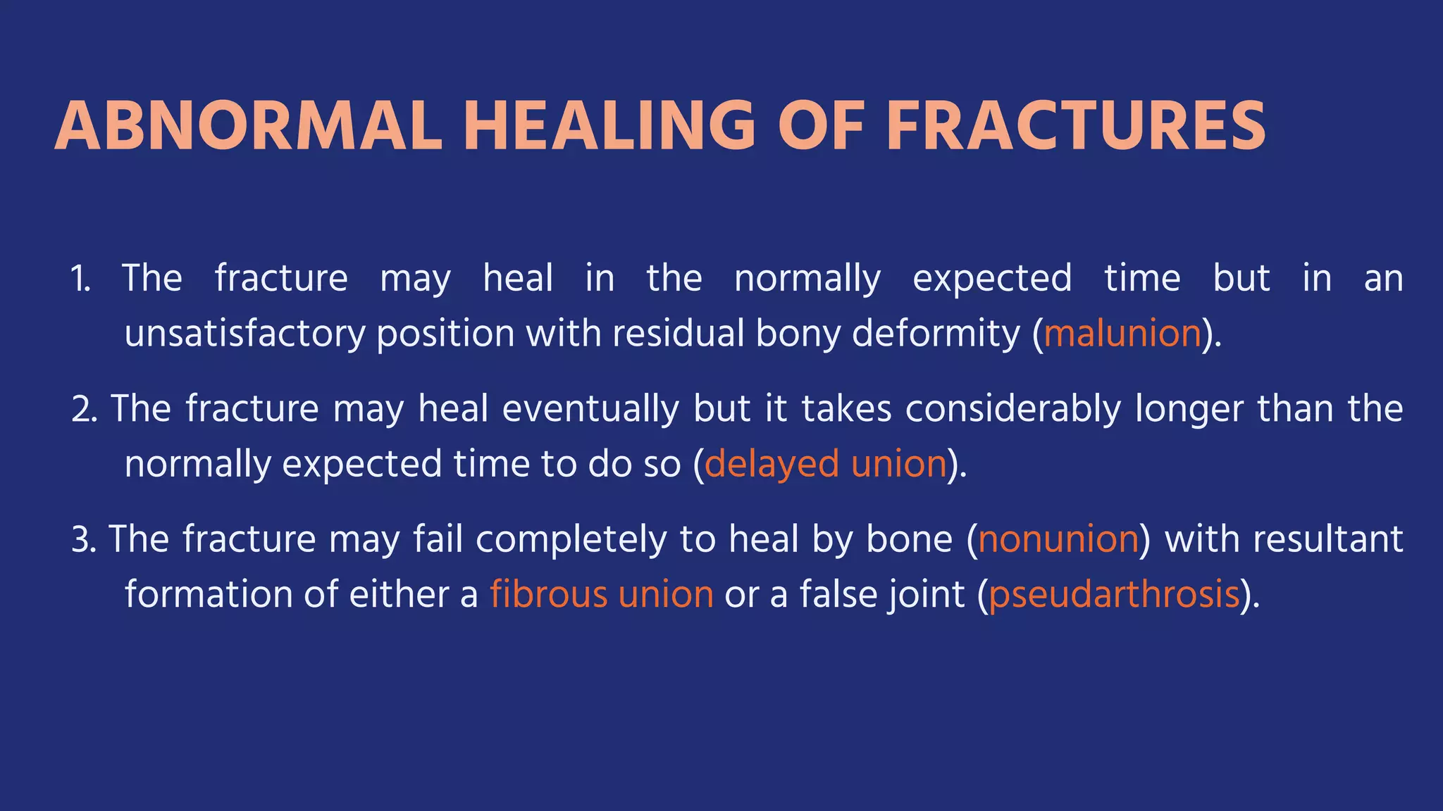
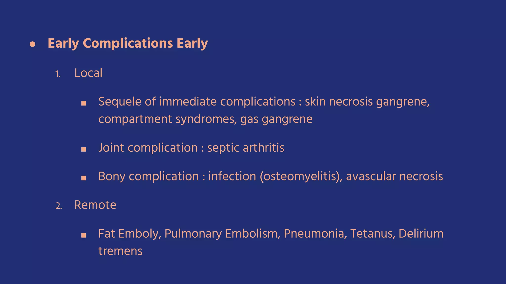
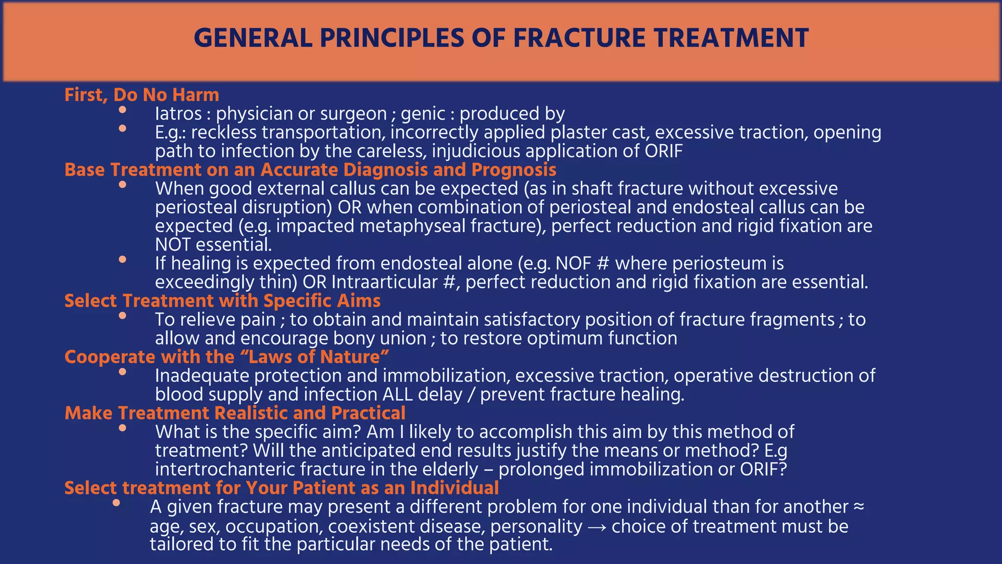
SlideShare a Scribd company logo
1 of 143
Fractures And Joint
Injuries - General Features
Kevin Pratama
GENERAL INCIDENCE AND
SIGNIFICANCE
High speed travel
Complex Industry
Competitive and recreational sports,
FracturesandJointInjuries–GENERALFEATURES
Age of Injury/ Age
of trauma
Trauma Remains the number one killer of young people in
North America
The estimated annual cost of trauma in North America
alone is over $160 billion. Approximately 10% of all
hospital beds are occupied at any given time by the
victims of trauma
IsolatedMuskuloskletalinjuries=
lowmortalitybutaHighmorbidity
Multipleinjuries=
HighmortalityandHighmorbidity
Old Age when Decreasing coordination frequent falls Osteoporosis Musculoskeletal
injury treated by prolonged bed rest Pathological processes Progessive deterioration
Death
● A Fracture, whether of a bone, an epiphyseal plate, or
cartilaginous joint surface, is simply :
“ Structural break in continuity ‘’.
● The physical forces that produce a fracture
● The associated injuries soft tissues injury
○ Brain
○ Spinal cord
○ Thoracic and abdominal viscera
○ Major artery or a peripheral nerve
FRACTURESANDASSOCIATEDINJURIES
may assume much greater clinical
significance than the fracture itself.
PHYSICAL FACTORS IN THE
PRODUCTION OF FRACTURES
• Normal living bone Has a degree of elasticity or flexibility and capable of being
bent slighly, like wood in a living tree.
• Cortical bone as a structure can withstand compression and shearing forces better
than it can withstand tension forces. The majority of fracture represent tension
failure of bending, twisting, and straight pull.
 A bending forces causes an almost explosive tension failure of the bone on the
convex side, the failure producing a tranverse fracture or an oblique fracture.
 In young children, cortical bone is like green wood, tension failure on the
convex side of the bend and only bending on the concave side of the
greenstick fracture
 Twisting Force, (Torsional/ Rotational) –, producing a spiraling fracture.
 Pulling force on small bone through attached ligaments or muscle  avulsion
fracture
 Involvement of articular cartilage : intraarticular fracture
 Cancellous bone, having a sponge like structure ( spongiosa), more susceptible to
crushing ( compression force)
 Sudden Compression – Compression Fracture
 In young children, a compression fracture merely “buckle” – the thin cortex
of metaphysis producing a buckle fracture/ torus fracture.
 Causatife Force that produces a fracture.
 Direct Injury
 Indirect Injury
Descriptive Terms Pertaining to Fractures
Important because they indicate the nature of the clinical problem and
the type of treatment.
 Site – diaphyseal, metaphyseal, epiphyseal, intra-articular,
fracture-dislocation
 Extent – complete, incomplete (hairline/crack, buckle, greenstick)
 Configuration – transverse, oblique, spiral, comminuted
 Relationship of fragments – displaced or undisplaced ;
translated, angulated, rotated, distracted, overriding, impacted
 Relationship to the external environment – closed vs open
 Complication – uncomplicated or complicated (local or systemic;
by the injury or by the treatment – iatrogenic).
Associated Injury to the Periosteum
 Periosteum in children is thicker,
stronger, more osteogenic ; easily
separated from the underlying bone
and not readily torn across, unlike in
adults ; usually remains intact on at
least one side (except in severely
displaced fracture), creating intact
periosteal hinge.
 Intact periosteal sleeve : advantage
in reducing the fracture, maintain it,
and aids fracture healing.
Diagnosis of Fracture and Associated Injuries
• May be obvious, or may not be all
apparent (due to communication
barrier, infancy, unconsciousness, or
mental confusion; in undisplaced
case, impacted and stable)
• Even when diagnosis of fracture is
obvious, overlook an associated soft
tissue injury, a visceral injury, a
coexistent dislocation, or even a second
fracture.
• History of fall, a twisting injury, a direct blow, a road accident
• Mechanism of injury is often lacking
• Localized pain, decreased function , “heard the bone break”
crepitus.
INSPECTION
 Facial expression, protect the injured part
 Swelling (except deep in the tissue), deformity, abnormal
movement, ecchymosis (discoloration)
PALPATION
 Sharply localized tenderness, aggravation of pain, muscle
spasm on passive movement
 DON’T feel /listen for the crepitus
 REMEMBER General condition , others – brain, spinal cord,
peripheral nerves, major vessels, skin, thoracic, abdomen
• Before examination, provide radioluscent splint for
immobilization
• Include entire length of injured bone + the joints at each end
• At least TWO projections (AP and lateral), often additional
oblique projection (small bone, ankle, pelvis,vertebrae)
• For the spine and pelvis: CT and MRI
• REMEMBER: the bone fragments are more widely displaced
during the moment of fracture due to immediate elastic recoil
of soft tissue
• Undisplaced fracture: will be apparent after 1 or 2 weeks as the
result of healing process.
• Primary / direct healing by internal
remodelling
• Secondary / indirect healing by callus
formation
• Primary Bone Healing
• Occurs only with absolute stability, is a
biological process of osteonal bone
remodelling.
• Secondary Bone Healing
• Inflammation
• Soft Callus Formation
• Hard Callus Formation
• Remodelling
• Inflammation
• 1-7 days after fracture
• Hematome formation  inflammatory exudation from ruptured vessels
• Bone necrosis at fracture ends; release of powerful cytokines causes
vasodilatation and hyperemia, migration and proliferation of PMN
neutrophils, macrophages.
• Hematome is graduallty replaced by granulation tissue
• Osteoclasts remove necrotic bone at fragment ends.
• Soft callus formation
• 2-3 weeks after fracture
• Progenitor cells at periosteum and endosteum are stimulated to become
osteoblast
• Intramembranous bone growth starts away from the fracture gap,
forming a woven bone.
• Next, ingrowth of capillaries into the callus and increased vascularity
follows.
• Mesenchymal progenitor cells migrate into callus differentiate into
fibroblasts or chondrocytes  replacing hematoma
• Hard callus formation
• After soft callus forms until 3-4 months
• Soft tissue within the gap undergoes endochondral ossification; callus is
converted into rigid calcified tissue
• Hard callus formation starts peripherally, moving towards the center of fracture
and fracture gap.
• Remodelling
• Few months to several years
• Starts after fracture has solidly united with woven bone
• Woven bone is slowly replaced by lamellar bone through surface erosion and
osteonal remodelling.
• It lasts until original morphology returns; including restoration of the medullary
canal.
HealingofaFractureinArticularCartilage
• Hyaline cartilage is extremely limited in its ability to
heal or regenerate.
• Scar will become local degenerative arthritis; joint
incongruity.
Age of The Patient
• Fastest at birth, less rapid with each year of childhood
• Early adult to old age : constant
Site and configuration of fracture
• Fractures surrounded by muscles : more rapid healing
• Cancellous bone heals more rapidly than cortical bone
• Epiphyseal separation heals twice as quickly as cancellous metaphyseal fracture of the
same bone
• Spiral / oblique fracture heals more readily than transverse – due to larger surface area
Initial displacement
• The greater the initial displacement  periosteal sleeve tearing is more extensive 
healing time is prolonged
Blood supply to the fragments
• Avascular fragments bony union cannot occur until revascularized
ASSESSMENTOFFRACTUREHEALINGINPATIENTS
● Clinical and radiographic examination
○ Bending, twisting, compression to the fracture  if both the examiner and the
patient cannot detect movement at the fracture site, it is clinically united
○ Clinical union precedes radiographic consolidation
○ At the time of clinical union, immobilization is no longer required. But it still needs to
be protected  until radiographic consolidation has been achieved
○ Radiographic consolidation : bony callus completely bridges the fracture, obliterates
fracture line.
○ Remodelling phase : medullary cavity seen in shaft / trabeculae in cancellous bone
1. The fracture may heal in the normally expected time but in an
unsatisfactory position with residual bony deformity (malunion).
2. The fracture may heal eventually but it takes considerably longer than the
normally expected time to do so (delayed union).
3. The fracture may fail completely to heal by bone (nonunion) with resultant
formation of either a fibrous union or a false joint (pseudarthrosis).
ComplicationsofFractures
● IMMEDIATE/ INITIAL
A. Lokal Complications
1. Skin Injuries
a) From Without : Abrasion,
Laceration, puncture
wound, penetrating missile
wound, avulsion, loss of
skin
b) From Within : penetration
of the skin by a facture
fragment
2. Vaskular Injuries
a) Major Artery :
Division, contusion, Arterial Spasm
b) Major Vein :
Division, contusion.
C) Local Hemorrhage
3. Neurological Injuries :
Brain, Spinal Cord, Peripheral Nerves.
4. Muscular Injuries
5. Visceral Injuries
B. Remote Complications
1. Multiple Injuries
2. Hemorragic Shock
● Early Complications Early
1. Local
■ Sequele of immediate complications : skin necrosis gangrene,
compartment syndromes, gas gangrene
■ Joint complication : septic arthritis
■ Bony complication : infection (osteomyelitis), avascular necrosis
2. Remote
■ Fat Emboly, Pulmonary Embolism, Pneumonia, Tetanus, Delirium
tremens
● Late
1. Local
■ Joint complications : stiffness,
posttraumatic degenerative
arthritis
■ Bony complications
● Abnormal healing (mal-,
delayed, non union)
● Growth disturbance
● Chronic osteomyelitis
● Posttraumatic
osteoporosis
● Sudeck’s posttraumatic
painful osteoporosis
● Refracture
■ Muscular complications : MO,
late rupture of tendon
■ Neurological complication :
tardy nerve palsy
2. Remote Complications
■ Renal calculi
■ Accident neurosis
GENERAL PRINCIPLES OF FRACTURE TREATMENT
First, Do No Harm
• Iatros : physician or surgeon ; genic : produced by
• E.g.: reckless transportation, incorrectly applied plaster cast, excessive traction, opening
path to infection by the careless, injudicious application of ORIF
Base Treatment on an Accurate Diagnosis and Prognosis
• When good external callus can be expected (as in shaft fracture without excessive
periosteal disruption) OR when combination of periosteal and endosteal callus can be
expected (e.g. impacted metaphyseal fracture), perfect reduction and rigid fixation are
NOT essential.
• If healing is expected from endosteal alone (e.g. NOF # where periosteum is
exceedingly thin) OR Intraarticular #, perfect reduction and rigid fixation are essential.
Select Treatment with Specific Aims
• To relieve pain ; to obtain and maintain satisfactory position of fracture fragments ; to
allow and encourage bony union ; to restore optimum function
Cooperate with the “Laws of Nature”
• Inadequate protection and immobilization, excessive traction, operative destruction of
blood supply and infection ALL delay / prevent fracture healing.
Make Treatment Realistic and Practical
• What is the specific aim? Am I likely to accomplish this aim by this method of
treatment? Will the anticipated end results justify the means or method? E.g
intertrochanteric fracture in the elderly – prolonged immobilization or ORIF?
Select treatment for Your Patient as an Individual
• A given fracture may present a different problem for one individual than for another ≈
age, sex, occupation, coexistent disease, personality → choice of treatment must be
tailored to fit the particular needs of the patient.
As An undergraduate student
• BLS
• ATLS
Preliminary Care For Patients with Fractures
This preliminary care for patients with fractures is best
considered in three phases:
1) immediate care outside a hospital (resuscitation and first
aid);
2) care during transportation to hospital;
3) emergency care in a hospital.
EmergencyLifeSupport
Immediate Care Outside a Hospital (First Aid)
• The summoning of emergency services-police,
firemen,ambulance
• A B C Priorities
• Airway : rolling the person to the prone position,
pulling the jaw and tongue forward, clearing the
pharynx with a finger, the patient’s neck should not
be moved.
• Breathing : mouth to mouth resuscitation
• Circulation : CPR, firm manual pressure on open
wound
• Shock : controlled hemorrhage and minimizing pain
• Fractures and Dislocations : splinting and traction
• Tranportation : stretcher to keep the spine straight,
cervical collar, helmet should not be removed
EmergencyCareinaHospital
• AMPLE history
• Airway
Suction, OPA, intubation, tracheostomy
• Breathing
Mechanically assisted respiration
• Circulation
Clamping, pressure on wounds,
• Shock
Blood typing and crossmatching, IV infusions
CVP monitoring, adequate analgesia.
EmergencyCareinaHospital
• The patient with multiple critical injuries
(polytrauma), one surgeon must serve as the team
captain.
• Vascular impairment and nerve injury should be
assessed. The patient must be carefully examined
for other fractures as well as for soft tissue
injuries and visceral lesions.
• Before radiographic examination, the limb should
be splinted. Is it important to move the tube and
film rather than to move the patient or the
fractured extremity.
ResponsibilitiesfortheCareoftheCriticallyInjured
• The patient with multiple critical injuries
(polytrauma), one surgeon must serve as the team
captain.
• Vascular impairment and nerve injury should be
assessed. The patient must be carefully examined
for other fractures as well as for soft tissue
injuries and visceral lesions.
• Before radiographic examination, the limb should
be splinted. Is it important to move the tube and
film rather than to move the patient or the
fractured extremity.
• In upper limb by a simple sling and in the lower
limb by relief of weightbearing with crutches.
• Indications
 Undisplaced, stable fractures of the ribs, phalanges,
metacarpals, and in children of the clavicle.
 Mild Compression fractures of the spine
 Impacted fractures of the upper end of the humerus.
 After clinical union, but before radiological
consolidation has established.
1. Protection Alone
Specific Methods of Treatment for Closed Fractures
REDUCTION IMMOBILIZATION
- -
Risks:
In a very young child or an
uncooperative adult, the
fracture may become
displaced. There is a need of
radiographic examinations at
regular intervals.
• It is only relative immobilization, as opposed to
rigid fixation, inasmuch as some motion can still
occur during early phases of healing.
• Indications
 Fractures that are relatively undisplaced, yet
unstable.
 Long bone with good contact, no significant
angulation or rotation
2. Immobilization by External Splinting
Specific Methods of Treatment for Closed Fractures
REDUCTION IMMOBILIZATION
- Plaster-of-Paris casts
Metallic or plastic splints
Risks:
 Subsequent muscle pull or
gravitation forces may cause further
displacement. Hence the need for
repeated radiographic examinations
during early stages.
 Improperly applied casts or splints
may cause local pressure sores over
bony prominences or constrictions
of a limb with resultant impairment
of venous or arterial circulation.
• Most common method
• Technique
 Placing the fracture fragments where they were at the time of
maximal displacement , then reversing the path of
displacement
 Applying plaster casts
• Indications
 Displaced fractures that require reduction, and
when it is predicted that sufficient accurate
reduction can be both obtained and maintained by
closed means.
3. Closed Reduction by Manipulation Followed by Immobilization
Specific Methods of Treatment for Closed Fractures
REDUCTION IMMOBILIZATION
Manipulation Plaster-of-Paris casts
Risks:
• Further damage to soft tissues
including blood vessels, nerves
and even periosteum.
• Impair Circulation
• Expressive Traction = Spasme
arteri particularly at the Elbow
and Knee = Volkmann’Ischemia
• Pressure sore
• Displacement especially oblique,
spiral and comminuted fracture
• Overcoming muscle pull and gravity
• When has become “sticky”  continuous traction can be
replaced by immobilization by appropriate casts
4. Closed Reduction by Continuous Traction Followed by Immobilization
Specific Methods of Treatment for Closed Fractures
REDUCTION IMMOBILIZATION
Continuous Traction Plaster-of-Paris casts
Indication
 Unstable oblique, spiral,
or comminuted fractures
of major long bones.
 Unstable spinal fractures.
 Fractures complicated by
vascular injuries,
excessive swelling, or
skin loss in which an
encircling bandage or
cast would be
dangerous.
Skin Traction Skeletal Traction
For fractures in young children For older children and adults,
traction is applied through the skin it is best applied through bone
by means of extension tape. by means of transverse rigid wire or
pin.
Risks:
• Excessive longitudinal traction, particularly if applied several
hours or longer after the fracture the fracture occured –
Arterial spasm with resultant Volkmann’s ischemia.
• Ineptly applied skin traction or excessive traction –
Superficial skin loss.
• Skeletal traction may become complicated by pin track
infection that reach the bone.
• If inaccurately applied and monitored, may fail to achieve and
maintain reduction
• Excessive traction – Distract the fracture fragment with
resultant delayed union or nonunion.
• Longer hospital stay.
Specific Methods of Treatment for Closed Fractures
4. Closed Reduction by Continuous Traction Followed by Immobilization
Principle is based on the following concepts :
• Rigid immobilization of fracture fragments is
unnecessary and undesirable for fracture healing.
• Function and the resultant controlled motion at the
fracture site actually stimulate healing through
abundant callus formation.
• Such function prevents iatrogenic disease.
• Somewhat less than perfect reduction of a fracture of
the shaft of a long bone does not create significant
problems concerning either function or appearance.
5. Closed Reduction Followed by Functional Fracture Bracing
Specific Methods of Treatment for Closed Fractures
REDUCTION IMMOBILIZATION
Manipulation
Continuous traction
Plaster casts followed by functional
fracture-bracing
• Benefits: Shorter period of hospitalization, no risk of
infection, and more rapid return to normal activities.
• Indications: Fractures of the shaft of the tibia, distal third of
femur, humerus, and ulna in adults.
• Contraindications: Fractures that can be more effectively
treated by open reduction and internal skeletal fixation,
including intertrochanteric, subtrochanteric, and mid-shaft
fractures of the femur, shaft fractures of the radius, and
intra-articular fractures.
• Risks: Although relatively risk-free, the method may fail to
maintain an acceptable position of the fracture fragments.
Specific Methods of Treatment for Closed Fractures
5. Closed Reduction Followed by Functional Fracture Bracing
Techniques
• Two or three metal pins are inserted percutaneously
through the bone above and below the fracture site, and
held together by external bars to provide firm (but not
rigid) fixation of the fracture “at a distance”
• More sophisticated: Hoffman type, Ilizarov frame
Indications
• Severely comminuted and unstable tibia/femur shaft
fracture
• Unstable fracture of pelvis, humerus, radius, metacarpals.
5. Closed Reduction By manipulation Followed by External Skeletal Fixation
Specific Methods of Treatment for Closed Fractures
REDUCTION IMMOBILIZATION
Manipulation External skeletal fixation
Risks :
• Pin track infection
with/without
Osteomyelitis
• High-speed power drills
 producing heat 
bone is burnt to death
 ring sequestrum
Specific Methods of Treatment for Closed Fractures
6. Closed Reduction By manipulation Followed by External Skeletal Fixation
Techniques
• Two or three metal pins are inserted percutaneously
through the bone above and below the fracture site, and
held together by external bars to provide firm (but not
rigid) fixation of the fracture “at a distance”
• More sophisticated: Hoffman type, Ilizarov frame
Indications
• Severely comminuted and unstable tibia/femur shaft
fracture
• Unstable fracture of pelvis, humerus, radius, metacarpals.
6. Closed Reduction By manipulation Followed by External Skeletal Fixation
Specific Methods of Treatment for Closed Fractures
REDUCTION IMMOBILIZATION
Manipulation External skeletal fixation
• Percutaneous insertion of metallic nails or intramedullary rods
across the fracture site for the purpose of providing internal skeletal
fixation. Both the closed manipulative reduction and the “blind”
insertion of the fixation are performed using.
• Indications:
Certain fractures in which accurate reduction can be obtained by
closed means but cannot and should not be maintained by external
immobilization. The most common indication is the unstable
fracture of the neck of the femur in both children and adults.
Fractures in the midshaft of the long bones that can be reduced by
closed means also lend themselves to this method.
7. Closed Reduction By manipulation Followed by Internal Skeletal Fixation
Specific Methods of Treatment for Closed Fractures
REDUCTION IMMOBILIZATION
Manipulation Internal skeletal fixation
Risks :
• Manipulative reduction
may fail to obtain a
satisfactory position.
• Internal skeletal fixation
may fail to achieve
sufficiently rigid fixation.
• Infection.
Specific Methods of Treatment for Closed Fractures
7. Closed Reduction By manipulation Followed by Internal Skeletal Fixation
• Internal skeletal fixation is achieved by using some type of metallic
device ≈ osteosynthesis, which include various types of transfixation
screws, onlay plates held by screws, intramedullary nails and rods,
smooth and threaded pins, encircling bands, and wire sutures.
Current thicking is that such devices should not be removed because
the risks of doing so > the risks of leaving them in place.
• Biodegradable screws are useful for fractures involving cancellous
bone, especially in children.
• AO/ASIF system: Rigid internal fixation ≈ “primary” fracture
healing → rapid recovery of function in the injured limb, and
thereby avoids “immobilization disease.”
8. Open Reduction Followed by Internal Fixation
Specific Methods of Treatment for Closed Fractures
REDUCTION IMMOBILIZATION
OPEN Internal skeletal fixation
Indications
• Failed / impossible closed reduction e.g. displaced avulsion ,
intraarticular , displaced epiphyseal in children, soft tissue
interposition.
• Grossly unstable fractures, which may be possible to obtain
reduction by closed means, but impossible to maintain the reduction
– Intertrochanteric fractures of the femur, fractures of both bones of
the forearm in adults, and displaced fractures of phalanges.
• Coexistent vascular injury that require exploration and repair.
• In order to facilitate nursing care and prevent serious complications
e.g. intertrochanteric femur fracture in the elderly, extremity
fractures with severe head injury, fracture dislocation of the spine
complicated by paraplegia
• Pathological fractures
8. Open Reduction Followed by Internal Fixation
Specific Methods of Treatment for Closed Fractures
Contraindications:
• Fractures of the shaft of the tibia
and shaft of the humerus, both of
which can usually be managed
either by closed nailing or by
functional fracture-bracing.
Risk :
• Infection
• Further Damage to the blood
supply of the fracture fragments =
Delayed Union or Non union
• Metal Failure
• Postoperative adhesions  joint stiffness
8. Open Reduction Followed by Internal Fixation
Specific Methods of Treatment for Closed Fractures
Indicates
• Displaced intracapsular fracture of NOF in the elderly
(high incidence of AVN)
• Comminuted fracture of radial head in adults  excision
of radial head. Excision of radial head is contraindicated in
children.
• Severely comminuted supracondylar fracture of humerus
in adult elbow prosthesis
• Severely comminuted fracture of patella excision of
entire patella and recon. of quadriceps mechanism.
• Comminuted humeral head  excision and
9. Excision of a Fracture Fragment and Replacement by an Endoprosthesis
Specific Methods of Treatment for Closed Fractures
REDUCTION IMMOBILIZATION
EXCISION OF FRACUTE
FRAGMENT
Joint Replacement
Risk :
• Infection
• Migration of
endoprosthesis
Spesial Consideration
• Extensive soft tissue Injury
• Bacterial contamination
Particular Emphasis
• Prevention of Infection
• Obtaining union of the fracture
Treatment For Open Fractures
• Instant photograph should be taken of every open fracture before dressing
Classification of Open Fractures
● Gustilo and Anderson
● Skin primary closure in
types 1 and 2 is
recommended, and
delayed primary closure in
type 3.
Aspects of treatment for Open Fractures
1. Cleansing the wound
● Gross dirt, bits of clothing, and other foreign material should be washed away by extensive
pulsating irrigation as well as mechanical cleansing with copious amounts of sterile water or
isotonic saline. Residual material should be carefully picked out of the wound. The wound may
even have to be opened further to allow adequate assessment of the degree of contamination and
to deal with it.Excision of devitalized tissue (Debridement)
2. Treatment of the Fracture
● Because tissues that have lost their blood supply prevent primary wound healing and enhance
infection, the meticulous surgical excision of all devitalized tissue, such as skin, subcutaneous fat,
fascia, muscle, and loose fragments of bone, is essential. It also is wise to obtain a culture of the
wound.
Aspects of treatment for Open Fractures
3. Treatment Of the Fracture
● Open wound is small: Can usually be treated by closed means, after the wound has
been cleansed, debrided, and left open.
● Open wound is extensive: The fracture may require either skeletal traction or open
reduction with skeletal fixation. External skeletal fixation is often of value.
4. Closure of the wound
● After 4-7 days, provided no infection has developed, delayed primary closure of the
wound is indicated.
● Loss of skin may necessitate the delayed application of split thickness skin grafts.
Suction drainage should be used to prevent accumulation of blood and serum in the
depths of the wound.
Aspects of treatment for Open Fractures
5. Antibacterial drugs
● To be effective in the prevention of infection, antibacterial drugs must be administered
must be administered in large doses before, during, and after treatment of the wound.
6. Prevention of tetanus
● If the patient has been previously immunized by tetanus toxoid → a booster dose of
tetanus toxoid.
● If there is no previous immunization, or inadequate information is available → tetanus
Ig + tetanus toxoid.
Anesthesia for Patients with Fractures
● First hour of fracture  numb , hence reduction without
anesthesia is possible.
● Infiltration of local anesthetic agent e.g. Colles’ fracture
● Regional anesthesia e.g. brachial plexus block for upper limb,
spinal anesthetic for the lower limb
● General anesthesia : majority of reduction are best treated under
general anesthesia
- Risk:aspiration of stomach content
After-care and Rehabilitation for Patients with Fractures
1. To restore optimum function! (4th and the most important aim of fracture treatment),
Begins with the immediate care of the patient’s injury, continues through emergency
treatment, definitive treatment, and beyond until the patient is restored to normal or as near
normal as the injury permits
2. Excessive and persistent soft tissue edema produce adhesions with resultant joint stiffness
>< Appropriate elevation of the limb during the early phase of fracture.
3. Active exercise of all regional muscle
• Active static (isometric) exercise: for muscles that control the immobilized joints
• Active dynamic (isotonic) exercise : all other muscle of limb and trunk.
4. Psychological consideration
5. Occupational therapy
6. Rehabilitation
Complication of Fracture Treatment
• Related to three main factors: excessive local pressure, excessive traction,
infection.
Classification:
1. Skin complication
• Tattoo effect from abrasions
• Pressure sores (Bed sore , cast sore)
2. Vascular complication (Traction and pressure, Volkmann’s ischemia, Gangrene
and gas gangrene, Venous thrombosis and pulmonary embolism)
3. Neurological complication (Traction and pressure)
4. Joint complication (Infection - septic arthritis after open operative treatment of a
closed injury)
5. Bony complication (Infection – osteomyelitis)
Classifications of the Complications of
the Original Injury
Initial and Early Complications
SKIN INJURIES
• Abrasion (friction burn) with particles of dirt having been ground into dermis, if not thoroughly cleansed →
residual pigmentation under the re-epithelized surface → tattoo effect.
• Lacerations, puncture wounds, penetrating missile wounds, avulsion of skin, and skin loss ≈ open fractures
• Gross swelling → stretching of the overlying skin → compromise circulation → blister or bleb formation.
• Area of skin compressed between a firm surface and bony prominence → bed sores (decubitus ulcers).
• Excessive local pressure from incorrectly applied plaster cast → pressure sores (cast ulcer).
Vascular Complications
Arterial Complications (Injury to a Major Artery)
1. Axillary a >< Fracture-dislocation or dislocation
of shoulder.
2. Brachial a >< Supracondylar fracture of
humerus.
3. Femoral a >< Fracture of the shaft of femur.
4. Popliteal a >< Fracture of the distal end of
femur, proximal end of tibia, or dislocation of
the knee.
5. Dorsalis pedis a >< Fracture of the forefoot.
Vascular Complications
Arterial Division
• Completely torn ≈ retracts, stops bleeding spontaneously.
• Incompletely torn ≈ tends to continue bleeding, may lead to pulsating
hematoma (false aneurysm).
• Hemorrhage
‾ External – Obvious.
‾ Internal – Progressively enlarging local swelling.
• Treatment: Direct suture; if not possible, continuity can be achieved by means
of an autogenous venous graft or a plastic arterial prosthesis.
Vascular Complications
Arterial Spasm
Sudden and severe traction of a major artery
(either at the time of fracture Or during its treatment)
Treatment
• Intimal tear – Resection of the damaged portion and restore its continuity by direct
suture, vein graft, or prosthesis.
• Persistent spasm – Local application or warm papaverine, or if unsuccessful, the
constricted portion can be permanently dilated by intra-arterial injection of saline.
• Severe arterial spasm – Meticulous microsurgical excision of the encircling adventitia
of the spastic portion.
Vascular Complications
Arterial Compression
• A major artery becomes trapped and compressed between fracture
fragments
• Excessively tight encircling plaster cast externally + Progressive swelling
internally
Treatment
Is released, and provided there is no associated spasm, the flow will be re-established.
Vascular Complications
Arterial Thrombosis
• The presence of pre-existing arteriosclerosis
• Any arterial injury that results in persistent occlusion
Treatment
Should be Removed.
Vascular Complications
Recognition of arterial complications
Complete arterial occlusion
• Initial –Pallor and coolness of the skin distally, loss of arterial pulse.
• Later – Mottled, dark discoloration that heralds gangrene.
• Investigation with Doppler ultrasound and angiography.
Treatment of arterial occlusion:
A. Any constricting cast or bandage must be completely removed.
B. Any distortion of the fractured limb or extreme position of anearby joint should be
lessened.
C. If the fracture is being treated by continuous traction, the amount of traction should
be decreased.
D. If these measures fail to restore circulation → emergency arteriogram.
E. No improvement within half an hour → surgical exploration.
Vascular Complications
Recognition of arterial complications
Sequelae of arterial complications:
A. Gangrene – Results from persistent total ischemia distal to an arterial lesion.
B. Compartment syndrome – Results from persistent occlusion of deep arteries > 6
hours.
C. Intermittent claudication – When an arterial lesion has not been sufficiently
severe or persistent.
D. Gas gangrene.
Vascular Complications
Definition
Increased pressure of progressive edema within a rigid
osteofascial compartment of either the forearm or the leg
threatens the circulation to the enclosed
(intracompartmental) muscles and nerves.
Compartment Syndrome
(Volkmann’s Ischemia)
Etiology and Risk Factors
Mechanism Of Injury
• High Speed Travel
• Work Accident
• Natural Disaster
Etiology
• Interna : Fracture (69%), Others (23%)
like Bleeding, Vaskular Injuries, Burn Injury
• External : Too Rigid Casting and Thigh
Splinting.
Compartment Syndrome
(Volkmann’s Ischemia)
Etiology and Risk Factors
Increased Volume of Compartement Content
• Fractures
• Soft Tissue Trauma
• Surgical Treatment
• Exercise
• Vascular Injury
• Hematoma
Decreased Compartment Size
• Fascial Defect/ Tight Dressings
• Burn Injury
• External Pressure Like Case, splints, Burn
Escar, lying on Limb For long Period,
Lithothomy position
Compartment Syndrome
(Volkmann’s Ischemia)
• MUSCLE
 3 – 4 Hours : Reversible
 6 Hourss : Variable
 8 Hours : Irreversible
• Nerve
 2 Hours : Lose Nerve
conduction
 4 Hours : Neurophaxia
 8 Hour : Irreversible
Pathophysiology
Compartment Syndrome
(Volkmann’s Ischemia)
Diagnosis
• ATLS
• Secondary Survey
• AMPLE
• Early Sign of Compartment
SindromPain out of Proportion
 Pain on passive Strecth
 Parasthesia (Two Point
Discrimination)
• Late signs : 5P ( Pain, Parasthesia,
Pallor, Paraliysis, Pulselessness)
If in Doubt , measure
intracompartment pressure
• Unconscious patient
• Children Patient
• Uncooperative patient
• Neurologic Injury
• Regional Anesthesia
Where the check
• All Compartments suspected to
have compartement syndrome
• In case of Fracture, close to
fracture site
• If not possibe, measurement can be
done at most accessible site.
Compartment Syndrome
(Volkmann’s Ischemia)
Intracompartmental pressure measurement
METHOD :
• Injection / Infusion Technique (Whitesides) =
equipment in expensive and readily available
in most hospitals, emergensi rooms = not
accurate.
• Wick Catheter (Mubarak)
• Slit Catheter (Rorabeck)
• Solid State tranducer intracomp cathether
(STIC) : Portable Device : Stryker App
• Fiber Optic tranducer tipped = very expensive
• Latest Device : Electronic Tranducer Tipped
Catheter = best device
Compartment Syndrome
(Volkmann’s Ischemia)
WHITESIDE TECHNIQUE
Compartment Syndrome
(Volkmann’s Ischemia)
INTERPRETATION OF INTRA COMPARTMENT
PRESSURE MEASUREMENTS
Normal Tissue Pressure
• 0 - 4 mmHg
• 8 – 10 with Exercise
Absolute Pressure Theory
• 30 mmHg – Mubarak
• 45 mmHg – Matsen
Compartment Syndrome
(Volkmann’s Ischemia)
LABORATORIUM
• Creatinin Kinase Serum
• Increases 2 hours - 12 hours post-injury,
peaks within 24 hours - 72 hours post-
injury, decreases slowly 7 days - 10 days
an increase in CSK indicates muscle
damage caused by chronic disease and
acute muscle injury 10x elevation =
muscle damage In Valdez et al's study,
the maximum CK was more than 4000
U/L, chloride level more than 104
MG/DL, Strong Indication Compartment
syndrome, rhabdomyolisys, CK range
(10,000 – 200.000 U/L)
• Myoglobulin
• Range Normal low serum level of 0 to
0.03 mg/dL3
• Myoglobulin > 0.5 to 1.5 mg/dL = urin
output
• Urin myoglobulin level of 100mg/Dl >
tea or cola
• LDL
• LDH Level > 5200 or more would be
alarming for suspicion of muscle
compartment syndrome.
Compartment Syndrome
(Volkmann’s Ischemia)
TREATMENT
Compartment Syndrome
(Volkmann’s Ischemia)
• Don’t elevate LIMB
• General condition stabilization like
shock, antibiotics, Analgesic, heparin,
anti oxcidant,
Operatif :
• Fasciotomy
• Wount treatment : VAC or shoe lace
suture.
COMPLICATION
Compartment Syndrome
(Volkmann’s Ischemia)
Acute
• Local
 Organ or compartment
infection
 Tissue Necrosis =
Amputation
• Sistemik
 Rhabdomiolisis
 Acute Renal Failure
 Crush Syndrome
Chronic
• Volkman Ischemic
Contracture
DIVISION OF A MAJOR VEIN
• A major vein may be completed or incompleted divided
• Either by
 The displacement of a fracture fragment from within
 An object or missile that has penetrated the deep tissues from without.
Treatment : Surgical Repair
VENOUS COMPLICATIONS
VENOUS TROMBOSIS AND PULMONARY EMBOLISM.
• DVT + PE is a common cause of morbidity and mortality in adult orthopaedic
patients.
• Local pressure from prolonged bed rest
• from a tight plaster cast or bandage
• Thrombosis in the calf: Local pain, tenderness in the midline posteriorly and
distal swelling due to congestion. Passive dorsiflexion of the ankle aggravates the
pain (Homan’s sign).
• Thrombosis in the thigh: The entire lower limb becomes swollen.
VENOUS COMPLICATIONS
VENOUS TROMBOSIS AND PULMONARY EMBOLISM.
Pulmonary embolism
• Small – Undetected or mild chest pain.
• Moderate – Sudden onset of chest pain, dyspnea, sometimes hemoptisis.
• Massive – Dramatic onset of severe chest pain, immediate blanches, dead.
Prevention:
• By avoiding constant local pressure on veins and by encouraging the patient to do
active movements.
• For adults confined to bed, the use of elastic compressive stocking, CPM, and
cyclic external pneumatic compression help in the prevention.
• Patient at high risk should be given prophylactic anticoagulant.
Treatment: Appropriate anticoagulant drugs. Recent thrombosis in the femoral vein
is best treated by surgical thrombectomy.
VENOUS COMPLICATIONS
Complicating injuries to
brain, spinal cord,
peripheral nerves.
NEUROLOGICAL COMPLICATIONS
VISCERAL COMPLICATIONS
Thoraco abdominal viscera may be injured at the time of accident
independent of any fractures; or may also be injured by penetration by a
sharp fracture fragment from a nearby bone.
• Displaced fractures of the ribs
‾ Damage the heart → hemopericardium, cardiac tamponade.
‾ Perforate the pleura → hemothorax.
‾ Perforate the lung → hemopneumothorax.
‾ Perforate the liver, spleen, or kidneys.
• Fractures of the thoracic and lumbar spine → paralytic ileus and
gastric dilatation.
• Displaced fractures of the pelvis → rupture the bladder or urethra.
VISCERA COMPLICATIONS
Infection of a joint (septic arthritis): After an open intra-articular
fracture, or less oftern after open operation on a closed intra-
articular fracture, septic arthritis may ensue (Chapter 10).
JOINT COMPLICATIONS
INFECTION OF BONE (OSTEOMYELITIS)
• From open fractures, a closed fracture after open operation, along the track of a
metal pin used for continuous skeletal traction or external skeletal fixation (pin
track osteomyelitis, may form a ring sequestrum).
BONY COMPLICATIONS
AVASCULAR NECROSIS OF BONE
Usually caused by disruption of nutrient vessels at the time of original injury, but may also be
iatrogenic as a result of excessive dissection during open reduction. It leads to delayed union
and to joint incongruity with resultant degenerative arthritis.
Usually occurs after certain fractures and dislocations because of the precarious blood supply
to bone at these sites :
• Femoral head >< Fractures of the femoral neck, dislocations of the hip.
• Lunate >< Dislocations of the lunate.
• Scaphoid >< Fractures of the scaphoid.
• Radial head >< Fractures of the neck of the radius.
• Lateral condyle >< Fractures of the lateral condyle (especially after excessive soft tissue dissection during open reduction)
• Middle segment of a comminuted fracture
• Body of the talus >< Fractures of the neck of the talus.
Posttraumatic avascular necrosis is also discussed in Chapter 13.
BONY COMPLICATIONS
BONY COMPLICATIONS
AVASCULAR NECROSIS OF BONE
Initial and Early Complications –
Remote Complications
INTRODUCTION
Fat globules can be found in the circulation in most adults after a major
fracture; only 9% develop detectable systemic fat embolization and a
significant respiratory distress syndrome + severe arterial hypoxia, the
combination of which constitutes fat embolism syndrome.
Most susceptible:
• Previously healthy young adults, especially when associated with other injuries
(polytrauma).
• Elderly who sustain fractures of the upper end of the femur.
• Children with pre-existing systemic collagen disease.
FAT EMBOLISM SYNDROME
ETIOLOGY AND PATHOGENESIS
Stress-induced changes in lipid metabolism and in blood
coagulation → coalescence of chylomicrons to form macroglobules
of fat → fat embolization → arterial hypoxia with metabolic and
respiratory acidosis → may pass to the lung or to the brain.
FAT EMBOLISM SYNDROME
CLINICAL FEATURES
Usually develops after 2-3 days, or within few hours in very severe cases.
• Pulmonary emboli :Dyspnea, hemoptysis, tachypnea, and
cyanosis.
• Cerebral emboli :Headache, confusion, and irritability followed
by delirium, stupor, and coma.
• Cardiac emboli :Tachycardia and a drop in blood pressure.
• Transient thrombocytopenia → multiple petechial hemorrhage
of the skin, particularly of the upper chest, axillae, and
conjunctivae.
• FEBRIS
FAT EMBOLISM SYNDROME
CLINICAL FEATURES
Usually develops after 2-3 days, or within few hours in very severe cases.
• Pulmonary emboli :Dyspnea, hemoptysis, tachypnea, and cyanosis.
• Cerebral emboli :Headache, confusion, and irritability followed by
delirium, stupor, and coma.
• Cardiac emboli :Tachycardia and a drop in blood pressure.
• Transient thrombocytopenia → multiple petechial hemorrhage of the skin,
particularly of the upper chest, axillae, and conjunctivae.
• FEBRIS
FAT EMBOLISM SYNDROME
RADIOGRAHIC FEATURES
• Multiple areas of consolidation (a “snow storm” appearance).
FAT EMBOLISM SYNDROME
LABORATORY FEATURES
• The serum fatty acids are elevated
• There is free fat in sputum and urine
• Hemoglobin usually drops sharply very early in the process
• PO2 is reduced (< 100 mm), sometimes ass low as 60 mm
• Thrombocytopenia
FAT EMBOLISM SYNDROME
PREVENTION OF FAT EMBOLISM
• High carbohydrate intake.
• Constant maintenance of fluid and electrolyte balance.
• Early operative fixation of associated fractures.
FAT EMBOLISM SYNDROME
TREATMENT
• Heparin – Increase the rate of hydrolysis and removal of emboli.
• Large dose corticosteroid – Decrease lung tissue injury.
• Restoration of blood volume and electrolytes.
• Low molecular weight dextran – Improve microcirculation in
involved organs.
• In the presence of respiratory distress, endotracheal intubation
or tracheostomy followed by mechanically assisted intermittent
positive pressure ventilation (IPPV).
• Constant monitoring of PO2, PCO2, and arterial pH.
FAT EMBOLISM SYNDROME
PROGNOSIS
• The prognosis in patients who exhibit pulmonary insufficiency
and coma is grave in that the mortality rate is approximately
20%, a fatal outcome usually being related to a combination of
pulmonary and cerebrallesions.
• Fat embolism syndrome has been estimated to be the major
cause of death in 20% fatalities associated with fractures.
FAT EMBOLISM SYNDROME
• Has been discussed, related to venous thrombosis
PULMONARY EMBOLISM
• With treatment involves complete and prolonged bed rest
(which should be avoided whenever possible), the patient may
become complicated by hypostatic pneumonia.
• Elderly are susceptible; painful fractures of the ribs with
associated limitation of respiratory excursion may lead to the
development of pneumonia.
• Treatment: Antibiotic therapy, deep breathing exercises,
frequent turning of the bedfast patient, and -if necessary-
bronchoscopic suction.
PNEUMONIA
DEFINITION
a neurological disorder characterized by increased muscle tone and
spasm, caused by pressure spasm (a strong protein toxin) produced by
clostridium tetani.
TETANUS
PATHOPHYSIOLOGY
Clostridium tetani is a gram-positive, anaerobic, obligate
bacterium, can form spores and form drum sticks which grow
on necrotic tissue that produces neurotoxins. Neurotoxins are
removed by lymph nodes and blood flow to the central nervous
system and then bound to the anterior horn which cannot be
neutralized by antitoxin. Incubation period 10 - 14 days.
TETANUS
Clinical Manifestasion
• Tonic and later clonic-contractions of skeletal muscles (tetanic
spasms)
• Spasm of the neck and trunk muscles → arched back posture
(opisthotonus).
• Spasm of the jaw muscles → trismus (“lock jaw”).
• Spasm in the fascial muscles (Risus Sardonikus)
• Eventual involvement of intercostal muscles and diaphragm →
fatal asphyxia.
TETANUS
CLASSIFICATION
• Mild tetanus = Trismus:
Not accompanied by seizures even when stimulated
• Moderate tetanus = Trismus < 3cm
Accompanied by seizures, but when stimulated
• Severe tetanus = Trismus > 1 cm
Accompanied by spontaneous generalized seizures
TETANUS
DIAGNOSIS
• Clinical Symptoms
• Presence of a wound
• Positive clostridium tetani culture
• Laboratory : SGOT increase, Myoglobinuria (+), CPK increase
• Spatula Test +
TETANUS
TREATMENT
• Human Tetanus Immunoglobin (HTIg 3000 – 6000 IU/IM)
• AB
• ATS 50.000 – 100.000 IU/IM
• Diazepam 0,5 – 1 mg/kgBB
• Endotracheal intubation and mechanically assisted respiration.
TETANUS
When a chronic alcoholic sustain a major injury, alcohol intake is
abruptly withdrawn. The patient may exhibit dramatic withdrawal
symptoms, characterized by disorientation, anxiety, agitation, and
disturbing visual hallucinations.
DELIRIUM TREMENS
LATE COMPLICATIONS
BONY COMPLICATIONS
ABNORMAL HEALING OF FRACTURES
MAL UNION
DEFINITION
• Union has occurred in an unsatisfactory position of
significant deformity.
ETIOLOGY
• Fracture without treatment
• Due to inadequate immobilization.
Treatment
• Treated with corrective osteotomy, prevented by
obtaining and maintaining an acceptable reduction
of the fracture.
BONY COMPLICATIONS
ABNORMAL HEALING OF FRACTURES
DELAYED UNION
• Healing of a fracture is much slower thatn the
estimated rate of healing for that particular fracture
• Due to metabolic disease, hyperthyroidism, malnutrition
• Occasionally, union must be encouraged by means of
autogenous bone graft.
BONY COMPLICATIONS
ABNORMAL HEALING OF FRACTURES
NON UNION
• Fibrous nonunion : have potential for bony union if
internally immobilized and infection eradicated
autogenous bone graft may be added
• False joint / pseudoarthrosis : requires bone grafting
Factors that favor delayed/nonunion:
• Severe disruption of periosteal sleeve
• Loss of blood supply
• Inadequate immobilization of the fracture
• Inadequate period of immobilization
• Distraction of fracture fragments by excessive traction
• Interposition of soft tissue
• Infection at the fracture site
• Local and progressive disease of bone
Persistent Infection of Bone
● It leads to delayed union or even nonunion (infected nonunion) , fracture cannot heal
until the infection is completely controlled.
Post traumatic Osteoporosis
● Immobilization  bones atrophy  bone resorption exceeds bone deposition ; Th/
intensive physiotherapy
Sudeck’s Posttraumatic Painful Osteoporosis (Reflex Sympathetic Dystrophy)
● Sympathetically mediated pain syndrome
● Severe pain of foot or hand, stiff joints, edematous , skin is moist, mottled, smooth,
and shiny.
● Ro: disuse osteoporosis.
● Th/ local warmth, active exercise
Refracture
● Between clinical union and complete consolidation  still susceptible.
● Site of screw : weaker than normal bone
Metal failure
● Implant: serves as temporary internal splint to maintain reduction; subjected to
diminishing stress as union occurs.
● In delayed/nonunion  persistent movement  repeated stress  metal fatigue 
metal cracks and breaks
MUSCULAR COMPLICATIONS
Traumatic myositis ossificans (posttraumatic ossification)
• Due to a fracture, dislocation, or muscle injury ; particularly near elbow, thigh
of children and young adults
• X-ray: extensive ossification
• Heterotopic ossification : new bone formation in an abnormal site
• Accompanied by limited motion
• Prevention : indocid, prophylactic radiation
• Treatment : local rest by splinting  resorbs spontaneously; Don’t : passive
stretching, excise in the early stage.
Late rupture of tendons
• Metaphyseal fracture  heals with irregularity  lost of smoothness of bony
grooves for tendons to glide (esp. near ankle and wrist)  frayed tendons 
rupture. E.g. EPL rupture after colles’ fracture.
NEUROLOGICAL COMPLICATIONS
Tardy nerve palsy
- Residual valgus deformity of the elbow excessive
stretching of ulnar nerve + friction between nerve and
distal end of humerus  intraneural fibrosis and
thickening after 10-20 years
- Th/ surgical transposition (relocation) to the anterior
aspect of elbow.
NEUROLOGICAL COMPLICATIONS
Renal calculi (calcium type)
- Due to inadequate drainage of urine , hypercalcemia
associated with generalized disuse osteoporosis
- Prevention : increase fluid intake (up to 4000 ml) ,
frequent turning , allowed out of bed ASAP
Accident neurosis
- Consistent denial of his/her ability to return to work in
the hope for compensation / insurance
- Th/ extensive rehabilitation , psychiatric assessment
Stress Fractures
• Repeated stress  small crack with no time to
become conditioned by the normal process of work
hypertrophy ; activities include long march, track
and field activity , ballet dancing on someone who
is out of condition.
• E.g. march fracture (on 2-4 metatarsals), lower end
of fibula in runners, upper third of tibia in jumpers
and ballet dancers.
Treatment: Stop activity, then gradually return to
activity
Special Types of Fractures
Pathological fractures
• Occurs through abnormal bone
• The bone may be so weak that is fractured by a trivial injury
or even normal use; also if the fracture is due to major
injury through a pathological bone.
Clinical Features and Diagnosis
• Related to the underlying condition
Prognosis
• Most pathological fractures will unite ; osteomyelitis-related
patho # will not usually unite until infection is controlled.
• In highly malignant primary neoplasm  union is markedly
delayed, and amputation is indicated
• MBD  needs internal fixation + irradiation
Special Types of Fractures
I. Congenital abnormalities
• Localized – congenital defect of tibia
(pseudoarthrosis)
• Disseminated – enchondromatosis
• Generalized – Osteogenesis Imperfecta,
osteopetrosis
II. Metabolic Bone Disease
• Rickets
• Osteomalacia
• Scurvy
• Osteoporosis
• Hyperparathyroidism
III. Disseminated bone disorders of
unknown etiology
• Polyostotic fibrous dysplasia
• Skeletal reticuloses
• Langerhans’ cell histiocytosis
• Gaucher’s disease
ClassficationDisorders that predispose Bone to Pathological Fracture
IV. Inflammatory disorders
• Hematogenous osteomyelitis
• Osteomyelitis secondary to wounds
• TB Osteomyelitis
• RA
V. Neuromuscular disorders with disuse osteoporosis
• Paralytic disorders (poliomyelitis, paraplegia)
• Disorders of muscle (muscular dystrophy)
VI. Avascular Necrosis of Bone
• Posttraumatic, postradiation
ClassficationDisorders that predispose Bone to Pathological Fracture
VII. Neoplasms of bone
◦ Neoplasm-like lesion of bone
◦ Osteogenic – do not weaken the bone
◦ Chondrogenic
◦ Enchondroma
◦ Multiple enchondromata
◦ Fibrogenic
◦ Subperiosteal cortical defect
◦ Nonosteogenic fibroma (NOF)
◦ Monostotic fibrous dysplasia
◦ Polyostotic fibrous dysplasia
◦ Osteofibrous dysplasia
◦ “Brown tumor” (hyperparathyroidism)
◦ Angiogenic
◦ Angioma of bone (hemangioma and lymphangioma)
◦ ABC
◦ Uncertain origin
◦ SBC
◦ True primary neoplasms of bone
◦ Osteogenic
◦ Osteosarcoma
◦ Surface osteosarcoma
◦ Chondrogenic
◦ Benign chondroblastoma
◦ Chondromyxoid fibroma
◦ Chondrosarcoma
◦ Fibrogenic
◦ Fibrosarcoma of bone
◦ Malignant Fibrous Histiocytoma of bone
◦ Angiogenic
◦ Angiosarcoma of bone
◦ Myelogenic
◦ Myeloma of bone (MM)
◦ Ewing’s sarcoma
◦ HL
◦ NHL (Reticulum Cell sarcoma)
◦ Skeletal reticuloses (Langerhans’ Cell histiocytoses)
◦ Leukemia
◦ Uncertain Origin
◦ GCT
◦ Metastatic neoplasm in bone
◦ Metastatic carcinoma
◦ Metastatic neuroblastoma
• Normal Joint Stability
• Three structural factors are responsible for preventing an abnormal range of
motion, for providing joint stability:
1) contours of the opposite joint surfaces
2) integrity of joint capsule and ligaments
3) the protective power of muscles that move the joint
• Physical Factors in the Production of Joint Injuries
• Dislocation of a joint = structural loss of its stability
• The physical factors that suddenly force a joint beyond its normal range of
motion  tension failure of either in bony component of joint or in fibrous
capsule and ligaments. ; the force is usually an indirect injury (the force is
transmitted through the bone)
DislocationsandAssociatedInjuries
• Ligamentous sprain : sudden tension on a ligament that causes
severe stretching, resulting in minor tears and some hemorrhage
without loss of joint stability
• Ligamentous tear : may be partial or complete, with loss of joint
stability
• Ligamentous avulsion : tearing of a fragment of bony attachment
of a ligament
• Ligamentous strain : gradual elongation of a ligament due to
repeated mild stretching over a prolonged period.
• Three degree of joint instability:
• Occult joint instability : unstable only under stress
• Subluxation : joint surfaces have lost normal relationship, but still
retain considerable contact
• Dislocation : lost contact of joint surfaces
• Fracture-dislocation: dislocation is accompanied by an
intraarticular or extraarticular fracture
Descriptive Terms Pertaining to
Joint Injuries
Associated Injury to the Fibrous Capsule
• Intracapsular dislocation: the fibrous capsule and contiguous periosteum may be stripped up
from the bony margin of the joint and stretched
• Extracapsular dislocation: fibrous capsule is torn and one bone end perforates
• Buttonhole dislocation : large bone end is trapped in the dislocated position by the small rent in
the capsule  impossible for closed reduction
•Occasionally, a flap of torn capsule becomes trapped between the joint surfaces , preventing
perfect reduction  residual subluxation  absolute indication for open reduction
Diagnosis of Joint Injuries
Symptoms :
◦ Because of normal proprioceptive sensation, the patient is usually aware that a given joint
◦ has “gone out of place.”
◦ Associated joint instability and stretching → pain and muscle spasm.
◦ Decreased function of the involved part.
Sign :
◦ Swelling – Unless the dislocated joint is deep as in the hip.
◦ Deformity – Angulation, rotation, loss of normal contour, shortening.
◦ Abnormal movement – Occuring through the unstable joint.
◦ Local tenderness over a sprained or torn ligament.
◦ Diligent search for any associated injuries to spinal cord, peripheral nerves, or major vessels.
Radiographic examination :
◦ Typical features of a dislocation or subluxation.
◦ Occult joint instability – Additional radiograph taken while the joint is being stressed (under
anesthesia
Normal Healing of Ligament
• Partial tears : heal reasonably well with protection
• Complete tears : gap heals by fibrous scar tissue  elongated and weak
Complications of Dislocations and Associated Injury
• Immediate local : injury to skin, blood vessels, peripheral nerves, spinal cord, multiple injuries
• Early local : infection, AVN
• Late : joint stiffness, persistent instability, recurrence, arthritis, osteoporosis, reflex sympathetic
dystrophy, posttraumatic MO
Specific Types of Joint Injuries
Contusion
• Direct blow to the joint  synovial membrane produces effusion , rupture of synovial vessels produce
hemarthrosis
Ligamentous sprain
• Sudden stretching of the ligament with minor, incomplete tear , with local hemorrhage but no loss of
continuity.
• Local swelling, tenderness, pain aggravated by joint movement, no joint instability
• Ro :exclude disloc, sublux, or fracture, or even occult joint instability.
• Treatment:protect, adhesive strapping to limit certain movement, active exercise.
Dislocations and Subluxations
• Perfect reduction must be achieved, either by closed or open means.
• Consideration must be given to the torn ligaments to prevent residual joint instability with resultant
recurrent dislocation of the joint.
Torn ligaments
• Certain types : repaired surgically ASAP , e.g. collateral ligaments of the knee
• Others : immobilization to protect from further injury e.g. lateral ligaments of ankle, collateral ligaments of
fingers
Muscle Injuries
• Occurs when sudden severe tension is applied
to an already contracted muscle  rupture of
muscle bundle
• Strain : chronic overstretching of a muscle or
its tendon due to overuse.
Tendon Injuries
Closed Tendon Injuries
• only after becomes degenerated or frayed 
ruptured even with normal activity.
• Th : reconstructive operations
• Sudden tension on normal tendon may avulse
bone fragment e.g mallet finger
Open Tendon Injuries
• Clean, open division  immediate surgical repair
Salter bab 15 ppt

More Related Content

Similar to Salter bab 15 ppt

fractures their classification and treatment.pptx
fractures their classification and treatment.pptxfractures their classification and treatment.pptx
fractures their classification and treatment.pptxUzairRashid2
 
P M S 2 Fractures And Healing
P M S 2  Fractures And  HealingP M S 2  Fractures And  Healing
P M S 2 Fractures And HealingAriff Anuar
 
Principles of fractures
Principles of fracturesPrinciples of fractures
Principles of fracturesAhmad Sulong
 
Principles_of_fractures-1.ppt
Principles_of_fractures-1.pptPrinciples_of_fractures-1.ppt
Principles_of_fractures-1.pptAhmedMufleh1
 
BONE FRACTURES.pptx
BONE FRACTURES.pptxBONE FRACTURES.pptx
BONE FRACTURES.pptxhopekalunga
 
Fractures and Dislocations. Of joints pptx
Fractures and Dislocations. Of joints pptxFractures and Dislocations. Of joints pptx
Fractures and Dislocations. Of joints pptxBharath Doltade
 
Fracture types - Plaster Of Paris tecniques and Complications
Fracture  types - Plaster  Of  Paris  tecniques  and  ComplicationsFracture  types - Plaster  Of  Paris  tecniques  and  Complications
Fracture types - Plaster Of Paris tecniques and ComplicationsVenkatesh Ghantasala
 
archfracture.pptx
archfracture.pptxarchfracture.pptx
archfracture.pptxsidra234490
 
Fracture healing
Fracture healingFracture healing
Fracture healingNafeeyabano
 
Fracture healing and wound healing
Fracture healing and wound healingFracture healing and wound healing
Fracture healing and wound healingShermil Sayd
 
fracture pp 2.ppt medical surgical nursing
fracture pp 2.ppt medical surgical nursingfracture pp 2.ppt medical surgical nursing
fracture pp 2.ppt medical surgical nursingMadhuriRaychura1
 
Paediatric orthopaedic fracture; dislocation -lec 9 (june 2016) n.c.
Paediatric orthopaedic  fracture; dislocation -lec 9 (june 2016) n.c.Paediatric orthopaedic  fracture; dislocation -lec 9 (june 2016) n.c.
Paediatric orthopaedic fracture; dislocation -lec 9 (june 2016) n.c.yasser Amr
 

Similar to Salter bab 15 ppt (20)

fractures their classification and treatment.pptx
fractures their classification and treatment.pptxfractures their classification and treatment.pptx
fractures their classification and treatment.pptx
 
Fracture ppt
Fracture pptFracture ppt
Fracture ppt
 
P M S 2 Fractures And Healing
P M S 2  Fractures And  HealingP M S 2  Fractures And  Healing
P M S 2 Fractures And Healing
 
Principles of fractures
Principles of fracturesPrinciples of fractures
Principles of fractures
 
Principles_of_fractures-1.ppt
Principles_of_fractures-1.pptPrinciples_of_fractures-1.ppt
Principles_of_fractures-1.ppt
 
BONE FRACTURES.pptx
BONE FRACTURES.pptxBONE FRACTURES.pptx
BONE FRACTURES.pptx
 
BONE FRACTURES.pptx
BONE FRACTURES.pptxBONE FRACTURES.pptx
BONE FRACTURES.pptx
 
Fractures and Dislocations. Of joints pptx
Fractures and Dislocations. Of joints pptxFractures and Dislocations. Of joints pptx
Fractures and Dislocations. Of joints pptx
 
Fracture types - Plaster Of Paris tecniques and Complications
Fracture  types - Plaster  Of  Paris  tecniques  and  ComplicationsFracture  types - Plaster  Of  Paris  tecniques  and  Complications
Fracture types - Plaster Of Paris tecniques and Complications
 
Fracture
FractureFracture
Fracture
 
archfracture.pptx
archfracture.pptxarchfracture.pptx
archfracture.pptx
 
Fracture (1)
Fracture  (1)Fracture  (1)
Fracture (1)
 
Fracture healing
Fracture healingFracture healing
Fracture healing
 
Fracture healing and wound healing
Fracture healing and wound healingFracture healing and wound healing
Fracture healing and wound healing
 
fracture.pptx
fracture.pptxfracture.pptx
fracture.pptx
 
fracture pp.ppt
fracture pp.pptfracture pp.ppt
fracture pp.ppt
 
fracture pp 2.ppt medical surgical nursing
fracture pp 2.ppt medical surgical nursingfracture pp 2.ppt medical surgical nursing
fracture pp 2.ppt medical surgical nursing
 
fracture pp.ppt
fracture pp.pptfracture pp.ppt
fracture pp.ppt
 
Paediatric orthopaedic fracture; dislocation -lec 9 (june 2016) n.c.
Paediatric orthopaedic  fracture; dislocation -lec 9 (june 2016) n.c.Paediatric orthopaedic  fracture; dislocation -lec 9 (june 2016) n.c.
Paediatric orthopaedic fracture; dislocation -lec 9 (june 2016) n.c.
 
Fractures
FracturesFractures
Fractures
 

Recently uploaded

Science 7 - LAND and SEA BREEZE and its Characteristics
Science 7 - LAND and SEA BREEZE and its CharacteristicsScience 7 - LAND and SEA BREEZE and its Characteristics
Science 7 - LAND and SEA BREEZE and its CharacteristicsKarinaGenton
 
URLs and Routing in the Odoo 17 Website App
URLs and Routing in the Odoo 17 Website AppURLs and Routing in the Odoo 17 Website App
URLs and Routing in the Odoo 17 Website AppCeline George
 
ECONOMIC CONTEXT - LONG FORM TV DRAMA - PPT
ECONOMIC CONTEXT - LONG FORM TV DRAMA - PPTECONOMIC CONTEXT - LONG FORM TV DRAMA - PPT
ECONOMIC CONTEXT - LONG FORM TV DRAMA - PPTiammrhaywood
 
The Most Excellent Way | 1 Corinthians 13
The Most Excellent Way | 1 Corinthians 13The Most Excellent Way | 1 Corinthians 13
The Most Excellent Way | 1 Corinthians 13Steve Thomason
 
Sanyam Choudhary Chemistry practical.pdf
Sanyam Choudhary Chemistry practical.pdfSanyam Choudhary Chemistry practical.pdf
Sanyam Choudhary Chemistry practical.pdfsanyamsingh5019
 
BASLIQ CURRENT LOOKBOOK LOOKBOOK(1) (1).pdf
BASLIQ CURRENT LOOKBOOK  LOOKBOOK(1) (1).pdfBASLIQ CURRENT LOOKBOOK  LOOKBOOK(1) (1).pdf
BASLIQ CURRENT LOOKBOOK LOOKBOOK(1) (1).pdfSoniaTolstoy
 
MENTAL STATUS EXAMINATION format.docx
MENTAL     STATUS EXAMINATION format.docxMENTAL     STATUS EXAMINATION format.docx
MENTAL STATUS EXAMINATION format.docxPoojaSen20
 
How to Configure Email Server in Odoo 17
How to Configure Email Server in Odoo 17How to Configure Email Server in Odoo 17
How to Configure Email Server in Odoo 17Celine George
 
Concept of Vouching. B.Com(Hons) /B.Compdf
Concept of Vouching. B.Com(Hons) /B.CompdfConcept of Vouching. B.Com(Hons) /B.Compdf
Concept of Vouching. B.Com(Hons) /B.CompdfUmakantAnnand
 
Employee wellbeing at the workplace.pptx
Employee wellbeing at the workplace.pptxEmployee wellbeing at the workplace.pptx
Employee wellbeing at the workplace.pptxNirmalaLoungPoorunde1
 
Organic Name Reactions for the students and aspirants of Chemistry12th.pptx
Organic Name Reactions  for the students and aspirants of Chemistry12th.pptxOrganic Name Reactions  for the students and aspirants of Chemistry12th.pptx
Organic Name Reactions for the students and aspirants of Chemistry12th.pptxVS Mahajan Coaching Centre
 
“Oh GOSH! Reflecting on Hackteria's Collaborative Practices in a Global Do-It...
“Oh GOSH! Reflecting on Hackteria's Collaborative Practices in a Global Do-It...“Oh GOSH! Reflecting on Hackteria's Collaborative Practices in a Global Do-It...
“Oh GOSH! Reflecting on Hackteria's Collaborative Practices in a Global Do-It...Marc Dusseiller Dusjagr
 
A Critique of the Proposed National Education Policy Reform
A Critique of the Proposed National Education Policy ReformA Critique of the Proposed National Education Policy Reform
A Critique of the Proposed National Education Policy ReformChameera Dedduwage
 
Accessible design: Minimum effort, maximum impact
Accessible design: Minimum effort, maximum impactAccessible design: Minimum effort, maximum impact
Accessible design: Minimum effort, maximum impactdawncurless
 
Class 11 Legal Studies Ch-1 Concept of State .pdf
Class 11 Legal Studies Ch-1 Concept of State .pdfClass 11 Legal Studies Ch-1 Concept of State .pdf
Class 11 Legal Studies Ch-1 Concept of State .pdfakmcokerachita
 
Introduction to ArtificiaI Intelligence in Higher Education
Introduction to ArtificiaI Intelligence in Higher EducationIntroduction to ArtificiaI Intelligence in Higher Education
Introduction to ArtificiaI Intelligence in Higher Educationpboyjonauth
 
Incoming and Outgoing Shipments in 1 STEP Using Odoo 17
Incoming and Outgoing Shipments in 1 STEP Using Odoo 17Incoming and Outgoing Shipments in 1 STEP Using Odoo 17
Incoming and Outgoing Shipments in 1 STEP Using Odoo 17Celine George
 
Contemporary philippine arts from the regions_PPT_Module_12 [Autosaved] (1).pptx
Contemporary philippine arts from the regions_PPT_Module_12 [Autosaved] (1).pptxContemporary philippine arts from the regions_PPT_Module_12 [Autosaved] (1).pptx
Contemporary philippine arts from the regions_PPT_Module_12 [Autosaved] (1).pptxRoyAbrique
 
Call Girls in Dwarka Mor Delhi Contact Us 9654467111
Call Girls in Dwarka Mor Delhi Contact Us 9654467111Call Girls in Dwarka Mor Delhi Contact Us 9654467111
Call Girls in Dwarka Mor Delhi Contact Us 9654467111Sapana Sha
 
Interactive Powerpoint_How to Master effective communication
Interactive Powerpoint_How to Master effective communicationInteractive Powerpoint_How to Master effective communication
Interactive Powerpoint_How to Master effective communicationnomboosow
 

Recently uploaded (20)

Science 7 - LAND and SEA BREEZE and its Characteristics
Science 7 - LAND and SEA BREEZE and its CharacteristicsScience 7 - LAND and SEA BREEZE and its Characteristics
Science 7 - LAND and SEA BREEZE and its Characteristics
 
URLs and Routing in the Odoo 17 Website App
URLs and Routing in the Odoo 17 Website AppURLs and Routing in the Odoo 17 Website App
URLs and Routing in the Odoo 17 Website App
 
ECONOMIC CONTEXT - LONG FORM TV DRAMA - PPT
ECONOMIC CONTEXT - LONG FORM TV DRAMA - PPTECONOMIC CONTEXT - LONG FORM TV DRAMA - PPT
ECONOMIC CONTEXT - LONG FORM TV DRAMA - PPT
 
The Most Excellent Way | 1 Corinthians 13
The Most Excellent Way | 1 Corinthians 13The Most Excellent Way | 1 Corinthians 13
The Most Excellent Way | 1 Corinthians 13
 
Sanyam Choudhary Chemistry practical.pdf
Sanyam Choudhary Chemistry practical.pdfSanyam Choudhary Chemistry practical.pdf
Sanyam Choudhary Chemistry practical.pdf
 
BASLIQ CURRENT LOOKBOOK LOOKBOOK(1) (1).pdf
BASLIQ CURRENT LOOKBOOK  LOOKBOOK(1) (1).pdfBASLIQ CURRENT LOOKBOOK  LOOKBOOK(1) (1).pdf
BASLIQ CURRENT LOOKBOOK LOOKBOOK(1) (1).pdf
 
MENTAL STATUS EXAMINATION format.docx
MENTAL     STATUS EXAMINATION format.docxMENTAL     STATUS EXAMINATION format.docx
MENTAL STATUS EXAMINATION format.docx
 
How to Configure Email Server in Odoo 17
How to Configure Email Server in Odoo 17How to Configure Email Server in Odoo 17
How to Configure Email Server in Odoo 17
 
Concept of Vouching. B.Com(Hons) /B.Compdf
Concept of Vouching. B.Com(Hons) /B.CompdfConcept of Vouching. B.Com(Hons) /B.Compdf
Concept of Vouching. B.Com(Hons) /B.Compdf
 
Employee wellbeing at the workplace.pptx
Employee wellbeing at the workplace.pptxEmployee wellbeing at the workplace.pptx
Employee wellbeing at the workplace.pptx
 
Organic Name Reactions for the students and aspirants of Chemistry12th.pptx
Organic Name Reactions  for the students and aspirants of Chemistry12th.pptxOrganic Name Reactions  for the students and aspirants of Chemistry12th.pptx
Organic Name Reactions for the students and aspirants of Chemistry12th.pptx
 
“Oh GOSH! Reflecting on Hackteria's Collaborative Practices in a Global Do-It...
“Oh GOSH! Reflecting on Hackteria's Collaborative Practices in a Global Do-It...“Oh GOSH! Reflecting on Hackteria's Collaborative Practices in a Global Do-It...
“Oh GOSH! Reflecting on Hackteria's Collaborative Practices in a Global Do-It...
 
A Critique of the Proposed National Education Policy Reform
A Critique of the Proposed National Education Policy ReformA Critique of the Proposed National Education Policy Reform
A Critique of the Proposed National Education Policy Reform
 
Accessible design: Minimum effort, maximum impact
Accessible design: Minimum effort, maximum impactAccessible design: Minimum effort, maximum impact
Accessible design: Minimum effort, maximum impact
 
Class 11 Legal Studies Ch-1 Concept of State .pdf
Class 11 Legal Studies Ch-1 Concept of State .pdfClass 11 Legal Studies Ch-1 Concept of State .pdf
Class 11 Legal Studies Ch-1 Concept of State .pdf
 
Introduction to ArtificiaI Intelligence in Higher Education
Introduction to ArtificiaI Intelligence in Higher EducationIntroduction to ArtificiaI Intelligence in Higher Education
Introduction to ArtificiaI Intelligence in Higher Education
 
Incoming and Outgoing Shipments in 1 STEP Using Odoo 17
Incoming and Outgoing Shipments in 1 STEP Using Odoo 17Incoming and Outgoing Shipments in 1 STEP Using Odoo 17
Incoming and Outgoing Shipments in 1 STEP Using Odoo 17
 
Contemporary philippine arts from the regions_PPT_Module_12 [Autosaved] (1).pptx
Contemporary philippine arts from the regions_PPT_Module_12 [Autosaved] (1).pptxContemporary philippine arts from the regions_PPT_Module_12 [Autosaved] (1).pptx
Contemporary philippine arts from the regions_PPT_Module_12 [Autosaved] (1).pptx
 
Call Girls in Dwarka Mor Delhi Contact Us 9654467111
Call Girls in Dwarka Mor Delhi Contact Us 9654467111Call Girls in Dwarka Mor Delhi Contact Us 9654467111
Call Girls in Dwarka Mor Delhi Contact Us 9654467111
 
Interactive Powerpoint_How to Master effective communication
Interactive Powerpoint_How to Master effective communicationInteractive Powerpoint_How to Master effective communication
Interactive Powerpoint_How to Master effective communication
 

Salter bab 15 ppt

  • 1. Fractures And Joint Injuries - General Features Kevin Pratama
  • 2. GENERAL INCIDENCE AND SIGNIFICANCE High speed travel Complex Industry Competitive and recreational sports, FracturesandJointInjuries–GENERALFEATURES Age of Injury/ Age of trauma Trauma Remains the number one killer of young people in North America The estimated annual cost of trauma in North America alone is over $160 billion. Approximately 10% of all hospital beds are occupied at any given time by the victims of trauma
  • 3. IsolatedMuskuloskletalinjuries= lowmortalitybutaHighmorbidity Multipleinjuries= HighmortalityandHighmorbidity Old Age when Decreasing coordination frequent falls Osteoporosis Musculoskeletal injury treated by prolonged bed rest Pathological processes Progessive deterioration Death
  • 4. ● A Fracture, whether of a bone, an epiphyseal plate, or cartilaginous joint surface, is simply : “ Structural break in continuity ‘’. ● The physical forces that produce a fracture ● The associated injuries soft tissues injury ○ Brain ○ Spinal cord ○ Thoracic and abdominal viscera ○ Major artery or a peripheral nerve FRACTURESANDASSOCIATEDINJURIES may assume much greater clinical significance than the fracture itself.
  • 5. PHYSICAL FACTORS IN THE PRODUCTION OF FRACTURES • Normal living bone Has a degree of elasticity or flexibility and capable of being bent slighly, like wood in a living tree. • Cortical bone as a structure can withstand compression and shearing forces better than it can withstand tension forces. The majority of fracture represent tension failure of bending, twisting, and straight pull.  A bending forces causes an almost explosive tension failure of the bone on the convex side, the failure producing a tranverse fracture or an oblique fracture.  In young children, cortical bone is like green wood, tension failure on the convex side of the bend and only bending on the concave side of the greenstick fracture  Twisting Force, (Torsional/ Rotational) –, producing a spiraling fracture.  Pulling force on small bone through attached ligaments or muscle  avulsion fracture  Involvement of articular cartilage : intraarticular fracture
  • 6.
  • 7.  Cancellous bone, having a sponge like structure ( spongiosa), more susceptible to crushing ( compression force)  Sudden Compression – Compression Fracture  In young children, a compression fracture merely “buckle” – the thin cortex of metaphysis producing a buckle fracture/ torus fracture.  Causatife Force that produces a fracture.  Direct Injury  Indirect Injury
  • 8. Descriptive Terms Pertaining to Fractures Important because they indicate the nature of the clinical problem and the type of treatment.  Site – diaphyseal, metaphyseal, epiphyseal, intra-articular, fracture-dislocation  Extent – complete, incomplete (hairline/crack, buckle, greenstick)  Configuration – transverse, oblique, spiral, comminuted  Relationship of fragments – displaced or undisplaced ; translated, angulated, rotated, distracted, overriding, impacted  Relationship to the external environment – closed vs open  Complication – uncomplicated or complicated (local or systemic; by the injury or by the treatment – iatrogenic).
  • 9.
  • 10. Associated Injury to the Periosteum  Periosteum in children is thicker, stronger, more osteogenic ; easily separated from the underlying bone and not readily torn across, unlike in adults ; usually remains intact on at least one side (except in severely displaced fracture), creating intact periosteal hinge.  Intact periosteal sleeve : advantage in reducing the fracture, maintain it, and aids fracture healing.
  • 11. Diagnosis of Fracture and Associated Injuries • May be obvious, or may not be all apparent (due to communication barrier, infancy, unconsciousness, or mental confusion; in undisplaced case, impacted and stable) • Even when diagnosis of fracture is obvious, overlook an associated soft tissue injury, a visceral injury, a coexistent dislocation, or even a second fracture.
  • 12. • History of fall, a twisting injury, a direct blow, a road accident • Mechanism of injury is often lacking • Localized pain, decreased function , “heard the bone break” crepitus.
  • 13. INSPECTION  Facial expression, protect the injured part  Swelling (except deep in the tissue), deformity, abnormal movement, ecchymosis (discoloration) PALPATION  Sharply localized tenderness, aggravation of pain, muscle spasm on passive movement  DON’T feel /listen for the crepitus  REMEMBER General condition , others – brain, spinal cord, peripheral nerves, major vessels, skin, thoracic, abdomen
  • 14.
  • 15. • Before examination, provide radioluscent splint for immobilization • Include entire length of injured bone + the joints at each end • At least TWO projections (AP and lateral), often additional oblique projection (small bone, ankle, pelvis,vertebrae) • For the spine and pelvis: CT and MRI • REMEMBER: the bone fragments are more widely displaced during the moment of fracture due to immediate elastic recoil of soft tissue • Undisplaced fracture: will be apparent after 1 or 2 weeks as the result of healing process.
  • 16.
  • 17.
  • 18. • Primary / direct healing by internal remodelling • Secondary / indirect healing by callus formation • Primary Bone Healing • Occurs only with absolute stability, is a biological process of osteonal bone remodelling. • Secondary Bone Healing • Inflammation • Soft Callus Formation • Hard Callus Formation • Remodelling
  • 19. • Inflammation • 1-7 days after fracture • Hematome formation  inflammatory exudation from ruptured vessels • Bone necrosis at fracture ends; release of powerful cytokines causes vasodilatation and hyperemia, migration and proliferation of PMN neutrophils, macrophages. • Hematome is graduallty replaced by granulation tissue • Osteoclasts remove necrotic bone at fragment ends. • Soft callus formation • 2-3 weeks after fracture • Progenitor cells at periosteum and endosteum are stimulated to become osteoblast • Intramembranous bone growth starts away from the fracture gap, forming a woven bone. • Next, ingrowth of capillaries into the callus and increased vascularity follows. • Mesenchymal progenitor cells migrate into callus differentiate into fibroblasts or chondrocytes  replacing hematoma
  • 20. • Hard callus formation • After soft callus forms until 3-4 months • Soft tissue within the gap undergoes endochondral ossification; callus is converted into rigid calcified tissue • Hard callus formation starts peripherally, moving towards the center of fracture and fracture gap. • Remodelling • Few months to several years • Starts after fracture has solidly united with woven bone • Woven bone is slowly replaced by lamellar bone through surface erosion and osteonal remodelling. • It lasts until original morphology returns; including restoration of the medullary canal.
  • 21.
  • 22. HealingofaFractureinArticularCartilage • Hyaline cartilage is extremely limited in its ability to heal or regenerate. • Scar will become local degenerative arthritis; joint incongruity.
  • 23. Age of The Patient • Fastest at birth, less rapid with each year of childhood • Early adult to old age : constant Site and configuration of fracture • Fractures surrounded by muscles : more rapid healing • Cancellous bone heals more rapidly than cortical bone • Epiphyseal separation heals twice as quickly as cancellous metaphyseal fracture of the same bone • Spiral / oblique fracture heals more readily than transverse – due to larger surface area Initial displacement • The greater the initial displacement  periosteal sleeve tearing is more extensive  healing time is prolonged Blood supply to the fragments • Avascular fragments bony union cannot occur until revascularized
  • 24. ASSESSMENTOFFRACTUREHEALINGINPATIENTS ● Clinical and radiographic examination ○ Bending, twisting, compression to the fracture  if both the examiner and the patient cannot detect movement at the fracture site, it is clinically united ○ Clinical union precedes radiographic consolidation ○ At the time of clinical union, immobilization is no longer required. But it still needs to be protected  until radiographic consolidation has been achieved ○ Radiographic consolidation : bony callus completely bridges the fracture, obliterates fracture line. ○ Remodelling phase : medullary cavity seen in shaft / trabeculae in cancellous bone
  • 25. 1. The fracture may heal in the normally expected time but in an unsatisfactory position with residual bony deformity (malunion). 2. The fracture may heal eventually but it takes considerably longer than the normally expected time to do so (delayed union). 3. The fracture may fail completely to heal by bone (nonunion) with resultant formation of either a fibrous union or a false joint (pseudarthrosis).
  • 26. ComplicationsofFractures ● IMMEDIATE/ INITIAL A. Lokal Complications 1. Skin Injuries a) From Without : Abrasion, Laceration, puncture wound, penetrating missile wound, avulsion, loss of skin b) From Within : penetration of the skin by a facture fragment 2. Vaskular Injuries a) Major Artery : Division, contusion, Arterial Spasm b) Major Vein : Division, contusion. C) Local Hemorrhage 3. Neurological Injuries : Brain, Spinal Cord, Peripheral Nerves. 4. Muscular Injuries 5. Visceral Injuries B. Remote Complications 1. Multiple Injuries 2. Hemorragic Shock
  • 27. ● Early Complications Early 1. Local ■ Sequele of immediate complications : skin necrosis gangrene, compartment syndromes, gas gangrene ■ Joint complication : septic arthritis ■ Bony complication : infection (osteomyelitis), avascular necrosis 2. Remote ■ Fat Emboly, Pulmonary Embolism, Pneumonia, Tetanus, Delirium tremens
  • 28. ● Late 1. Local ■ Joint complications : stiffness, posttraumatic degenerative arthritis ■ Bony complications ● Abnormal healing (mal-, delayed, non union) ● Growth disturbance ● Chronic osteomyelitis ● Posttraumatic osteoporosis ● Sudeck’s posttraumatic painful osteoporosis ● Refracture ■ Muscular complications : MO, late rupture of tendon ■ Neurological complication : tardy nerve palsy 2. Remote Complications ■ Renal calculi ■ Accident neurosis
  • 29. GENERAL PRINCIPLES OF FRACTURE TREATMENT First, Do No Harm • Iatros : physician or surgeon ; genic : produced by • E.g.: reckless transportation, incorrectly applied plaster cast, excessive traction, opening path to infection by the careless, injudicious application of ORIF Base Treatment on an Accurate Diagnosis and Prognosis • When good external callus can be expected (as in shaft fracture without excessive periosteal disruption) OR when combination of periosteal and endosteal callus can be expected (e.g. impacted metaphyseal fracture), perfect reduction and rigid fixation are NOT essential. • If healing is expected from endosteal alone (e.g. NOF # where periosteum is exceedingly thin) OR Intraarticular #, perfect reduction and rigid fixation are essential. Select Treatment with Specific Aims • To relieve pain ; to obtain and maintain satisfactory position of fracture fragments ; to allow and encourage bony union ; to restore optimum function Cooperate with the “Laws of Nature” • Inadequate protection and immobilization, excessive traction, operative destruction of blood supply and infection ALL delay / prevent fracture healing. Make Treatment Realistic and Practical • What is the specific aim? Am I likely to accomplish this aim by this method of treatment? Will the anticipated end results justify the means or method? E.g intertrochanteric fracture in the elderly – prolonged immobilization or ORIF? Select treatment for Your Patient as an Individual • A given fracture may present a different problem for one individual than for another ≈ age, sex, occupation, coexistent disease, personality → choice of treatment must be tailored to fit the particular needs of the patient.
  • 30. As An undergraduate student • BLS • ATLS Preliminary Care For Patients with Fractures This preliminary care for patients with fractures is best considered in three phases: 1) immediate care outside a hospital (resuscitation and first aid); 2) care during transportation to hospital; 3) emergency care in a hospital. EmergencyLifeSupport
  • 31. Immediate Care Outside a Hospital (First Aid) • The summoning of emergency services-police, firemen,ambulance • A B C Priorities • Airway : rolling the person to the prone position, pulling the jaw and tongue forward, clearing the pharynx with a finger, the patient’s neck should not be moved. • Breathing : mouth to mouth resuscitation • Circulation : CPR, firm manual pressure on open wound • Shock : controlled hemorrhage and minimizing pain • Fractures and Dislocations : splinting and traction • Tranportation : stretcher to keep the spine straight, cervical collar, helmet should not be removed
  • 32. EmergencyCareinaHospital • AMPLE history • Airway Suction, OPA, intubation, tracheostomy • Breathing Mechanically assisted respiration • Circulation Clamping, pressure on wounds, • Shock Blood typing and crossmatching, IV infusions CVP monitoring, adequate analgesia.
  • 33. EmergencyCareinaHospital • The patient with multiple critical injuries (polytrauma), one surgeon must serve as the team captain. • Vascular impairment and nerve injury should be assessed. The patient must be carefully examined for other fractures as well as for soft tissue injuries and visceral lesions. • Before radiographic examination, the limb should be splinted. Is it important to move the tube and film rather than to move the patient or the fractured extremity.
  • 34. EmergencyCareinaHospital • The patient with multiple critical injuries (polytrauma), one surgeon must serve as the team captain. • Vascular impairment and nerve injury should be assessed. The patient must be carefully examined for other fractures as well as for soft tissue injuries and visceral lesions. • Before radiographic examination, the limb should be splinted. Is it important to move the tube and film rather than to move the patient or the fractured extremity.
  • 35.
  • 36. ResponsibilitiesfortheCareoftheCriticallyInjured • The patient with multiple critical injuries (polytrauma), one surgeon must serve as the team captain. • Vascular impairment and nerve injury should be assessed. The patient must be carefully examined for other fractures as well as for soft tissue injuries and visceral lesions. • Before radiographic examination, the limb should be splinted. Is it important to move the tube and film rather than to move the patient or the fractured extremity.
  • 37.
  • 38. ResponsibilitiesfortheCareoftheCriticallyInjured • The patient with multiple critical injuries (polytrauma), one surgeon must serve as the team captain. • Vascular impairment and nerve injury should be assessed. The patient must be carefully examined for other fractures as well as for soft tissue injuries and visceral lesions. • Before radiographic examination, the limb should be splinted. Is it important to move the tube and film rather than to move the patient or the fractured extremity.
  • 39. • In upper limb by a simple sling and in the lower limb by relief of weightbearing with crutches. • Indications  Undisplaced, stable fractures of the ribs, phalanges, metacarpals, and in children of the clavicle.  Mild Compression fractures of the spine  Impacted fractures of the upper end of the humerus.  After clinical union, but before radiological consolidation has established. 1. Protection Alone Specific Methods of Treatment for Closed Fractures REDUCTION IMMOBILIZATION - - Risks: In a very young child or an uncooperative adult, the fracture may become displaced. There is a need of radiographic examinations at regular intervals.
  • 40.
  • 41. • It is only relative immobilization, as opposed to rigid fixation, inasmuch as some motion can still occur during early phases of healing. • Indications  Fractures that are relatively undisplaced, yet unstable.  Long bone with good contact, no significant angulation or rotation 2. Immobilization by External Splinting Specific Methods of Treatment for Closed Fractures REDUCTION IMMOBILIZATION - Plaster-of-Paris casts Metallic or plastic splints Risks:  Subsequent muscle pull or gravitation forces may cause further displacement. Hence the need for repeated radiographic examinations during early stages.  Improperly applied casts or splints may cause local pressure sores over bony prominences or constrictions of a limb with resultant impairment of venous or arterial circulation.
  • 42.
  • 43. • Most common method • Technique  Placing the fracture fragments where they were at the time of maximal displacement , then reversing the path of displacement  Applying plaster casts • Indications  Displaced fractures that require reduction, and when it is predicted that sufficient accurate reduction can be both obtained and maintained by closed means. 3. Closed Reduction by Manipulation Followed by Immobilization Specific Methods of Treatment for Closed Fractures REDUCTION IMMOBILIZATION Manipulation Plaster-of-Paris casts Risks: • Further damage to soft tissues including blood vessels, nerves and even periosteum. • Impair Circulation • Expressive Traction = Spasme arteri particularly at the Elbow and Knee = Volkmann’Ischemia • Pressure sore • Displacement especially oblique, spiral and comminuted fracture
  • 44.
  • 45. • Overcoming muscle pull and gravity • When has become “sticky”  continuous traction can be replaced by immobilization by appropriate casts 4. Closed Reduction by Continuous Traction Followed by Immobilization Specific Methods of Treatment for Closed Fractures REDUCTION IMMOBILIZATION Continuous Traction Plaster-of-Paris casts Indication  Unstable oblique, spiral, or comminuted fractures of major long bones.  Unstable spinal fractures.  Fractures complicated by vascular injuries, excessive swelling, or skin loss in which an encircling bandage or cast would be dangerous. Skin Traction Skeletal Traction For fractures in young children For older children and adults, traction is applied through the skin it is best applied through bone by means of extension tape. by means of transverse rigid wire or pin.
  • 46. Risks: • Excessive longitudinal traction, particularly if applied several hours or longer after the fracture the fracture occured – Arterial spasm with resultant Volkmann’s ischemia. • Ineptly applied skin traction or excessive traction – Superficial skin loss. • Skeletal traction may become complicated by pin track infection that reach the bone. • If inaccurately applied and monitored, may fail to achieve and maintain reduction • Excessive traction – Distract the fracture fragment with resultant delayed union or nonunion. • Longer hospital stay. Specific Methods of Treatment for Closed Fractures 4. Closed Reduction by Continuous Traction Followed by Immobilization
  • 47.
  • 48. Principle is based on the following concepts : • Rigid immobilization of fracture fragments is unnecessary and undesirable for fracture healing. • Function and the resultant controlled motion at the fracture site actually stimulate healing through abundant callus formation. • Such function prevents iatrogenic disease. • Somewhat less than perfect reduction of a fracture of the shaft of a long bone does not create significant problems concerning either function or appearance. 5. Closed Reduction Followed by Functional Fracture Bracing Specific Methods of Treatment for Closed Fractures REDUCTION IMMOBILIZATION Manipulation Continuous traction Plaster casts followed by functional fracture-bracing
  • 49. • Benefits: Shorter period of hospitalization, no risk of infection, and more rapid return to normal activities. • Indications: Fractures of the shaft of the tibia, distal third of femur, humerus, and ulna in adults. • Contraindications: Fractures that can be more effectively treated by open reduction and internal skeletal fixation, including intertrochanteric, subtrochanteric, and mid-shaft fractures of the femur, shaft fractures of the radius, and intra-articular fractures. • Risks: Although relatively risk-free, the method may fail to maintain an acceptable position of the fracture fragments. Specific Methods of Treatment for Closed Fractures 5. Closed Reduction Followed by Functional Fracture Bracing
  • 50.
  • 51. Techniques • Two or three metal pins are inserted percutaneously through the bone above and below the fracture site, and held together by external bars to provide firm (but not rigid) fixation of the fracture “at a distance” • More sophisticated: Hoffman type, Ilizarov frame Indications • Severely comminuted and unstable tibia/femur shaft fracture • Unstable fracture of pelvis, humerus, radius, metacarpals. 5. Closed Reduction By manipulation Followed by External Skeletal Fixation Specific Methods of Treatment for Closed Fractures REDUCTION IMMOBILIZATION Manipulation External skeletal fixation
  • 52. Risks : • Pin track infection with/without Osteomyelitis • High-speed power drills  producing heat  bone is burnt to death  ring sequestrum Specific Methods of Treatment for Closed Fractures 6. Closed Reduction By manipulation Followed by External Skeletal Fixation
  • 53. Techniques • Two or three metal pins are inserted percutaneously through the bone above and below the fracture site, and held together by external bars to provide firm (but not rigid) fixation of the fracture “at a distance” • More sophisticated: Hoffman type, Ilizarov frame Indications • Severely comminuted and unstable tibia/femur shaft fracture • Unstable fracture of pelvis, humerus, radius, metacarpals. 6. Closed Reduction By manipulation Followed by External Skeletal Fixation Specific Methods of Treatment for Closed Fractures REDUCTION IMMOBILIZATION Manipulation External skeletal fixation
  • 54. • Percutaneous insertion of metallic nails or intramedullary rods across the fracture site for the purpose of providing internal skeletal fixation. Both the closed manipulative reduction and the “blind” insertion of the fixation are performed using. • Indications: Certain fractures in which accurate reduction can be obtained by closed means but cannot and should not be maintained by external immobilization. The most common indication is the unstable fracture of the neck of the femur in both children and adults. Fractures in the midshaft of the long bones that can be reduced by closed means also lend themselves to this method. 7. Closed Reduction By manipulation Followed by Internal Skeletal Fixation Specific Methods of Treatment for Closed Fractures REDUCTION IMMOBILIZATION Manipulation Internal skeletal fixation Risks : • Manipulative reduction may fail to obtain a satisfactory position. • Internal skeletal fixation may fail to achieve sufficiently rigid fixation. • Infection.
  • 55. Specific Methods of Treatment for Closed Fractures 7. Closed Reduction By manipulation Followed by Internal Skeletal Fixation
  • 56. • Internal skeletal fixation is achieved by using some type of metallic device ≈ osteosynthesis, which include various types of transfixation screws, onlay plates held by screws, intramedullary nails and rods, smooth and threaded pins, encircling bands, and wire sutures. Current thicking is that such devices should not be removed because the risks of doing so > the risks of leaving them in place. • Biodegradable screws are useful for fractures involving cancellous bone, especially in children. • AO/ASIF system: Rigid internal fixation ≈ “primary” fracture healing → rapid recovery of function in the injured limb, and thereby avoids “immobilization disease.” 8. Open Reduction Followed by Internal Fixation Specific Methods of Treatment for Closed Fractures REDUCTION IMMOBILIZATION OPEN Internal skeletal fixation
  • 57. Indications • Failed / impossible closed reduction e.g. displaced avulsion , intraarticular , displaced epiphyseal in children, soft tissue interposition. • Grossly unstable fractures, which may be possible to obtain reduction by closed means, but impossible to maintain the reduction – Intertrochanteric fractures of the femur, fractures of both bones of the forearm in adults, and displaced fractures of phalanges. • Coexistent vascular injury that require exploration and repair. • In order to facilitate nursing care and prevent serious complications e.g. intertrochanteric femur fracture in the elderly, extremity fractures with severe head injury, fracture dislocation of the spine complicated by paraplegia • Pathological fractures 8. Open Reduction Followed by Internal Fixation Specific Methods of Treatment for Closed Fractures Contraindications: • Fractures of the shaft of the tibia and shaft of the humerus, both of which can usually be managed either by closed nailing or by functional fracture-bracing. Risk : • Infection • Further Damage to the blood supply of the fracture fragments = Delayed Union or Non union • Metal Failure • Postoperative adhesions  joint stiffness
  • 58. 8. Open Reduction Followed by Internal Fixation Specific Methods of Treatment for Closed Fractures
  • 59. Indicates • Displaced intracapsular fracture of NOF in the elderly (high incidence of AVN) • Comminuted fracture of radial head in adults  excision of radial head. Excision of radial head is contraindicated in children. • Severely comminuted supracondylar fracture of humerus in adult elbow prosthesis • Severely comminuted fracture of patella excision of entire patella and recon. of quadriceps mechanism. • Comminuted humeral head  excision and 9. Excision of a Fracture Fragment and Replacement by an Endoprosthesis Specific Methods of Treatment for Closed Fractures REDUCTION IMMOBILIZATION EXCISION OF FRACUTE FRAGMENT Joint Replacement Risk : • Infection • Migration of endoprosthesis
  • 60. Spesial Consideration • Extensive soft tissue Injury • Bacterial contamination Particular Emphasis • Prevention of Infection • Obtaining union of the fracture Treatment For Open Fractures • Instant photograph should be taken of every open fracture before dressing
  • 61. Classification of Open Fractures ● Gustilo and Anderson ● Skin primary closure in types 1 and 2 is recommended, and delayed primary closure in type 3.
  • 62. Aspects of treatment for Open Fractures 1. Cleansing the wound ● Gross dirt, bits of clothing, and other foreign material should be washed away by extensive pulsating irrigation as well as mechanical cleansing with copious amounts of sterile water or isotonic saline. Residual material should be carefully picked out of the wound. The wound may even have to be opened further to allow adequate assessment of the degree of contamination and to deal with it.Excision of devitalized tissue (Debridement) 2. Treatment of the Fracture ● Because tissues that have lost their blood supply prevent primary wound healing and enhance infection, the meticulous surgical excision of all devitalized tissue, such as skin, subcutaneous fat, fascia, muscle, and loose fragments of bone, is essential. It also is wise to obtain a culture of the wound.
  • 63. Aspects of treatment for Open Fractures 3. Treatment Of the Fracture ● Open wound is small: Can usually be treated by closed means, after the wound has been cleansed, debrided, and left open. ● Open wound is extensive: The fracture may require either skeletal traction or open reduction with skeletal fixation. External skeletal fixation is often of value. 4. Closure of the wound ● After 4-7 days, provided no infection has developed, delayed primary closure of the wound is indicated. ● Loss of skin may necessitate the delayed application of split thickness skin grafts. Suction drainage should be used to prevent accumulation of blood and serum in the depths of the wound.
  • 64. Aspects of treatment for Open Fractures 5. Antibacterial drugs ● To be effective in the prevention of infection, antibacterial drugs must be administered must be administered in large doses before, during, and after treatment of the wound. 6. Prevention of tetanus ● If the patient has been previously immunized by tetanus toxoid → a booster dose of tetanus toxoid. ● If there is no previous immunization, or inadequate information is available → tetanus Ig + tetanus toxoid.
  • 65. Anesthesia for Patients with Fractures ● First hour of fracture  numb , hence reduction without anesthesia is possible. ● Infiltration of local anesthetic agent e.g. Colles’ fracture ● Regional anesthesia e.g. brachial plexus block for upper limb, spinal anesthetic for the lower limb ● General anesthesia : majority of reduction are best treated under general anesthesia - Risk:aspiration of stomach content
  • 66. After-care and Rehabilitation for Patients with Fractures 1. To restore optimum function! (4th and the most important aim of fracture treatment), Begins with the immediate care of the patient’s injury, continues through emergency treatment, definitive treatment, and beyond until the patient is restored to normal or as near normal as the injury permits 2. Excessive and persistent soft tissue edema produce adhesions with resultant joint stiffness >< Appropriate elevation of the limb during the early phase of fracture. 3. Active exercise of all regional muscle • Active static (isometric) exercise: for muscles that control the immobilized joints • Active dynamic (isotonic) exercise : all other muscle of limb and trunk. 4. Psychological consideration 5. Occupational therapy 6. Rehabilitation
  • 67. Complication of Fracture Treatment • Related to three main factors: excessive local pressure, excessive traction, infection. Classification: 1. Skin complication • Tattoo effect from abrasions • Pressure sores (Bed sore , cast sore) 2. Vascular complication (Traction and pressure, Volkmann’s ischemia, Gangrene and gas gangrene, Venous thrombosis and pulmonary embolism) 3. Neurological complication (Traction and pressure) 4. Joint complication (Infection - septic arthritis after open operative treatment of a closed injury) 5. Bony complication (Infection – osteomyelitis)
  • 68. Classifications of the Complications of the Original Injury
  • 69. Initial and Early Complications
  • 70. SKIN INJURIES • Abrasion (friction burn) with particles of dirt having been ground into dermis, if not thoroughly cleansed → residual pigmentation under the re-epithelized surface → tattoo effect. • Lacerations, puncture wounds, penetrating missile wounds, avulsion of skin, and skin loss ≈ open fractures • Gross swelling → stretching of the overlying skin → compromise circulation → blister or bleb formation. • Area of skin compressed between a firm surface and bony prominence → bed sores (decubitus ulcers). • Excessive local pressure from incorrectly applied plaster cast → pressure sores (cast ulcer).
  • 71. Vascular Complications Arterial Complications (Injury to a Major Artery) 1. Axillary a >< Fracture-dislocation or dislocation of shoulder. 2. Brachial a >< Supracondylar fracture of humerus. 3. Femoral a >< Fracture of the shaft of femur. 4. Popliteal a >< Fracture of the distal end of femur, proximal end of tibia, or dislocation of the knee. 5. Dorsalis pedis a >< Fracture of the forefoot.
  • 72. Vascular Complications Arterial Division • Completely torn ≈ retracts, stops bleeding spontaneously. • Incompletely torn ≈ tends to continue bleeding, may lead to pulsating hematoma (false aneurysm). • Hemorrhage ‾ External – Obvious. ‾ Internal – Progressively enlarging local swelling. • Treatment: Direct suture; if not possible, continuity can be achieved by means of an autogenous venous graft or a plastic arterial prosthesis.
  • 73. Vascular Complications Arterial Spasm Sudden and severe traction of a major artery (either at the time of fracture Or during its treatment) Treatment • Intimal tear – Resection of the damaged portion and restore its continuity by direct suture, vein graft, or prosthesis. • Persistent spasm – Local application or warm papaverine, or if unsuccessful, the constricted portion can be permanently dilated by intra-arterial injection of saline. • Severe arterial spasm – Meticulous microsurgical excision of the encircling adventitia of the spastic portion.
  • 74. Vascular Complications Arterial Compression • A major artery becomes trapped and compressed between fracture fragments • Excessively tight encircling plaster cast externally + Progressive swelling internally Treatment Is released, and provided there is no associated spasm, the flow will be re-established.
  • 75. Vascular Complications Arterial Thrombosis • The presence of pre-existing arteriosclerosis • Any arterial injury that results in persistent occlusion Treatment Should be Removed.
  • 76. Vascular Complications Recognition of arterial complications Complete arterial occlusion • Initial –Pallor and coolness of the skin distally, loss of arterial pulse. • Later – Mottled, dark discoloration that heralds gangrene. • Investigation with Doppler ultrasound and angiography. Treatment of arterial occlusion: A. Any constricting cast or bandage must be completely removed. B. Any distortion of the fractured limb or extreme position of anearby joint should be lessened. C. If the fracture is being treated by continuous traction, the amount of traction should be decreased. D. If these measures fail to restore circulation → emergency arteriogram. E. No improvement within half an hour → surgical exploration.
  • 77. Vascular Complications Recognition of arterial complications
  • 78. Sequelae of arterial complications: A. Gangrene – Results from persistent total ischemia distal to an arterial lesion. B. Compartment syndrome – Results from persistent occlusion of deep arteries > 6 hours. C. Intermittent claudication – When an arterial lesion has not been sufficiently severe or persistent. D. Gas gangrene. Vascular Complications
  • 79. Sequelae of arterial complications: A. Gangrene – Results from persistent total ischemia distal to an arterial lesion. B. Compartment syndrome – Results from persistent occlusion of deep arteries > 6 hours. C. Intermittent claudication – When an arterial lesion has not been sufficiently severe or persistent. D. Gas gangrene. Vascular Complications
  • 80. Definition Increased pressure of progressive edema within a rigid osteofascial compartment of either the forearm or the leg threatens the circulation to the enclosed (intracompartmental) muscles and nerves. Compartment Syndrome (Volkmann’s Ischemia)
  • 81. Etiology and Risk Factors Mechanism Of Injury • High Speed Travel • Work Accident • Natural Disaster Etiology • Interna : Fracture (69%), Others (23%) like Bleeding, Vaskular Injuries, Burn Injury • External : Too Rigid Casting and Thigh Splinting. Compartment Syndrome (Volkmann’s Ischemia)
  • 82. Etiology and Risk Factors Increased Volume of Compartement Content • Fractures • Soft Tissue Trauma • Surgical Treatment • Exercise • Vascular Injury • Hematoma Decreased Compartment Size • Fascial Defect/ Tight Dressings • Burn Injury • External Pressure Like Case, splints, Burn Escar, lying on Limb For long Period, Lithothomy position Compartment Syndrome (Volkmann’s Ischemia)
  • 83. • MUSCLE  3 – 4 Hours : Reversible  6 Hourss : Variable  8 Hours : Irreversible • Nerve  2 Hours : Lose Nerve conduction  4 Hours : Neurophaxia  8 Hour : Irreversible Pathophysiology Compartment Syndrome (Volkmann’s Ischemia)
  • 84. Diagnosis • ATLS • Secondary Survey • AMPLE • Early Sign of Compartment SindromPain out of Proportion  Pain on passive Strecth  Parasthesia (Two Point Discrimination) • Late signs : 5P ( Pain, Parasthesia, Pallor, Paraliysis, Pulselessness) If in Doubt , measure intracompartment pressure • Unconscious patient • Children Patient • Uncooperative patient • Neurologic Injury • Regional Anesthesia Where the check • All Compartments suspected to have compartement syndrome • In case of Fracture, close to fracture site • If not possibe, measurement can be done at most accessible site. Compartment Syndrome (Volkmann’s Ischemia)
  • 85. Intracompartmental pressure measurement METHOD : • Injection / Infusion Technique (Whitesides) = equipment in expensive and readily available in most hospitals, emergensi rooms = not accurate. • Wick Catheter (Mubarak) • Slit Catheter (Rorabeck) • Solid State tranducer intracomp cathether (STIC) : Portable Device : Stryker App • Fiber Optic tranducer tipped = very expensive • Latest Device : Electronic Tranducer Tipped Catheter = best device Compartment Syndrome (Volkmann’s Ischemia)
  • 87. INTERPRETATION OF INTRA COMPARTMENT PRESSURE MEASUREMENTS Normal Tissue Pressure • 0 - 4 mmHg • 8 – 10 with Exercise Absolute Pressure Theory • 30 mmHg – Mubarak • 45 mmHg – Matsen Compartment Syndrome (Volkmann’s Ischemia)
  • 88. LABORATORIUM • Creatinin Kinase Serum • Increases 2 hours - 12 hours post-injury, peaks within 24 hours - 72 hours post- injury, decreases slowly 7 days - 10 days an increase in CSK indicates muscle damage caused by chronic disease and acute muscle injury 10x elevation = muscle damage In Valdez et al's study, the maximum CK was more than 4000 U/L, chloride level more than 104 MG/DL, Strong Indication Compartment syndrome, rhabdomyolisys, CK range (10,000 – 200.000 U/L) • Myoglobulin • Range Normal low serum level of 0 to 0.03 mg/dL3 • Myoglobulin > 0.5 to 1.5 mg/dL = urin output • Urin myoglobulin level of 100mg/Dl > tea or cola Compartment Syndrome (Volkmann’s Ischemia)
  • 89. LABORATORIUM • Creatinin Kinase Serum • Increases 2 hours - 12 hours post-injury, peaks within 24 hours - 72 hours post- injury, decreases slowly 7 days - 10 days an increase in CSK indicates muscle damage caused by chronic disease and acute muscle injury 10x elevation = muscle damage In Valdez et al's study, the maximum CK was more than 4000 U/L, chloride level more than 104 MG/DL, Strong Indication Compartment syndrome, rhabdomyolisys, CK range (10,000 – 200.000 U/L) • Myoglobulin • Range Normal low serum level of 0 to 0.03 mg/dL3 • Myoglobulin > 0.5 to 1.5 mg/dL = urin output • Urin myoglobulin level of 100mg/Dl > tea or cola • LDL • LDH Level > 5200 or more would be alarming for suspicion of muscle compartment syndrome. Compartment Syndrome (Volkmann’s Ischemia)
  • 90. TREATMENT Compartment Syndrome (Volkmann’s Ischemia) • Don’t elevate LIMB • General condition stabilization like shock, antibiotics, Analgesic, heparin, anti oxcidant, Operatif : • Fasciotomy • Wount treatment : VAC or shoe lace suture.
  • 91. COMPLICATION Compartment Syndrome (Volkmann’s Ischemia) Acute • Local  Organ or compartment infection  Tissue Necrosis = Amputation • Sistemik  Rhabdomiolisis  Acute Renal Failure  Crush Syndrome Chronic • Volkman Ischemic Contracture
  • 92. DIVISION OF A MAJOR VEIN • A major vein may be completed or incompleted divided • Either by  The displacement of a fracture fragment from within  An object or missile that has penetrated the deep tissues from without. Treatment : Surgical Repair VENOUS COMPLICATIONS
  • 93. VENOUS TROMBOSIS AND PULMONARY EMBOLISM. • DVT + PE is a common cause of morbidity and mortality in adult orthopaedic patients. • Local pressure from prolonged bed rest • from a tight plaster cast or bandage • Thrombosis in the calf: Local pain, tenderness in the midline posteriorly and distal swelling due to congestion. Passive dorsiflexion of the ankle aggravates the pain (Homan’s sign). • Thrombosis in the thigh: The entire lower limb becomes swollen. VENOUS COMPLICATIONS
  • 94. VENOUS TROMBOSIS AND PULMONARY EMBOLISM. Pulmonary embolism • Small – Undetected or mild chest pain. • Moderate – Sudden onset of chest pain, dyspnea, sometimes hemoptisis. • Massive – Dramatic onset of severe chest pain, immediate blanches, dead. Prevention: • By avoiding constant local pressure on veins and by encouraging the patient to do active movements. • For adults confined to bed, the use of elastic compressive stocking, CPM, and cyclic external pneumatic compression help in the prevention. • Patient at high risk should be given prophylactic anticoagulant. Treatment: Appropriate anticoagulant drugs. Recent thrombosis in the femoral vein is best treated by surgical thrombectomy. VENOUS COMPLICATIONS
  • 95. Complicating injuries to brain, spinal cord, peripheral nerves. NEUROLOGICAL COMPLICATIONS
  • 96. VISCERAL COMPLICATIONS Thoraco abdominal viscera may be injured at the time of accident independent of any fractures; or may also be injured by penetration by a sharp fracture fragment from a nearby bone. • Displaced fractures of the ribs ‾ Damage the heart → hemopericardium, cardiac tamponade. ‾ Perforate the pleura → hemothorax. ‾ Perforate the lung → hemopneumothorax. ‾ Perforate the liver, spleen, or kidneys. • Fractures of the thoracic and lumbar spine → paralytic ileus and gastric dilatation. • Displaced fractures of the pelvis → rupture the bladder or urethra. VISCERA COMPLICATIONS
  • 97. Infection of a joint (septic arthritis): After an open intra-articular fracture, or less oftern after open operation on a closed intra- articular fracture, septic arthritis may ensue (Chapter 10). JOINT COMPLICATIONS
  • 98. INFECTION OF BONE (OSTEOMYELITIS) • From open fractures, a closed fracture after open operation, along the track of a metal pin used for continuous skeletal traction or external skeletal fixation (pin track osteomyelitis, may form a ring sequestrum). BONY COMPLICATIONS
  • 99. AVASCULAR NECROSIS OF BONE Usually caused by disruption of nutrient vessels at the time of original injury, but may also be iatrogenic as a result of excessive dissection during open reduction. It leads to delayed union and to joint incongruity with resultant degenerative arthritis. Usually occurs after certain fractures and dislocations because of the precarious blood supply to bone at these sites : • Femoral head >< Fractures of the femoral neck, dislocations of the hip. • Lunate >< Dislocations of the lunate. • Scaphoid >< Fractures of the scaphoid. • Radial head >< Fractures of the neck of the radius. • Lateral condyle >< Fractures of the lateral condyle (especially after excessive soft tissue dissection during open reduction) • Middle segment of a comminuted fracture • Body of the talus >< Fractures of the neck of the talus. Posttraumatic avascular necrosis is also discussed in Chapter 13. BONY COMPLICATIONS
  • 101. Initial and Early Complications – Remote Complications
  • 102. INTRODUCTION Fat globules can be found in the circulation in most adults after a major fracture; only 9% develop detectable systemic fat embolization and a significant respiratory distress syndrome + severe arterial hypoxia, the combination of which constitutes fat embolism syndrome. Most susceptible: • Previously healthy young adults, especially when associated with other injuries (polytrauma). • Elderly who sustain fractures of the upper end of the femur. • Children with pre-existing systemic collagen disease. FAT EMBOLISM SYNDROME
  • 103. ETIOLOGY AND PATHOGENESIS Stress-induced changes in lipid metabolism and in blood coagulation → coalescence of chylomicrons to form macroglobules of fat → fat embolization → arterial hypoxia with metabolic and respiratory acidosis → may pass to the lung or to the brain. FAT EMBOLISM SYNDROME
  • 104. CLINICAL FEATURES Usually develops after 2-3 days, or within few hours in very severe cases. • Pulmonary emboli :Dyspnea, hemoptysis, tachypnea, and cyanosis. • Cerebral emboli :Headache, confusion, and irritability followed by delirium, stupor, and coma. • Cardiac emboli :Tachycardia and a drop in blood pressure. • Transient thrombocytopenia → multiple petechial hemorrhage of the skin, particularly of the upper chest, axillae, and conjunctivae. • FEBRIS FAT EMBOLISM SYNDROME
  • 105. CLINICAL FEATURES Usually develops after 2-3 days, or within few hours in very severe cases. • Pulmonary emboli :Dyspnea, hemoptysis, tachypnea, and cyanosis. • Cerebral emboli :Headache, confusion, and irritability followed by delirium, stupor, and coma. • Cardiac emboli :Tachycardia and a drop in blood pressure. • Transient thrombocytopenia → multiple petechial hemorrhage of the skin, particularly of the upper chest, axillae, and conjunctivae. • FEBRIS FAT EMBOLISM SYNDROME
  • 106. RADIOGRAHIC FEATURES • Multiple areas of consolidation (a “snow storm” appearance). FAT EMBOLISM SYNDROME
  • 107. LABORATORY FEATURES • The serum fatty acids are elevated • There is free fat in sputum and urine • Hemoglobin usually drops sharply very early in the process • PO2 is reduced (< 100 mm), sometimes ass low as 60 mm • Thrombocytopenia FAT EMBOLISM SYNDROME
  • 108. PREVENTION OF FAT EMBOLISM • High carbohydrate intake. • Constant maintenance of fluid and electrolyte balance. • Early operative fixation of associated fractures. FAT EMBOLISM SYNDROME
  • 109. TREATMENT • Heparin – Increase the rate of hydrolysis and removal of emboli. • Large dose corticosteroid – Decrease lung tissue injury. • Restoration of blood volume and electrolytes. • Low molecular weight dextran – Improve microcirculation in involved organs. • In the presence of respiratory distress, endotracheal intubation or tracheostomy followed by mechanically assisted intermittent positive pressure ventilation (IPPV). • Constant monitoring of PO2, PCO2, and arterial pH. FAT EMBOLISM SYNDROME
  • 110. TREATMENT • Heparin – Increase the rate of hydrolysis and removal of emboli. • Large dose corticosteroid – Decrease lung tissue injury. • Restoration of blood volume and electrolytes. • Low molecular weight dextran – Improve microcirculation in involved organs. • In the presence of respiratory distress, endotracheal intubation or tracheostomy followed by mechanically assisted intermittent positive pressure ventilation (IPPV). • Constant monitoring of PO2, PCO2, and arterial pH. FAT EMBOLISM SYNDROME
  • 111. PROGNOSIS • The prognosis in patients who exhibit pulmonary insufficiency and coma is grave in that the mortality rate is approximately 20%, a fatal outcome usually being related to a combination of pulmonary and cerebrallesions. • Fat embolism syndrome has been estimated to be the major cause of death in 20% fatalities associated with fractures. FAT EMBOLISM SYNDROME
  • 112. • Has been discussed, related to venous thrombosis PULMONARY EMBOLISM
  • 113. • With treatment involves complete and prolonged bed rest (which should be avoided whenever possible), the patient may become complicated by hypostatic pneumonia. • Elderly are susceptible; painful fractures of the ribs with associated limitation of respiratory excursion may lead to the development of pneumonia. • Treatment: Antibiotic therapy, deep breathing exercises, frequent turning of the bedfast patient, and -if necessary- bronchoscopic suction. PNEUMONIA
  • 114. DEFINITION a neurological disorder characterized by increased muscle tone and spasm, caused by pressure spasm (a strong protein toxin) produced by clostridium tetani. TETANUS
  • 115. PATHOPHYSIOLOGY Clostridium tetani is a gram-positive, anaerobic, obligate bacterium, can form spores and form drum sticks which grow on necrotic tissue that produces neurotoxins. Neurotoxins are removed by lymph nodes and blood flow to the central nervous system and then bound to the anterior horn which cannot be neutralized by antitoxin. Incubation period 10 - 14 days. TETANUS
  • 116. Clinical Manifestasion • Tonic and later clonic-contractions of skeletal muscles (tetanic spasms) • Spasm of the neck and trunk muscles → arched back posture (opisthotonus). • Spasm of the jaw muscles → trismus (“lock jaw”). • Spasm in the fascial muscles (Risus Sardonikus) • Eventual involvement of intercostal muscles and diaphragm → fatal asphyxia. TETANUS
  • 117. CLASSIFICATION • Mild tetanus = Trismus: Not accompanied by seizures even when stimulated • Moderate tetanus = Trismus < 3cm Accompanied by seizures, but when stimulated • Severe tetanus = Trismus > 1 cm Accompanied by spontaneous generalized seizures TETANUS
  • 118. DIAGNOSIS • Clinical Symptoms • Presence of a wound • Positive clostridium tetani culture • Laboratory : SGOT increase, Myoglobinuria (+), CPK increase • Spatula Test + TETANUS
  • 119. TREATMENT • Human Tetanus Immunoglobin (HTIg 3000 – 6000 IU/IM) • AB • ATS 50.000 – 100.000 IU/IM • Diazepam 0,5 – 1 mg/kgBB • Endotracheal intubation and mechanically assisted respiration. TETANUS
  • 120. When a chronic alcoholic sustain a major injury, alcohol intake is abruptly withdrawn. The patient may exhibit dramatic withdrawal symptoms, characterized by disorientation, anxiety, agitation, and disturbing visual hallucinations. DELIRIUM TREMENS
  • 122. BONY COMPLICATIONS ABNORMAL HEALING OF FRACTURES MAL UNION DEFINITION • Union has occurred in an unsatisfactory position of significant deformity. ETIOLOGY • Fracture without treatment • Due to inadequate immobilization. Treatment • Treated with corrective osteotomy, prevented by obtaining and maintaining an acceptable reduction of the fracture.
  • 123. BONY COMPLICATIONS ABNORMAL HEALING OF FRACTURES DELAYED UNION • Healing of a fracture is much slower thatn the estimated rate of healing for that particular fracture • Due to metabolic disease, hyperthyroidism, malnutrition • Occasionally, union must be encouraged by means of autogenous bone graft.
  • 124. BONY COMPLICATIONS ABNORMAL HEALING OF FRACTURES NON UNION • Fibrous nonunion : have potential for bony union if internally immobilized and infection eradicated autogenous bone graft may be added • False joint / pseudoarthrosis : requires bone grafting Factors that favor delayed/nonunion: • Severe disruption of periosteal sleeve • Loss of blood supply • Inadequate immobilization of the fracture • Inadequate period of immobilization • Distraction of fracture fragments by excessive traction • Interposition of soft tissue • Infection at the fracture site • Local and progressive disease of bone
  • 125. BONY COMPLICATIONS ABNORMAL HEALING OF FRACTURES NON UNION • Fibrous nonunion : have potential for bony union if internally immobilized and infection eradicated autogenous bone graft may be added • False joint / pseudoarthrosis : requires bone grafting Factors that favor delayed/nonunion: • Severe disruption of periosteal sleeve • Loss of blood supply • Inadequate immobilization of the fracture • Inadequate period of immobilization • Distraction of fracture fragments by excessive traction • Interposition of soft tissue • Infection at the fracture site • Local and progressive disease of bone
  • 126. Persistent Infection of Bone ● It leads to delayed union or even nonunion (infected nonunion) , fracture cannot heal until the infection is completely controlled. Post traumatic Osteoporosis ● Immobilization  bones atrophy  bone resorption exceeds bone deposition ; Th/ intensive physiotherapy Sudeck’s Posttraumatic Painful Osteoporosis (Reflex Sympathetic Dystrophy) ● Sympathetically mediated pain syndrome ● Severe pain of foot or hand, stiff joints, edematous , skin is moist, mottled, smooth, and shiny. ● Ro: disuse osteoporosis. ● Th/ local warmth, active exercise Refracture ● Between clinical union and complete consolidation  still susceptible. ● Site of screw : weaker than normal bone Metal failure ● Implant: serves as temporary internal splint to maintain reduction; subjected to diminishing stress as union occurs. ● In delayed/nonunion  persistent movement  repeated stress  metal fatigue  metal cracks and breaks
  • 127.
  • 128. MUSCULAR COMPLICATIONS Traumatic myositis ossificans (posttraumatic ossification) • Due to a fracture, dislocation, or muscle injury ; particularly near elbow, thigh of children and young adults • X-ray: extensive ossification • Heterotopic ossification : new bone formation in an abnormal site • Accompanied by limited motion • Prevention : indocid, prophylactic radiation • Treatment : local rest by splinting  resorbs spontaneously; Don’t : passive stretching, excise in the early stage. Late rupture of tendons • Metaphyseal fracture  heals with irregularity  lost of smoothness of bony grooves for tendons to glide (esp. near ankle and wrist)  frayed tendons  rupture. E.g. EPL rupture after colles’ fracture.
  • 129. NEUROLOGICAL COMPLICATIONS Tardy nerve palsy - Residual valgus deformity of the elbow excessive stretching of ulnar nerve + friction between nerve and distal end of humerus  intraneural fibrosis and thickening after 10-20 years - Th/ surgical transposition (relocation) to the anterior aspect of elbow.
  • 130. NEUROLOGICAL COMPLICATIONS Renal calculi (calcium type) - Due to inadequate drainage of urine , hypercalcemia associated with generalized disuse osteoporosis - Prevention : increase fluid intake (up to 4000 ml) , frequent turning , allowed out of bed ASAP Accident neurosis - Consistent denial of his/her ability to return to work in the hope for compensation / insurance - Th/ extensive rehabilitation , psychiatric assessment
  • 131. Stress Fractures • Repeated stress  small crack with no time to become conditioned by the normal process of work hypertrophy ; activities include long march, track and field activity , ballet dancing on someone who is out of condition. • E.g. march fracture (on 2-4 metatarsals), lower end of fibula in runners, upper third of tibia in jumpers and ballet dancers. Treatment: Stop activity, then gradually return to activity Special Types of Fractures
  • 132. Pathological fractures • Occurs through abnormal bone • The bone may be so weak that is fractured by a trivial injury or even normal use; also if the fracture is due to major injury through a pathological bone. Clinical Features and Diagnosis • Related to the underlying condition Prognosis • Most pathological fractures will unite ; osteomyelitis-related patho # will not usually unite until infection is controlled. • In highly malignant primary neoplasm  union is markedly delayed, and amputation is indicated • MBD  needs internal fixation + irradiation Special Types of Fractures
  • 133. I. Congenital abnormalities • Localized – congenital defect of tibia (pseudoarthrosis) • Disseminated – enchondromatosis • Generalized – Osteogenesis Imperfecta, osteopetrosis II. Metabolic Bone Disease • Rickets • Osteomalacia • Scurvy • Osteoporosis • Hyperparathyroidism III. Disseminated bone disorders of unknown etiology • Polyostotic fibrous dysplasia • Skeletal reticuloses • Langerhans’ cell histiocytosis • Gaucher’s disease ClassficationDisorders that predispose Bone to Pathological Fracture IV. Inflammatory disorders • Hematogenous osteomyelitis • Osteomyelitis secondary to wounds • TB Osteomyelitis • RA V. Neuromuscular disorders with disuse osteoporosis • Paralytic disorders (poliomyelitis, paraplegia) • Disorders of muscle (muscular dystrophy) VI. Avascular Necrosis of Bone • Posttraumatic, postradiation ClassficationDisorders that predispose Bone to Pathological Fracture
  • 134. VII. Neoplasms of bone ◦ Neoplasm-like lesion of bone ◦ Osteogenic – do not weaken the bone ◦ Chondrogenic ◦ Enchondroma ◦ Multiple enchondromata ◦ Fibrogenic ◦ Subperiosteal cortical defect ◦ Nonosteogenic fibroma (NOF) ◦ Monostotic fibrous dysplasia ◦ Polyostotic fibrous dysplasia ◦ Osteofibrous dysplasia ◦ “Brown tumor” (hyperparathyroidism) ◦ Angiogenic ◦ Angioma of bone (hemangioma and lymphangioma) ◦ ABC ◦ Uncertain origin ◦ SBC ◦ True primary neoplasms of bone ◦ Osteogenic ◦ Osteosarcoma ◦ Surface osteosarcoma ◦ Chondrogenic ◦ Benign chondroblastoma ◦ Chondromyxoid fibroma ◦ Chondrosarcoma ◦ Fibrogenic ◦ Fibrosarcoma of bone ◦ Malignant Fibrous Histiocytoma of bone ◦ Angiogenic ◦ Angiosarcoma of bone ◦ Myelogenic ◦ Myeloma of bone (MM) ◦ Ewing’s sarcoma ◦ HL ◦ NHL (Reticulum Cell sarcoma) ◦ Skeletal reticuloses (Langerhans’ Cell histiocytoses) ◦ Leukemia ◦ Uncertain Origin ◦ GCT ◦ Metastatic neoplasm in bone ◦ Metastatic carcinoma ◦ Metastatic neuroblastoma
  • 135. • Normal Joint Stability • Three structural factors are responsible for preventing an abnormal range of motion, for providing joint stability: 1) contours of the opposite joint surfaces 2) integrity of joint capsule and ligaments 3) the protective power of muscles that move the joint • Physical Factors in the Production of Joint Injuries • Dislocation of a joint = structural loss of its stability • The physical factors that suddenly force a joint beyond its normal range of motion  tension failure of either in bony component of joint or in fibrous capsule and ligaments. ; the force is usually an indirect injury (the force is transmitted through the bone) DislocationsandAssociatedInjuries
  • 136. • Ligamentous sprain : sudden tension on a ligament that causes severe stretching, resulting in minor tears and some hemorrhage without loss of joint stability • Ligamentous tear : may be partial or complete, with loss of joint stability • Ligamentous avulsion : tearing of a fragment of bony attachment of a ligament • Ligamentous strain : gradual elongation of a ligament due to repeated mild stretching over a prolonged period. • Three degree of joint instability: • Occult joint instability : unstable only under stress • Subluxation : joint surfaces have lost normal relationship, but still retain considerable contact • Dislocation : lost contact of joint surfaces • Fracture-dislocation: dislocation is accompanied by an intraarticular or extraarticular fracture Descriptive Terms Pertaining to Joint Injuries
  • 137. Associated Injury to the Fibrous Capsule • Intracapsular dislocation: the fibrous capsule and contiguous periosteum may be stripped up from the bony margin of the joint and stretched • Extracapsular dislocation: fibrous capsule is torn and one bone end perforates • Buttonhole dislocation : large bone end is trapped in the dislocated position by the small rent in the capsule  impossible for closed reduction •Occasionally, a flap of torn capsule becomes trapped between the joint surfaces , preventing perfect reduction  residual subluxation  absolute indication for open reduction
  • 138. Diagnosis of Joint Injuries Symptoms : ◦ Because of normal proprioceptive sensation, the patient is usually aware that a given joint ◦ has “gone out of place.” ◦ Associated joint instability and stretching → pain and muscle spasm. ◦ Decreased function of the involved part. Sign : ◦ Swelling – Unless the dislocated joint is deep as in the hip. ◦ Deformity – Angulation, rotation, loss of normal contour, shortening. ◦ Abnormal movement – Occuring through the unstable joint. ◦ Local tenderness over a sprained or torn ligament. ◦ Diligent search for any associated injuries to spinal cord, peripheral nerves, or major vessels. Radiographic examination : ◦ Typical features of a dislocation or subluxation. ◦ Occult joint instability – Additional radiograph taken while the joint is being stressed (under anesthesia
  • 139. Normal Healing of Ligament • Partial tears : heal reasonably well with protection • Complete tears : gap heals by fibrous scar tissue  elongated and weak Complications of Dislocations and Associated Injury • Immediate local : injury to skin, blood vessels, peripheral nerves, spinal cord, multiple injuries • Early local : infection, AVN • Late : joint stiffness, persistent instability, recurrence, arthritis, osteoporosis, reflex sympathetic dystrophy, posttraumatic MO
  • 140. Specific Types of Joint Injuries Contusion • Direct blow to the joint  synovial membrane produces effusion , rupture of synovial vessels produce hemarthrosis Ligamentous sprain • Sudden stretching of the ligament with minor, incomplete tear , with local hemorrhage but no loss of continuity. • Local swelling, tenderness, pain aggravated by joint movement, no joint instability • Ro :exclude disloc, sublux, or fracture, or even occult joint instability. • Treatment:protect, adhesive strapping to limit certain movement, active exercise. Dislocations and Subluxations • Perfect reduction must be achieved, either by closed or open means. • Consideration must be given to the torn ligaments to prevent residual joint instability with resultant recurrent dislocation of the joint. Torn ligaments • Certain types : repaired surgically ASAP , e.g. collateral ligaments of the knee • Others : immobilization to protect from further injury e.g. lateral ligaments of ankle, collateral ligaments of fingers
  • 141. Muscle Injuries • Occurs when sudden severe tension is applied to an already contracted muscle  rupture of muscle bundle • Strain : chronic overstretching of a muscle or its tendon due to overuse.
  • 142. Tendon Injuries Closed Tendon Injuries • only after becomes degenerated or frayed  ruptured even with normal activity. • Th : reconstructive operations • Sudden tension on normal tendon may avulse bone fragment e.g mallet finger Open Tendon Injuries • Clean, open division  immediate surgical repair