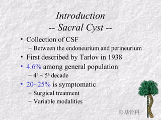
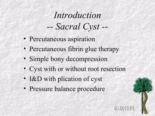
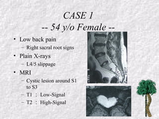
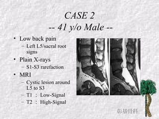
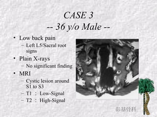
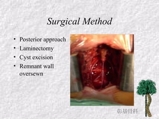
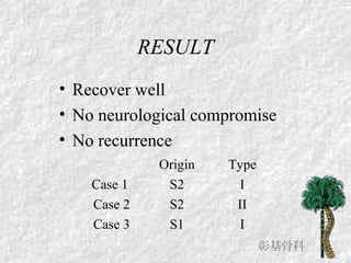
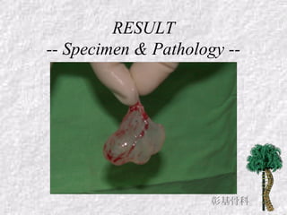
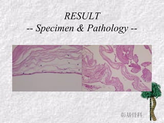
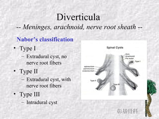
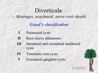
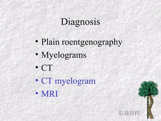
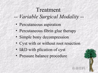
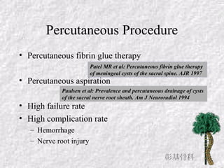
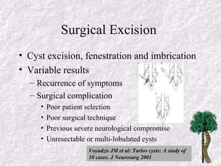
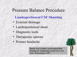
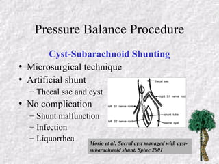
Surgical Treatment of
Symptomatic Sacral Cyst :
Three Cases Report and Literature
Review
JUI-KUO HUNG 洪瑞國 MD&MHA
Department of Orthopaedic Surgery
Changhua Christian Hospital
Introduction
-- Sacral Cyst --
• Collection of CSF
– Between the endoneurium and perineurium
• First described by Tarlov in 1938
• 4.6% among general population
– 4th
~ 5th
decade
• 20~25% is symptomatic
– Surgical treatment
– Variable modalities
彰基骨科
Introduction
-- Sacral Cyst --
Introduction
-- Sacral Cyst --
• Percutaneous aspiration
• Percutaneous fibrin glue therapy
• Simple bony decompression
• Cyst with or without root resection
• I&D with plication of cyst
• Pressure balance procedure
彰基骨科
CASE REPORT
CASE 1
-- 54 y/o Female --
• Low back pain
– Right sacral root signs
• Plain X-rays
– L4/5 slippage
• MRI
– Cystic lesion around S1
to S3
– T1 : Low-Signal
– T2 : High-Signal
CASE 2
-- 41 y/o Male --
• Low back pain
– Left L5/sacral root
signs
• Plain X-rays
– S1-S3 rarefaction
• MRI
– Cystic lesion around
L5 to S3
– T1 : Low-Signal
– T2 : High-Signal
•
彰基骨科
CASE 3
-- 36 y/o Male --
• Low back pain
– Left L5/Sacral root
signs
• Plain X-rays
– No significant finding
• MRI
– Cystic lesion around
S1 to S3
– T1 : Low-Signal
– T2 : High-Signal
彰基骨科
Surgical Method
• Posterior approach
• Laminectomy
• Cyst excision
• Remnant wall
oversewn
彰基骨科
RESULT
• Recover well
• No neurological compromise
• No recurrence
Origin Type
Case 1 S2 I
Case 2 S2 II
Case 3 S1 I
彰基骨科
RESULT
-- Specimen & Pathology --
彰基骨科
RESULT
-- Specimen & Pathology --
彰基骨科
DISCUSSION
Sacral Cyst
• First described by Tarlov in 1938
• 4.6% prevalence in general population
– 4th
to 5th
decade
– ♀ >♂
– 20% is symptomatic
• Compression S/S
• Low back pain, sensory impairment
• Bladder dysfunction, motor weakness, or
impotence
彰基骨科
Diverticula
-- Meninges, arachnoid, nerve root sheath --
Nabor’s classification
• Type I
– Extradural cyst, no
nerve root fibers
• Type II
– Extradural cyst, with
nerve root fibers
• Type III
– Intradural cyst
彰基骨科
Diverticula
-- Meninges, arachnoid, nerve root sheath
Goyal’s classification
I Perineural cysts
II Root sleeve dilatations
III Intradural and extradural arachnoid
cysts
IV Traumatic root cysts
V Extradural ganglion cysts
彰基骨科
Diagnosis
• Plain roentgenography
• Myelograms
• CT
• CT myelogram
• MRI
彰基骨科
Treatment
-- Variable Surgical Modality --
• Percutaneous aspiration
• Percutaneous fibrin glue therapy
• Simple bony decompression
• Cyst with or without root resection
• I&D with plication of cyst
• Pressure balance procedure
彰基骨科
Percutaneous Procedure
• Percutaneous fibrin glue therapy
• Percutaneous aspiration
• High failure rate
• High complication rate
– Hemorrhage
– Nerve root injury
Patel MR et al: Percutaneous fibrin glue therapy
of meningeal cysts of the sacral spine. AJR 1997
Paulsen et al: Prevalence and percutaneous drainage of cysts
of the sacral nerve root sheath. Am J Neuroradiol 1994
彰基骨科
Surgical Excision
• Cyst excision, fenestration and imbrication
• Variable results
– Recurrence of symptoms
– Surgical complication
• Poor patient selection
• Poor surgical technique
• Previous severe neurological compromise
• Unresectable or multi-lobulated cysts
Voyadzis JM et al: Tarlov cysts: A study of
10 cases. J Neurosurg 2001
Pressure Balance Procedure
Lumboperitoneal CSF Shunting
• External drainage
• Lumboperitoneal shunt
• Diagnostic tools
• Therapeutic options
• Posture headache
Bartels et al: Lumbar cerebrospinal fluid
drainage for symptomatic sacral nerve root
cysts. Neurosurgery, 1997
Pressure Balance Procedure
Cyst-Subarachnoid Shunting
• Microsurgical technique
• Artificial shunt
– Thecal sac and cyst
• No complication
– Shunt malfunction
– Infection
– Liquorrhea Morio et al: Sacral cyst managed with cyst-
subarachnoid shunt. Spine 2001
SUMMARY
Sacral cysts
• Uncommon but not rare lesions
• Variable symptoms and signs
• Differential diagnosis
– Sacral radiculopathy
– Sacral pain syndrome
• Diagnostic tools
– CT myelogram
– MRI
彰基骨科
Surgical Intervention
• Percutaneous procedure
– No longer used
• Surgical excision and cyst plication
– Well-encapsulated, easily dissected lesion
– Better to preserve nerve roots
• Pressure balance procedure
– Large, multi-lobulated cysts
– Complete excision is impossible
– Nerve root will be sacrified
彰基骨科
Surgical Treatment of Symptomatic Sacral Cysts

Surgical Treatment of Symptomatic Sacral Cysts

  • 1.
    Surgical Treatment of SymptomaticSacral Cyst : Three Cases Report and Literature Review JUI-KUO HUNG 洪瑞國 MD&MHA Department of Orthopaedic Surgery Changhua Christian Hospital
  • 2.
    Introduction -- Sacral Cyst-- • Collection of CSF – Between the endoneurium and perineurium • First described by Tarlov in 1938 • 4.6% among general population – 4th ~ 5th decade • 20~25% is symptomatic – Surgical treatment – Variable modalities 彰基骨科
  • 3.
  • 4.
    Introduction -- Sacral Cyst-- • Percutaneous aspiration • Percutaneous fibrin glue therapy • Simple bony decompression • Cyst with or without root resection • I&D with plication of cyst • Pressure balance procedure 彰基骨科
  • 5.
  • 6.
    CASE 1 -- 54y/o Female -- • Low back pain – Right sacral root signs • Plain X-rays – L4/5 slippage • MRI – Cystic lesion around S1 to S3 – T1 : Low-Signal – T2 : High-Signal
  • 7.
    CASE 2 -- 41y/o Male -- • Low back pain – Left L5/sacral root signs • Plain X-rays – S1-S3 rarefaction • MRI – Cystic lesion around L5 to S3 – T1 : Low-Signal – T2 : High-Signal • 彰基骨科
  • 8.
    CASE 3 -- 36y/o Male -- • Low back pain – Left L5/Sacral root signs • Plain X-rays – No significant finding • MRI – Cystic lesion around S1 to S3 – T1 : Low-Signal – T2 : High-Signal 彰基骨科
  • 9.
    Surgical Method • Posteriorapproach • Laminectomy • Cyst excision • Remnant wall oversewn 彰基骨科
  • 10.
    RESULT • Recover well •No neurological compromise • No recurrence Origin Type Case 1 S2 I Case 2 S2 II Case 3 S1 I 彰基骨科
  • 11.
    RESULT -- Specimen &Pathology -- 彰基骨科
  • 12.
    RESULT -- Specimen &Pathology -- 彰基骨科
  • 13.
  • 14.
    Sacral Cyst • Firstdescribed by Tarlov in 1938 • 4.6% prevalence in general population – 4th to 5th decade – ♀ >♂ – 20% is symptomatic • Compression S/S • Low back pain, sensory impairment • Bladder dysfunction, motor weakness, or impotence 彰基骨科
  • 15.
    Diverticula -- Meninges, arachnoid,nerve root sheath -- Nabor’s classification • Type I – Extradural cyst, no nerve root fibers • Type II – Extradural cyst, with nerve root fibers • Type III – Intradural cyst 彰基骨科
  • 16.
    Diverticula -- Meninges, arachnoid,nerve root sheath Goyal’s classification I Perineural cysts II Root sleeve dilatations III Intradural and extradural arachnoid cysts IV Traumatic root cysts V Extradural ganglion cysts 彰基骨科
  • 17.
    Diagnosis • Plain roentgenography •Myelograms • CT • CT myelogram • MRI 彰基骨科
  • 18.
    Treatment -- Variable SurgicalModality -- • Percutaneous aspiration • Percutaneous fibrin glue therapy • Simple bony decompression • Cyst with or without root resection • I&D with plication of cyst • Pressure balance procedure 彰基骨科
  • 19.
    Percutaneous Procedure • Percutaneousfibrin glue therapy • Percutaneous aspiration • High failure rate • High complication rate – Hemorrhage – Nerve root injury Patel MR et al: Percutaneous fibrin glue therapy of meningeal cysts of the sacral spine. AJR 1997 Paulsen et al: Prevalence and percutaneous drainage of cysts of the sacral nerve root sheath. Am J Neuroradiol 1994 彰基骨科
  • 20.
    Surgical Excision • Cystexcision, fenestration and imbrication • Variable results – Recurrence of symptoms – Surgical complication • Poor patient selection • Poor surgical technique • Previous severe neurological compromise • Unresectable or multi-lobulated cysts Voyadzis JM et al: Tarlov cysts: A study of 10 cases. J Neurosurg 2001
  • 21.
    Pressure Balance Procedure LumboperitonealCSF Shunting • External drainage • Lumboperitoneal shunt • Diagnostic tools • Therapeutic options • Posture headache Bartels et al: Lumbar cerebrospinal fluid drainage for symptomatic sacral nerve root cysts. Neurosurgery, 1997
  • 22.
    Pressure Balance Procedure Cyst-SubarachnoidShunting • Microsurgical technique • Artificial shunt – Thecal sac and cyst • No complication – Shunt malfunction – Infection – Liquorrhea Morio et al: Sacral cyst managed with cyst- subarachnoid shunt. Spine 2001
  • 23.
  • 24.
    Sacral cysts • Uncommonbut not rare lesions • Variable symptoms and signs • Differential diagnosis – Sacral radiculopathy – Sacral pain syndrome • Diagnostic tools – CT myelogram – MRI 彰基骨科
  • 25.
    Surgical Intervention • Percutaneousprocedure – No longer used • Surgical excision and cyst plication – Well-encapsulated, easily dissected lesion – Better to preserve nerve roots • Pressure balance procedure – Large, multi-lobulated cysts – Complete excision is impossible – Nerve root will be sacrified 彰基骨科